


1. Background
Acute respiratory infections (ARIs) are complex infections that affect all anatomical sites of the respiratory tract. As a major global public health concern, they are among the main reasons for outpatient and inpatient care in children (1). To date, acute lower respiratory tract infection (ALRI) remains the leading cause of morbidity and mortality in children younger than 5 years worldwide (2). Various pathogens can cause ARIs; viruses are believed to account for 32.3% - 70% of cases (3, 4) and may be associated with up to 80% of upper respiratory tract infections (3). Influenza A and B viruses, rhinoviruses, coronaviruses, respiratory syncytial viruses (RSVs), parainfluenza viruses, adenoviruses, enteroviruses, human metapneumovirus (HMPV), and human bocavirus (HBoV) are among the most common viruses infecting the human respiratory tract. By the end of the 20th century, SARS-CoV-2 had taken its place among other important respiratory viruses, further expanding this list (4).
Several studies have demonstrated substantial changes in the pattern of respiratory infections throughout the COVID-19 pandemic. Following nonpharmaceutical interventions (NPIs) implemented by governments during the COVID-19 pandemic, changes in health behaviors, viral interference, and other factors (5), a significant reduction in the burden of non-SARS-CoV-2 respiratory infections, including influenza virus and RSV, and some bacterial infections, such as Streptococcus pneumoniae, was reported (5). The extent of this tendency varied during the SARS-CoV-2 pandemic depending on the viral strain. For adenovirus, only minimal changes in prevalence were observed during the COVID-19 pandemic compared with the pre-COVID-19 era. For rhinovirus and bocavirus, an increasing trend was reported during the COVID-19 pandemic (6, 7).
The viral coinfection rate in patients with COVID-19 has been reported to be 3%, which is lower than the bacterial coinfection rate of 7% (8). The most common symptoms of COVID-19 are fever, cough, shortness of breath, fatigue, muscle ache, and diarrhea, which largely align with the definition of influenza-like illness (ILI) (9). Notably, limited data are available on the impact of SARS-CoV-2 coinfection on disease severity or clinical outcomes, and multiple viral infections have not been reported to predict worse disease outcomes (10, 11).
In Iran, only a limited number of epidemiological studies have evaluated the main ARI-related viruses in hospitalized children during the COVID-19 period, particularly during the post-COVID-19 period, largely because of diagnostic constraints. Across these studies, the observed prevalence of viral infections in this population has aligned with global patterns (12, 13).
Recent studies have reported new patterns of respiratory infections in neighboring countries and worldwide (14). Clinical evidence also indicates changes in the prevalence of respiratory infections in our country.
2. Objectives
Therefore, we investigated trends in viral respiratory infections during autumn and winter 2022, a period that coincided with the end of the COVID-19 pandemic. In addition to SARS-CoV-2 and rhinovirus, influenza and RSV, the main treatable viruses causing ARIs in children, were evaluated. Using a multiplex real-time polymerase chain reaction (PCR) assay in hospitalized pediatric patients, we assessed circulation trends of major respiratory viral infections, including SARS-CoV-2, and the clinical features of such coinfections.
This study aimed to address a key gap in the Iranian pediatric literature by highlighting the lack of post-COVID surveillance data in children. It also aimed to provide a multivirus perspective on post-pandemic dynamics in this population.
3. Methods
3.1. Study Design and Setting
This prospective epidemiological study evaluated the prevalence and characteristics of respiratory viruses among patients hospitalized with ARI from October 2022 to April 2023 in the pediatric wards of Namazi tertiary hospital, which is affiliated with Shiraz University of Medical Sciences. All children hospitalized in the pediatric wards whose guardians provided consent on their behalf and who were residents of Fars province were included.
3.2. Sample Collection and Viral Detection
Samples collected from individuals diagnosed with acute respiratory illness included nasopharyngeal aspirates, nose and throat swabs, sputum, and bronchoalveolar lavage specimens. Real-time PCR was used to detect respiratory viruses, including influenza A and B viruses, RSV, rhinovirus, and SARS-CoV-2, within 4 to 16 hours of sample receipt at the Professor Alborzi Clinical Microbiology Research Center virology laboratory.
A total of 200 μL of each clinical sample was extracted using the Sina Pure Viral kit (EX6061, Tehran, Iran) according to the manufacturer’s instructions. Extracted RNA was stored at -80°C until use.
TaqMan real-time PCR (RT-qPCR) was performed for each patient using the Genova commercial kit (GA-SARSFLURSV.100 kit, GenovA Co., Tehran, Iran) for the simultaneous detection of SARS-CoV-2, influenza A/B, and RSV.
Rhinovirus was detected by TaqMan RT-qPCR using the following primers and probe: forward primer, CY(+A)GCC(+t)GCGTGGY; reverse primer, GAAACACGGACACCCAAAGTA; and probe, 6-fam-TCCTCCCGCCCCTGAATGYGGC-BHQ1. Each 25-μL reaction contained 12.5 μL of 2× reaction mix (PCR Biosystems Ltd), 0.25 μL of 100 μM forward primer, 0.25 μL of 100 μM reverse primer, 0.25 μL of 10 μM probe, 0.5 μL of reverse transcriptase, 6.25 μL of nuclease-free water, and 5 μL of nucleic acid extract.
3.3. Variables and Eligibility
Demographic data, symptom evolution, and clinical features of hospitalized children diagnosed with ARI were extracted from the hospital information system (HIS). Age, sex, and clinical signs and symptoms were the main variables analyzed after diagnosis on admission.
Duplicate samples from the same patient collected within one week of another sample were identified and excluded if the results were the same. Samples with missing information, such as an unknown date or place of birth, and samples not tested for the relevant panels were also excluded.
Patient data were categorized into five age groups: 1 day to 12 months, 1 - 3 years (toddlers), 3 - 5 years (preschool), 5 - 12 years (school-age children), and 12 - 18 years (adolescents). Sex distribution was also considered. The research protocol was reviewed and approved by the Ethics Committee of Shiraz University of Medical Sciences (approval ID: IR.SUMS.REC.1401.694).
3.4. Statistical Analysis
Data were analyzed using SPSS version 25.0. Categorical data were presented as frequency and percentage, and continuous data were described as the mean and standard deviation. The chi-square and Fisher exact tests were used to determine statistical differences in qualitative variables. The significance level was set at 0.05.
4. Results
Out of more than 3,827 specimens evaluated between 11 October 2022 and 9 April 2023, 1,530 met the study inclusion criteria. The mean age of the participants was 5.15 ± 4.67 years, and the female/male ratio was 45/55. Of the 1,530 samples, 974 (63.7%) were free of respiratory viruses, 503 (32.9%) were positive for one viral agent, and 53 (3.4%) were positive for more than one virus.
The numbers of positive samples for influenza, RSV, SARS-CoV-2, and rhinovirus were 190, 178, 165, and 80, respectively. Although the prevalence of influenza and rhinovirus infections was lower in girls than in boys, the rates of different viral infections did not differ significantly between the sexes. Real-time PCR analysis of weekly samples revealed a peak in week 42 of 2022, followed by week 7 of 2023. The average positivity rate among samples was 36.3%, ranging from 16.7% in week 8, beginning on 21 February 2023, to 76.7% in week 42, beginning on 18 October.
4.1. Temporal Distribution of Respiratory Viruses
The prevalence of influenza, RSV, and COVID-19 each reached a peak during three distinct periods, whereas rhinovirus did not show a clear peak and exhibited a steady distribution throughout the study period. The study began on 11 October 2022, which coincided with the peak of influenza cases, followed by a decrease in early December. No influenza cases were detected by mid-January (Table 1).
Table 1.The Proportion of Positive Specimens in Each Age Bracket of Iranian Pediatric Patients Who Tested Positive for a Virus from Samples Retrieved Between October 11, 2022, and April 9, 2023 a
| Pediatric Patient Age Group | No. (%) | Negative | Positive for One Virus | Positive for More Than One Virus | COVID-19 | Influenza | RSV | Rhinovirus |
|---|---|---|---|---|---|---|---|---|
| Infants: 0 - 12 mo | 456 (29.5) | 282 (62) | 156 (34) | 18 (4) | 24 | 19 | 47 | 10 |
| Toddlers: 1 - 3 y | 265 (17.1) | 179 (67) | 76 (29) | 10 (4) | 24 | 28 | 33 | 16 |
| Preschool: 3 - 5 y | 187 (12.1) | 121 (65) | 63 (33) | 3 (2) | 27 | 40 | 17 | 16 |
| School-age: 5 - 12 y | 469 (30.3) | 303 (64) | 153 (33) | 13 (3) | 30 | 39 | 20 | 11 |
| Adolescents: 12 - 18 y | 153 (10) | 89 (58) | 55 (36) | 9 (6) | 33 | 45 | 9 | 13 |
| Total | 1530 (100) | 974 (64) | 503 (33) | 53 (3) | 27 | 32 | 29 | 12 |
a Values are expressed as No. (%) or percent.
The prevalence of RSV began to increase in early December, when a significant decline in influenza prevalence was observed. RSV prevalence peaked in the last week of December and then declined until early February, when RSV cases were rarely detected (Figure 1).
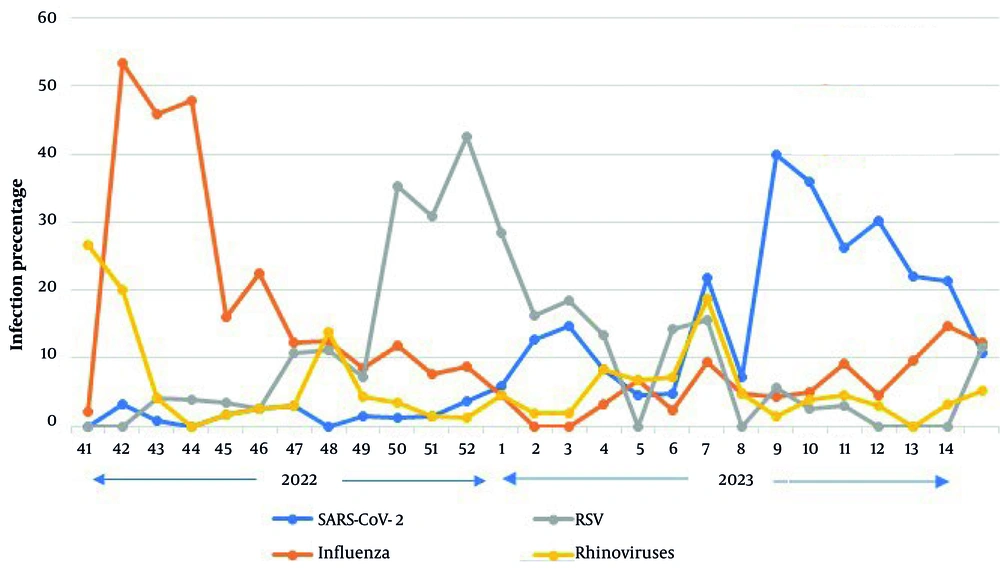
Figure 1.
The prevalence of respiratory specimens received from October 11, 2022, through April 9, 2023, as indicated by week of receipt.
SARS-CoV-2 prevalence increased from early January, with some variation in late February, and eventually peaked in the ninth week of early March. The downward trend continued until 9 April, the end of the study period (Figure 1).
4.2. Age Distribution of Respiratory Viral Infection
The highest numbers of specimens were received from the < 1-year and 5 - 12-year age groups. No statistically significant differences in positivity rates were observed among age groups (P ≥ 0.05). As shown in Table 1 and Figure 2, the 12 - 18-year age group had the fewest collected samples.
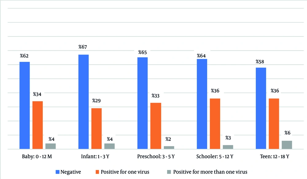
Figure 2.
Prevalence of respiratory infections by age group in Iranian pediatric samples collected between October 11, 2022, and April 9, 2023.
RSV was the most common virus detected in children younger than 3 years (98/145), whereas influenza was the most common virus in children aged 3 - 18 years (110/160). In contrast to influenza and RSV, COVID-19 and rhinovirus were equally prevalent across different age groups (Table 1 and Figure 3).
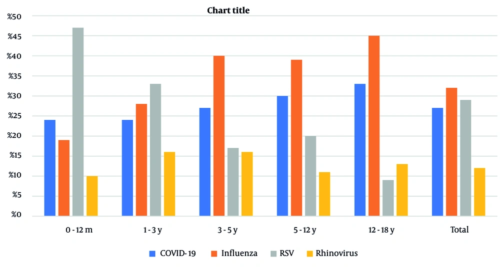
Figure 3.
Prevalence of diverse viral infections across age-stratified cohorts of Iranian infected patients from October 11, 2022, to April 9, 2023.
4.3. Monoinfection and Multiple Respiratory Viral Infections
A total of 503 samples (90.5%) were positive for a single respiratory virus, whereas 53 (9.5%) had multiple infections; among these, 89% involved two viruses and 11% involved three viruses. Rhinovirus was the predominant virus detected in coinfections with other viruses, at 23.7%, followed by RSV at 18.5%. Influenza had the lowest codetection rate with other viruses, at 15.8% (Table 2).
Table 2.The Frequency of Single and/or Coinfected Respiratory Viral Infections in Samples Received from October 11, 2022, to April 9, 2023, Among the Study Participants a
| Virus | Total | As Monoinfected Virus | As coinfected Virus |
|---|---|---|---|
| COVID-19 | 165 (11) | 137 (83) | 28 (17) |
| Influenza | 190 (12) | 160 (84) | 30 (16) |
| RSV | 178 (12) | 145 (82) | 33 (18) |
| Rhinovirus | 80 (5) | 61 (76) | 19 (24) |
a Values are expressed as No. (%).
4.4. Clinical Description of Patients with Respiratory Viral Infections
From a clinical perspective, data analysis revealed statistically significant variation in the rates of cough, sore throat, rhinorrhea, fatigue, and body pain among patients infected with different viruses. As shown in Table 3, cough was significantly more frequent in RSV-infected children than in children with other viral infections (P ≤ 0.001). Sore throat, rhinorrhea, and fatigue were most prevalent in pediatric patients with influenza. Body pain was notably higher among children affected by COVID-19 (P ≤ 0.05). Table 3 also shows that intensive care unit (ICU) admission was more frequent among hospitalized children with SARS-CoV-2 infection.
Table 3.Comparison of Symptoms in Acute Respiratory Infections by Viral Cause Among Hospitalized Children in Shiraz, Iran a
| Clinical presentation | SARS-CoV-2, 137 | Influenza, 161 | RSV, 146 | Rhinovirus, 63 | Dual infection, 49 | Triple infection, 3 | Negative for respiratory viruses, 971 | P-Value |
|---|---|---|---|---|---|---|---|---|
| Fever | 73 (53) | 100 (62) | 72 (50) | 34 (55) | 32 (65) | 1 (33) | 577 (59) | 0.204 b |
| Cough | 63 (46) | 108 (67) | 109 (75) | 44 (71) | 27 (55) | 2 (67) | 606 (62) | 0.001 b |
| Sore throat | 1 (1) | 17 (11) | 9 (6) | 2 (3) | 3 (6) | 0 (0) | 70 (7) | 0.034 c |
| Rhinorrhea | 5 (4) | 30 (19) | 22 (15) | 10 (16) | 6 (12) | 0 (0) | 107 (11) | 0.003 b |
| Headache | 0 (0) | 3 (2) | 1 (1) | 2 (3) | 2 (4) | 0 (0) | 25 (3) | 0.367 c |
| Dyspnea | 30 (22) | 55 (34) | 52 (36) | 23 (37) | 14 (28) | 1 (33) | 283 (29) | 0.110 b |
| Vomiting | 6 (4) | 9 (6) | 8 (6) | 0 (0) | 0 (0) | 0 (0) | 71 (7.) | 0.111 c |
| Diarrhea | 4 (3) | 10 (6.) | 5 (3) | 1 (2) | 1 (2) | 0 (0) | 66 (7) | 0.198 b |
| Arthralgia | 0 (0) | 2 (1) | 0 (0) | 0 (0) | 0 (0) | 0 (0) | 12 (1) | 0.954 c |
| General body weakness | 5 (4) | 11 (7) | 8 (6) | 3 (5) | 1 (2) | 1 (33) | 53 (6) | 0.283 c |
| Fatigue | 0 (0) | 14 (9) | 7 (5) | 3 (5) | 1 (2) | 0 (0) | 44 (5) | 0.029 c |
| Chest pain | 0 (0) | 0 (1) | 0 (0) | 1 (2) | 0 (0) | 0 (0) | 12 (1) | 0.397 c |
| Mild pneumonia | 0 (0) | 1 (1) | 0 (0) | 1 (2) | 0 (0) | 0 (0) | 11 (1) | 0.643 c |
| Severe pneumonia | 0 (0) | 1 (1) | 0 (0) | 0 (0) | 0 (0) | 0 (0) | 1 (0) | 0.890 c |
| Stomach ache | 1 (1) | 7 (4) | 0 (0) | 0 (0) | 1 (6) | 0 (0) | 7 (1) | 0.002 c |
| Body pain | 16 (12) | 18 (11) | 4 (3) | 2 (3) | 4 (8) | 0 (0) | 47 (5) | 0.001 c |
| Respiratory distress syndrome | 3 (2) | 2 (1) | 1 (1) | 0 (0) | 0 (0) | 0 (0) | 11 (1) | 0.804 c |
| ICU admission | 12 (9) | 4 (3) | 4 (3) | 0 (0) | 3 (6) | 0 (0) | 41 (4) | 0.021 c |
a Values are expressed as No. (%).
b Chi-square test.
c Fisher exact test.
5. Discussion
Among specimens received from pediatric inpatients with ARI, 36% showed the presence of at least one virus. ARIs are among the most common diagnoses in hospitalized children. Statistically, no variation was observed between males and females in either specimen count or positivity rate. Studies of hospitalized patients have reported respiratory viral infection rates of 36% - 85% before the onset of the COVID-19 pandemic (3, 14, 15). This rate shifted markedly during the COVID-19 pandemic. In general, many studies have shown a significant decrease in the prevalence of enveloped respiratory viruses during the pandemic, but not of nonenveloped viruses (12, 16-20). By mid-2020, most countries had introduced high-level infection control measures and experienced a dramatic year-on-year decline in influenza (21). Many temperate countries in both hemispheres exhibited pronounced reductions in seasonal influenza activity during the pandemic (22-25). These declines cannot be explained by viral interference due to SARS-CoV-2 spread in serologically nonendemic areas (24).
RSV cases declined markedly across geographic regions and climatic zones during 2020 - 2021, with notable reductions in Australia, South Africa, New Zealand, France, the United States, and Japan during the first two winters after COVID-19 emerged (18). In Iran, RSV prevalence dropped from 19% in the pre-pandemic era to undetectable levels during the pandemic (12, 26). Non-SARS-CoV-2 respiratory viruses exhibited multifactorial changes: widespread and sustained NPIs, shifts in health behaviors, and reduced international travel likely contributed to overall decreases in circulation. Nonenveloped viruses tended to persist more than enveloped viruses, which showed greater reductions.
In the United States, rhinoviruses remained the leading cause of ARIs, accounting for roughly 75% of viral diagnoses in both inpatient and outpatient pediatric settings during the pre-pandemic and pandemic periods (21).
During the winter of 2022 - 2023, influenza and RSV infections increased in different countries in the northern hemisphere, with varying incidence rates, hospitalization rates, and epidemic patterns, while COVID-19 remained a threat, according to the World Health Organization.
Research on respiratory illness trends after the pandemic remains sparse. Our study demonstrated a 2-year decline followed by a rebound in influenza and RSV among hospitalized children in Iran during the 2022 - 2023 autumn and winter seasons. Rhinoviruses, among the most frequent nonenveloped respiratory viruses, showed low and stable prevalence. Consistent with our study, Kandeel et al. in Egypt reported a rebound of influenza and RSV in the post-COVID-19 era (22), and Rankin et al. found that rhinovirus infection prevalence did not differ in the presence or absence of COVID-19 (20).
Analysis of viral respiratory infection trends indicated that Iran experienced earlier influenza surges than before the pandemic, as the World Health Organization had announced the risk of early influenza epidemics in Europe and the United States.
One notable feature of the data from the second half of the 2022 - 2023 season was that each respiratory virus exhibited distinct peaks at specific intervals against a background of rhinovirus circulation. This finding contrasts with studies by Tanner and Núñez-Samudio, which showed overlapping patterns of respiratory infections (3, 23).
Notably, in the first week of our study, which coincided with the beginning of autumn, a sudden decline in rhinovirus was accompanied by a sudden increase and peak in influenza viruses. This pattern may be explained by the viral interference phenomenon documented in various studies during the fall of the 2009 influenza pandemic (24-28).
Analysis of respiratory virus data from the second half of 2022 - 2023 by patient age showed that children younger than 3 years represented a different epidemiological group than older children. Although overall specimen positivity rates generally did not vary by age group, viral patterns differed among age groups. As in other studies, influenza infection rates were higher among older children, predominantly school-age children, than among younger children. This may be due to differences in social behavior, school attendance, peer contact, and sports activities (29-31). Biological, environmental, and sociological differences may also explain the varying levels of influenza infectivity among children of different ages (31).
Madaniyazi et al. in Japan (32) and Baker et al. in the United States (33) predicted a high prevalence of RSV, partly because of an increased number of individuals susceptible to the virus, according to their simulation and epidemiological models, respectively. In Iran, following the reduction of nonpharmaceutical and public health interventions and waning RSV antibodies, RSV prevalence increased to more than 42% in January.
With respiratory viruses surging nationwide among children in autumn 2022, influenza cases rising, and RSV and COVID-19 simmering in the background, some medical experts were concerned about a possible “dual- and/or tripledemic.” Fortunately, this did not occur, and coinfections or triple infections were observed in only 3.4% of cases. This low rate may be due to the limited number of ARI-causing viruses tested. Our result is consistent with that of Kandeel et al. (22), who reported a 2.8% coinfection rate, but contrasts with Tanner et al. (23), who reported a coinfection rate greater than 13%. In the latter study, the high coinfection rate was due to the large number of respiratory viruses included.
Rhinovirus is widespread and present throughout the year, with peak seasons in spring and autumn. Its consistently high prevalence increases the likelihood of simultaneous detection with other respiratory pathogens through statistical overlap; in this study, rhinovirus was also identified as the primary accompanying infection.
In our study, coinfection was observed across all age groups, whereas in other studies it was observed more prominently in infants and young children (3, 22).
In the present study, rhinovirus was the most frequently codetected virus, followed by RSV, whereas influenza was the least frequently codetected virus, consistent with the study by Tanner et al. (23). An important finding of this study is that the symptoms of respiratory viral infections were almost identical among patients infected with these viruses. Given the substantial overlap in the symptoms of respiratory viral infections, clinical suspicion alone is insufficient to identify patients as positive for influenza, COVID-19, RSV, or rhinovirus.
To accurately determine the presence of these viruses, screening patients with acute respiratory infections in hospitals, especially children, is necessary. In addition, acute respiratory agents should be identified to avoid inadequate treatment and antibiotic regimens, which may contribute to poor patient outcomes.
Overall, because ARI surveillance in Iran has been successfully adapted to the COVID-19 pandemic and has effectively characterized the clinical features and severity of circulating viruses, rigorous implementation of comprehensive health programs, such as IPPP for influenza, and monitoring of other respiratory viral activity to guide clinical management, including preventive and control measures, are necessary and recommended.
Multi-year follow-up after a pandemic is essential to detect delayed or evolving patterns in respiratory pathogens, including potential biennial cycles, waning immunity, and shifts in age susceptibility.
5.1. Limitations
Several limitations of this study should be noted. First, this research was conducted at a tertiary care hospital. Although many children with ARI visited this treatment center, the results cannot be generalized to all pediatric patients in our region, and patients from a single tertiary care center may not reflect the broader regional or national pediatric population. The second limitation was related to financial resources. If a broader range of respiratory viruses, such as human bocavirus, OC43, HKU-1, parainfluenza, and adenovirus, or bacterial infections, such as Mycoplasma and Chlamydia, had been examined, the trends of these infections would have been more clearly defined. Finally, the study was conducted in hospitalized pediatric patients and did not include outpatients, although outpatients are less affected by severe forms of ARI.
Broader, multicenter surveillance is recommended to improve generalizability across settings. Public health systems should adopt integrated surveillance to prepare for future viral resurges, including sentinel hospital networks for real-time monitoring and genomic sequencing to track viral evolution.
