
1. Introduction
Dengue is a self-limiting, systemic arboviral infection caused by one of four single-stranded RNA viruses (dengue virus types 1 - 4) of the genus Flavivirus (1). This mosquito-borne disease, transmitted by Aedes aegypti and Aedes albopictus, is endemic in more than 100 tropical and subtropical countries worldwide, and its transmission is strongly influenced by climatic factors such as rainfall, temperature, and urbanization (2, 3). Most patients present with fever, nonspecific constitutional symptoms, and signs of plasma leakage.
There is currently no specific antiviral therapy for dengue. The World Health Organization (WHO) emphasizes the development of a tetravalent dengue vaccine as a key preventive measure. Most cases of dengue fever are self-limiting and require only supportive care, including fluid replacement, pain relief, and rest. Fever can be managed with acetaminophen; however, other medications, such as methylprednisolone, are not recommended because of potential side effects and a lack of proven mortality benefit. In severe dengue, careful supportive management is essential to prevent complications and death (4).
Although most patients recover with supportive care, some develop atypical or severe manifestations that require heightened clinical awareness. Fever remains the hallmark of dengue’s broad clinical spectrum; however, progression to dengue hemorrhagic fever (DHF) requires prompt management to prevent fatal complications. With increasing clinical awareness, previously underrecognized atypical presentations, including neurological, gastrointestinal, respiratory, cardiac, renal, and ocular involvement, are now considered part of the expanded spectrum of dengue and are collectively termed expanded dengue syndrome.
International and regional guidelines are beginning to incorporate these expanded clinical presentations; however, standardized management protocols for complications remain limited (5). The atypical and rare manifestations of expanded dengue syndrome are diverse and affect multiple organs, including the liver, brain, heart, kidneys, and lungs (6). Dengue fever can affect the respiratory system, leading to complications such as pleural effusion (bilateral or unilateral), acute respiratory distress syndrome (ARDS), non-cardiogenic pulmonary edema, and pulmonary hemorrhage, all of which are related to increased vascular permeability and plasma leakage (7). Shortness of breath and cough are more commonly observed among older adults (8).
Dengue fever may rarely lead to permanent visual loss, although most ocular manifestations resolve with appropriate treatment. Severe ocular complications, such as macular edema, optic neuritis, and retinal vasculitis, can cause blurred vision or central vision loss. Delayed treatment may result in residual visual deficits (9). Cardiac complications of dengue include myocarditis, arrhythmias, pericarditis, and electrocardiographic (ECG) abnormalities, such as ST–T changes and other nonspecific findings suggestive of cardiac involvement (10). Uncommon manifestations of dengue include expanded dengue syndrome; neurological involvement such as encephalopathy, encephalitis, Guillain–Barré syndrome, and stroke; spontaneous bacterial peritonitis (SBP); and oral manifestations such as oral candidiasis and transient lingual papillitis (11, 12).
Dengue virus infection can be diagnosed using viral culture, serological assays for dengue-specific antibodies, nonstructural protein 1 (NS1) antigen detection, and polymerase chain reaction (PCR)-based viral RNA detection. NS1 assays improve early diagnosis and are available as rapid point-of-care tests, whereas PCR provides higher sensitivity but requires specialized laboratory resources. Diagnostic accuracy depends on the timing of specimen collection and the selection of appropriate tests, whereas newer molecular approaches, such as metagenomic sequencing, may support epidemiological surveillance (13).
In this systematic review, we summarize and analyze less recognized complications of dengue virus infection, including gastrointestinal, cardiovascular, ocular, and pulmonary manifestations, to highlight these presentations and support timely and appropriate clinical management.
2. Methods
Reports included in this review comprised case reports and case series. Only reports describing at least one of the four aforementioned clinical manifestations in patients with dengue virus infection were included in this systematic review.
2.1. Search Strategy and Study Selection
A systematic literature search was conducted in the electronic databases PubMed, EMBASE, and Web of Science for articles published from January 2000 to December 2024. The search strategy was designed to identify studies reporting atypical manifestations of dengue virus infection. The following Boolean query was used to ensure a comprehensive literature search by combining MeSH terms and Title/Abstract keywords: ("dengue virus" OR "dengue fever") AND ("expanded dengue syndrome"[Title/Abstract] OR "atypical manifestations"[Title/Abstract] OR "rare manifestations"[Title/Abstract] OR "Gastrointestinal Diseases"[Mesh] OR "Cardiovascular Diseases"[Mesh] OR "Eye Diseases"[Mesh] OR "Respiratory Tract Diseases"[Mesh]) (14) (Table 1 in the Supplementary File).
Table 1.
Summary of Gastrointestinal Manifestations of Dengue Virus Infection (N = 30) a
| Variables | Number | Percentage |
|---|---|---|
| Gender | ||
| Male | 19 | 63.3 |
| Female | 10 | 33.3 |
| NR | 1 | 3.4 |
| Outcome | ||
| Survived | 26 | 86.7 |
| Deceased | 1 | 3.3 |
| NR | 3 | 10.0 |
| Age | ||
| Mean age (years) | 33.8 | |
| Gastrointestinal involvements | ||
| Pancreatitis | 22 | 73.3 |
| Hepatitis | 1 | 3.3 |
| Vomiting | 15 | 50.0 |
| Nausea | 2 | 6.7 |
| Diarrhea | 6 | 20.0 |
| Hemoperitoneum | 1 | 3.3 |
| Ascites | 4 | 13.3 |
| Ileocecal intussusception | 1 | 3.3 |
| Gastrointestinal bleeding | 1 | 3.3 |
| Acute gastroenteritis | 1 | 3.3 |
| Acalculous cholecystitis | 1 | 3.3 |
| Underlying disease | ||
| Sickle cell anemia | 1 | 3.3 |
| Diabetes | 5 | 16.7 |
| Hemodialysis | 1 | 3.3 |
| Obesity | 1 | 3.3 |
| Alcoholism | 1 | 3.3 |
| ex-smoker | 1 | 3.3 |
| Diagnostic methods | ||
| Serology | 20 | 66.7 |
| NS1 | 11 | 36.7 |
| ELISA | 2 | 6.7 |
| RT-PCR | 1 | 3.3 |
| Clinical manifestations | ||
| Fever | 22 | 73.3 |
| Myalgia | 7 | 23.3 |
| Headache | 13 | 43.3 |
| Abdominal pain | 19 | 63.3 |
| Arthralgia | 4 | 13.3 |
| Seizure | 1 | 3.3 |
| Hematemesis | 1 | 3.3 |
| Rectal bleeding | 1 | 3.3 |
| Metrorrhagia | 1 | 3.3 |
| Fatigue | 2 | 6.7 |
| Pleural effusion | 1 | 3.3 |
| Laboratory findings | ||
| Plt: L | 24 | 80.0 |
| Amylase: H | 18 | 60.0 |
| Lipase: H | 14 | 46.7 |
| AST: H | 10 | 33.3 |
| ALT: H | 10 | 33.3 |
| ALP: H | 2 | 6.7 |
| GGT: H | 1 | 3.3 |
| TG: H | 1 | 3.3 |
| LDH: H | 2 | 6.7 |
| Urea: H | 1 | 3.3 |
| Ca: L | 1 | 3.3 |
| HG: L | 1 | 3.3 |
| Treatment | ||
| Intravenous fluid therapy | 3 | 10.0 |
| Proton pump inhibitor | 1 | 3.3 |
| Opioid analgesic | 1 | 3.3 |
| Parenteral nutrition | 1 | 3.3 |
a Abbreviations: NR, not reported; Plt, Platelet count; L, Low; H, High; AST, Aspartate aminotransferase; ALT, Alanine aminotransferase; ALP, Alkaline phosphatase; GGT, Gamma-glutamyl transferase; TG, Triglycerides; LDH, Lactate dehydrogenase; Ca, Calcium; HG, Hemoglobin; NS1, Nonstructural protein 1 antigen; ELISA, Enzyme-linked immunosorbent assay; RT-PCR, Reverse transcription polymerase chain reaction.
The search strategy was intentionally designed to capture all potentially relevant observational evidence related to atypical dengue manifestations, including prevalence studies and retrospective observational reports. However, during screening, only case reports and case series were retained as eligible studies to ensure methodological consistency and comparability across all manifestation domains.
The inclusion criteria were as follows: (1) case reports and case series; (2) patients with confirmed dengue infection; (3) documentation of atypical organ involvement (e.g., gastrointestinal, cardiac, ocular, or pulmonary); and (4) studies published in English. A confirmed case of dengue was defined according to WHO criteria and included patients presenting with compatible clinical features, such as acute febrile illness, headache, myalgia, rash, or bleeding tendency, together with laboratory confirmation by at least one of the following: detection of dengue-specific IgM antibodies, a positive NS1 antigen test, or reverse transcription–polymerase chain reaction (RT-PCR) for dengue virus.
Studies were excluded if they were animal studies, review articles, duplicates, or focused solely on common manifestations, such as fever or thrombocytopenia, without describing atypical organ involvement.
2.2. Data Quality Assessment
Two reviewers (MA and KK) independently screened titles and abstracts, assessed full-text eligibility, performed data extraction, and conducted quality assessment in accordance with the Preferred Reporting Items for Systematic Reviews and Meta-Analyses (PRISMA) guidelines (15). Disagreements were resolved through discussion, and unresolved discrepancies were adjudicated by a third reviewer (FS). The quality of the included studies was assessed using the Joanna Briggs Institute (JBI) Critical Appraisal Checklist (16).
2.3. Data Extraction
Information extracted from the published articles included geographical location, age, gender, outcomes, concurrent infections, underlying diseases, dengue virus diagnostic methods, clinical presentations, treatment, and dengue serotype, when available.
2.4. Data Synthesis
Owing to heterogeneity among the included studies in terms of study design and reported outcomes, a quantitative meta-analysis was not performed. Therefore, a narrative, descriptive synthesis was conducted. Data extracted from case reports and case series were systematically summarized and grouped according to organ system involvement, including gastrointestinal, cardiovascular, ocular, and pulmonary manifestations. Where appropriate, frequencies and proportions were calculated to describe the distribution of clinical features, complications, and outcomes across the included cases.
3. Results
3.1. Search Results
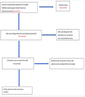
A comprehensive systematic search was conducted across the four predefined research domains, yielding a total of 1,562 records. All retrieved references were systematically organized and managed using EndNote version 20. Following a rigorous deduplication process, 493 duplicate entries were identified and removed, leaving 1,069 unique records for title and abstract screening. After the initial assessment, 437 records were excluded as irrelevant, and 632 full-text articles were subsequently evaluated for eligibility. Ultimately, 120 studies met the inclusion criteria and were incorporated into the final systematic review. These 120 studies represented unique articles; however, some patients in these studies presented with more than one atypical organ-system involvement.
The complete identification, screening, and inclusion process is illustrated in the PRISMA flow diagram (Figure 1), which provides a clear and transparent overview of the study selection pathway.
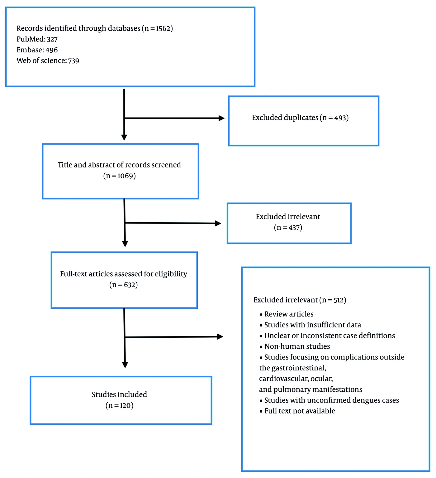
Figure 1.
Flow chart of study selection for inclusion in the systematic review
Regarding the methodological quality of the included studies, most were rated as medium quality (73/120, 60.8%), whereas high-quality studies comprised 22 (18.3%) and low-quality studies accounted for 25 (20.8%) (Table 2 in the Supplementary File). Across the four organ-system categories, medium-quality studies predominated in the gastrointestinal (71.4%), cardiovascular (52.2%), ocular (55.6%), and respiratory (63.6%) manifestation groups.
Table 2.
Summary of Cardiovascular Manifestations of Dengue Virus Infection (N = 26) a
| Variables | Number | Percentage |
|---|---|---|
| Gender | ||
| Male | 12 | 46.2 |
| Female | 14 | 53.8 |
| Outcome | ||
| Live | 21 | 80.8 |
| Dead | 4 | 15.4 |
| NR | 1 | 3.8 |
| Age | ||
| Mean age (years) | 33.06 | |
| DENV-serotypes | ||
| Serotype-2 | 3 | 11.5 |
| Serotype-3 | 3 | 11.5 |
| NR | 20 | 76.9 |
| Myocardial disorders (34.61) | ||
| Myocarditis | 8 | 30.8 |
| Severe Dilated Cardiomyopathy | 1 | 3.8 |
| Pericardial disorders (11.53) | ||
| Cardiac tamponade | 2 | 7.7 |
| Pericardial Effusion | 1 | 3.8 |
| Conduction disturbances (73.07) | ||
| AV dissociation | 1 | 3.8 |
| Junctional Rhythm | 1 | 3.8 |
| widespread T wave inversions | 1 | 3.8 |
| Atrial Fibrillation | 4 | 15.4 |
| Cardiac Arrest | 1 | 3.8 |
| Atrioventricular Block | 2 | 7.7 |
| ST elevation | 2 | 7.7 |
| Tachycardia | 5 | 19.2 |
| ST depression in inferior leads | 2 | 7.7 |
| Vascular diseases (7.6) | ||
| STEMI | 1 | 3.8 |
| Coronary Artery Spasms | 1 | 3.8 |
| Concurrent infections | ||
| Aspergillus | 1 | 3.8 |
| Underlying disease | ||
| Diabetes | 1 | 3.8 |
| Diagnostic methods | ||
| Serology | 15 | 57.7 |
| NS1 | 11 | 42.3 |
| ELISA | 1 | 3.8 |
| RT-PCR | 7 | 26.9 |
| Clinical manifestations | ||
| Fever | 22 | 84.6 |
| Headache | 11 | 42.3 |
| Photophobia | 1 | 3.8 |
| Myalgia | 6 | 23.1 |
| Retro-orbital pain | 3 | 11.5 |
| Chest pain | 1 | 3.8 |
| Bone pain | 1 | 3.8 |
| Joint pain | 1 | 3.8 |
| Abdominal pain | 2 | 7.7 |
| Arthralgia | 7 | 26.9 |
| Malaise | 4 | 15.4 |
| Vomiting | 5 | 19.2 |
| Hematochezia | 1 | 3.8 |
| Dizziness | 1 | 3.8 |
| Nausea | 1 | 3.8 |
| Diarrhea | 1 | 3.8 |
| Laboratory findings | ||
| Plt:L | 12 | 46.2 |
| AST:H | 9 | 34.6 |
| ALT:H | 10 | 38.5 |
| LDH:H | 2 | 7.7 |
| CPK:H | 1 | 3.8 |
| CK-MB:H | 3 | 11.5 |
| Trop-I: positive | 9 | 34.6 |
| Ca:L | 1 | 3.8 |
| BUN:H | 1 | 3.8 |
| Treatment | ||
| Inotropic support | 1 | 3.8 |
| Antipyretics | 1 | 3.8 |
| Prednisolone | 1 | 3.8 |
a Abbreviations: NR, Not reported; Plt, Platelet count; L, Low; H, High; AST, Aspartate aminotransferase; ALT, Alanine aminotransferase; LDH, Lactate dehydrogenase; CPK, Creatine phosphokinase; CK-MB, Creatine kinase-MB; Trop-I, Troponin I; BUN, Blood urea nitrogen; NS1, Nonstructural protein 1 antigen; ELISA, Enzyme-linked immunosorbent assay; RT-PCR, Reverse transcription polymerase chain reaction.
3.2. Gastrointestinal Manifestations
The gastrointestinal manifestations included in this study are summarized in Table 1, along with their reported prevalence among cases. A total of 30 dengue-associated gastrointestinal cases were reported from 13 countries across Asia and the Americas, with India (33.3%) and Sri Lanka (16.7%) contributing the most cases (Figure 2, Table 2A in the Supplementary File). No studies originated from Europe, Africa, or Oceania. The mean patient age was 33.8 years.
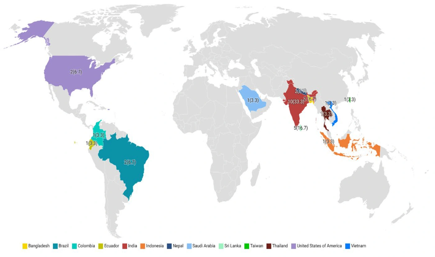
Figure 2.
Distribution of gastrointestinal findings in dengue cases
Among patients whose sex was reported (n = 28), 65% were males and 35% were females. The remaining patients (3.4% of the total) had unknown sex. One fatality (3.3%) was reported, occurring in a patient with acute pancreatitis. Among 12 distinct gastrointestinal manifestations, the most common were pancreatitis (73.3%), vomiting (50.0%), and diarrhea (20.0%). No co-infections were identified. Diabetes was the most prevalent underlying condition (16.7%). Diagnosis relied mainly on serological testing (66.7%), while no study specified the viral serotype.
Fever (73.3%), abdominal pain (63.3%), and headache (43.3%) were the most frequent clinical findings. Laboratory findings commonly included thrombocytopenia (80%) and elevated amylase (60%) or lipase (46.7%). All patients received supportive management only (Table 1).
The predominance of pancreatitis suggests possible pancreatic tropism of the dengue virus or secondary immune-mediated injury. The high proportion of Asian cases, particularly from India, may indicate regional reporting bias or a higher disease burden. Moreover, the association with diabetes highlights a potential vulnerability to severe systemic complications.
3.3. Cardiovascular Manifestations
The cardiovascular manifestations comprised a spectrum of disorders categorized as myocardial disorders (such as myocarditis and severe dilated cardiomyopathy), pericardial disorders (including cardiac tamponade and pericardial effusion), conduction disturbances (such as atrioventricular dissociation, atrial fibrillation, and tachycardia), and vascular diseases (including ST-elevation myocardial infarction and coronary artery spasms). A detailed summary of these manifestations is presented in Table 2.
A total of 26 cases of dengue-associated cardiovascular manifestations were identified across 11 countries, with the highest frequencies in India (34.6%) and Pakistan (15.4%) (Figure 3, Table 2B in the Supplementary File). No reports originated from Africa.
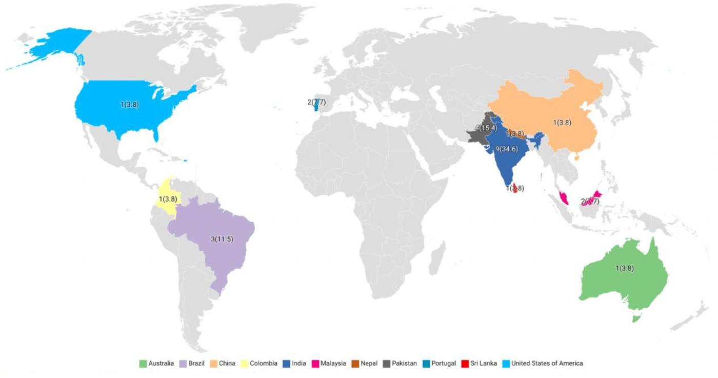
Figure 3.
Distribution of cardiovascular findings in dengue cases
The mean patient age was 33.1 years, and females (53.8%) were slightly more affected than males (46.2%). Four deaths (15.4%) were recorded, all in male patients presenting with myocarditis or conduction abnormalities.
Conduction disturbances (73.1%) were the most common category, followed by myocardial disorders (34.6%), pericardial disorders (11.5%), and vascular diseases (7.6%). Specific findings included myocarditis (30.8%), junctional rhythm (19.2%), and atrial fibrillation (15.4%). Among cases with serotyping, only DENV-2 and DENV-3 were identified, each in 11.5% of patients. One patient had concurrent aspergillosis, and another had diabetes as a comorbidity.
Fever (84.6%), headache (42.3%), and arthralgia (26.9%) were the most common systemic symptoms, and thrombocytopenia (46.2%) was the main laboratory abnormality. Treatment remained supportive, with occasional use of corticosteroids or inotropic agents.
The predominance of conduction disturbances and myocarditis underscores the cardiotropic potential of the dengue virus, likely driven by immune-mediated myocardial inflammation. Notably, mortality occurred only in males, suggesting possible sex-related differences in cardiovascular response or disease severity.
3.4. Ocular Manifestations
Ocular manifestations included a wide range of retinal, uveal, conjunctival, eyelid, neuropathic, scleral, and other eye-related disorders (Table 3). Ocular involvement associated with dengue virus infection has been reported across three continents—Asia, the Americas, and Oceania—with most cases originating from India (43.1%), China (15.5%), and the United States (6.9%) (Figure 4, Table 2C in the Supplementary File). No cases were reported from Europe or Africa.
Table 3.
Summary of Ocular Manifestations of Dengue Virus Infection (N = 58) a
| Variables | Number | Percentage |
|---|---|---|
| Gender | ||
| Male | 19 | 32.8 |
| Female | 29 | 50.0 |
| NR | 10 | 17.2 |
| Outcome | ||
| Live | 42 | 72.4 |
| Dead | 0 | |
| NR | 16 | 27.6 |
| Age | ||
| Mean age (years) | 31.7 | |
| DENV-serotypes | ||
| Serotype-1 | 2 | 3.4 |
| NR | 56 | 96.6 |
| Retinal diseases (87.93) | ||
| Superficial retinal hemorrhages | 1 | 1.7 |
| Pre-retinal hemorrhage | 1 | 1.7 |
| Macula cotton wool spots | 1 | 1.7 |
| Acute macular neuroretinopathy | 8 | 13.8 |
| Subtle hypopigmented macular lesions | 1 | 1.7 |
| Cystoid macular edema | 1 | 1.7 |
| Retinal oedema | 1 | 1.7 |
| Retinal vasculitis | 1 | 1.7 |
| Retinochoroiditis | 3 | 5.2 |
| Pallid disc edema with obliteration of the cup | 1 | 1.7 |
| Maculopathy | 12 | 20.7 |
| Ellipsoid zone (EZ) infiltration | 1 | 1.7 |
| Foveolitis | 6 | 10.3 |
| Branch Retinal Artery Occlusion | 1 | 1.7 |
| Retinitis | 1 | 1.7 |
| Viral retinitis | 1 | 1.7 |
| Optic disc swelling | 3 | 5.2 |
| Optic neuritis | 2 | 3.4 |
| Yellowish-orange lesion at the fovea | 1 | 1.7 |
| Multiple retinal hemorrhages | 1 | 1.7 |
| Retinal hemorrhages | 2 | 3.4 |
| Foveal lesions and hemorrhages | 1 | 1.7 |
| Uveal diseases (20.68) | ||
| Chronic bilateral panuveitis | 1 | 1.7 |
| Uveitis glaucoma | 1 | 1.7 |
| Uveitis | 6 | 10.3 |
| Chorioretinitis | 1 | 1.7 |
| Bilateral acute anterior uveitis | 1 | 1.7 |
| Bilateral Ciliochoroidal Effusion with Secondary Angle Closure | 1 | 1.7 |
| Choriocapillaris involvement | 1 | 1.7 |
| Conjunctival disorders (8.62) | ||
| Chemosis | 1 | 1.7 |
| Subconjunctival hemorrhage | 2 | 3.4 |
| Severe chemosis with lid edema with corneal haze and ring stromal infiltrates in the cornea | 2 | 3.4 |
| Eyelid disorders (1.72) | ||
| Bilateral eyelid ecchymosis | 1 | 1.7 |
| Neuropathic diseases (13.79) | ||
| Neuro-Ophthalmic | 1 | 1.7 |
| Bilateral non-arthritic ischemic optic neuropathy | 1 | 1.7 |
| Anterior ischemic optic neuropathy | 1 | 1.7 |
| Optic Neuropathy | 4 | 6.9 |
| Scleral diseases (1.72) | ||
| Scleral necrosis | 1 | 1.7 |
| Others (36.2) | ||
| Stromal keratitis | 1 | 1.7 |
| Terson’s syndrome | 1 | 1.7 |
| Panophthalmitis | 6 | 10.3 |
| Eviscerated eye | 1 | 1.7 |
| Myopic Shift | 1 | 1.7 |
| Unilateral lateral rectus palsy | 1 | 1.7 |
| Central serous chorioretinopathy | 1 | 1.7 |
| Bilateral macula oedema with yellow subretinal deposits and dot hemorrhages | 1 | 1.7 |
| Acute Posterior Multifocal Placoid Pigment Epitheliopathy | 2 | 3.4 |
| Bilateral acute angle closure glaucoma | 1 | 1.7 |
| Retrobulbar hemorrhage | 2 | 3.4 |
| Central scotoma | 2 | 3.4 |
| Bilateral Intraocular Hemorrhage | 1 | 1.7 |
| Concurrent infections | ||
| Norovirus | 1 | 1.7 |
| Diagnostic methods | ||
| Serology | 31 | 53.4 |
| NS1 | 8 | 13.8 |
| ELISA | 1 | 1.7 |
| RT-PCR | 3 | 5.2 |
| Clinical manifestations | ||
| Fever | 15 | 25.9 |
| Headache | 8 | 13.8 |
| Nausea | 2 | 3.4 |
| Vomiting | 3 | 5.2 |
| Diarrhea | 1 | 1.7 |
| Arthralgia | 1 | 1.7 |
| Malaise | 5 | 8.6 |
| Pyrexia | 1 | 1.7 |
| Severe dehydration | 1 | 1.7 |
| Joint pain | 1 | 1.7 |
| Abdominal pain | 2 | 3.4 |
| Seizures | 2 | 3.4 |
| Ocular pain | 1 | 1.7 |
| Muscle pain | 1 | 1.7 |
| Fatigue | 1 | 1.7 |
| Laboratory findings | ||
| Plt: L | 16 | 27.6 |
| AST: H | 3 | 5.2 |
| ALT: H | 2 | 3.4 |
| BUN: H | 1 | 1.7 |
| Cr: H | 1 | 1.7 |
| Pt: H | 1 | 1.7 |
| Glu: L | 1 | 1.7 |
| Treatment | ||
| Oral corticosteroids | 13 | 22.4 |
| Timolol eyedrops | 6 | 10.3 |
| Oral acetazolamide | 6 | 10.3 |
| Intravenous fluid | 2 | 3.4 |
| intravenous methylprednisolone | 1 | 1.7 |
| Intravenous colloid | 1 | 1.7 |
| Platelet transfusions | 1 | 1.7 |
a Abbreviations: NR, not reported; Plt, Platelet count; L, Low; H, High; AST, Aspartate aminotransferase; ALT, Alanine aminotransferase; BUN, Blood urea nitrogen; Cr, Creatinine; Pt, Prothrombin time; Glu, Glucose; NS1, Nonstructural protein 1 antigen; ELISA, Enzyme-linked immunosorbent assay; RT-PCR, Reverse transcription polymerase chain reaction.
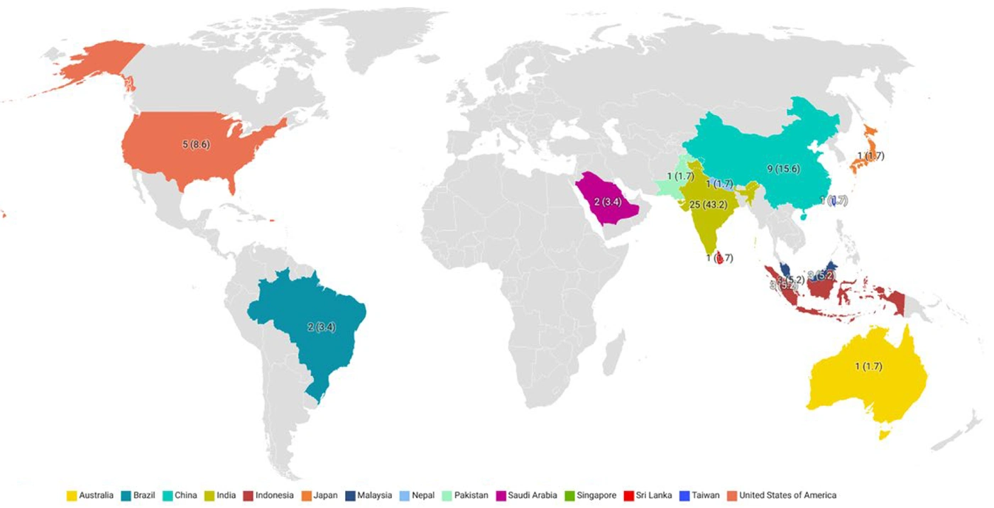
Figure 4.
Distribution of ocular findings in dengue cases
Among studies that provided demographic data, the mean age of patients was 31.7 years, with a slight female predominance. No fatalities were documented in this subgroup.
A total of 55 distinct ocular manifestations were identified, categorized mainly as retinal (87.9%), uveal (20.6%), neuropathic (13.7%), conjunctival (8.6%), eyelid (1.7%), and scleral disorders (1.7%). Retinal manifestations included maculopathy, acute macular neuroretinopathy, foveolitis, retinal hemorrhages, and optic disc edema. Uveal involvement primarily encompassed uveitis and chorioretinitis, whereas neuropathic disorders comprised optic neuropathy and anterior ischemic optic neuropathy. A comprehensive list of all ocular findings is presented in Table 3.
Other less common ocular conditions, including stromal keratitis, panophthalmitis, Terson’s syndrome, central serous chorioretinopathy, and unilateral lateral rectus palsy, collectively accounted for 36.2% of all ocular manifestations.
Concurrent norovirus infection was documented in one patient. Diagnostic confirmation of dengue infection was predominantly based on serological methods (53.4%), while the dengue viral serotype was reported in only two studies, both indicating serotype 1.
Treatment was largely supportive, with oral corticosteroids (22.4%), acetazolamide (10.3%), and timolol eye drops (10.3%) representing the most commonly administered interventions.
The predominance of retinal involvement may reflect the microvascular tropism of the dengue virus, underscoring the vulnerability of the retinal and choroidal vasculature to immune-mediated endothelial injury. These observations suggest that ocular manifestations might serve as early indicators of systemic vascular dysfunction in dengue infection.
3.5. Respiratory Manifestations
Pulmonary manifestations included radiological findings and respiratory symptoms such as consolidation, ARDS, and pleural effusion (Table 4).
Table 4.
Summary of Pulmonary Manifestations of Dengue Virus Infection (N = 45) a
| Variables | Number | Percentage |
|---|---|---|
| Gender | ||
| Male | 22 | 48.9 |
| Female | 22 | 48.9 |
| NR | 1 | 2.2 |
| Outcome | ||
| Live | 32 | 71.1 |
| Dead | 4 | 8.9 |
| NR | 9 | 20.0 |
| Age | ||
| Mean age (years) | 33.5 | |
| DENV-serotypes | ||
| Serotype-1 | 3 | 6.7 |
| Serotype-2 | 4 | 8.9 |
| Serotype-3 | 1 | 2.2 |
| Serotype-4 | 1 | 2.2 |
| NR | 36 | 80.0 |
| Radiological manifestations | ||
| Consolidation | 1 | 2.2 |
| GGO | 11 | 24.4 |
| pleural‐based opacity | 1 | 2.2 |
| Bilateral diffuse opacities | 1 | 2.2 |
| Nonspecific Respiratory manifestations | ||
| Pulmonary hemorrhage | 6 | 13.3 |
| Hemothorax | 2 | 4.4 |
| Bilateral pneumonia | 1 | 2.2 |
| ARDS | 11 | 24.4 |
| Respiratory depression | 1 | 2.2 |
| Pulmonary infiltrations | 1 | 2.2 |
| Breathlessness | 5 | 11.1 |
| Interstitial syndrome | 1 | 2.2 |
| Respiratory discomfort | 1 | 2.2 |
| Pulmonary edema | 3 | 6.7 |
| Pleural effusion | 22 | 48.9 |
| Respiratory distress | 3 | 6.7 |
| Concurrent infections | ||
| H1N1 | 1 | 2.2 |
| COVID-19 | 1 | 2.2 |
| Malaria | 3 | 6.7 |
| Legionella pneumophila | 1 | 2.2 |
| Plasmodium knowlesi | 1 | 2.2 |
| Human metapneumovirus | 1 | 2.2 |
| Tuberculosis | 1 | 2.2 |
| Underlying disease | ||
| Autoimmune disease | 1 | 2.2 |
| Diabetes | 1 | 2.2 |
| Asthma | 1 | 2.2 |
| Kidney transplantation | 1 | 2.2 |
| Hypertension | 2 | 4.4 |
| Diagnostic methods | ||
| Serology | 30 | 66.7 |
| NS1 | 1 | 2.2 |
| ELISA | 6 | 13.3 |
| RT-PCR | 10 | 22.2 |
| Clinical manifestations | ||
| Fever | 18 | 40.0 |
| Takotsubo syndrome | 1 | 2.2 |
| Vomiting | 6 | 13.3 |
| Diarrhea | 3 | 6.7 |
| Pericardial effusion | 2 | 4.4 |
| Cardiomegaly | 1 | 2.2 |
| Ascites | 10 | 22.2 |
| Hepatomegaly | 3 | 6.7 |
| Palpable axillary | 1 | 2.2 |
| Hemoptysis | 1 | 2.2 |
| Anemia | 1 | 2.2 |
| Hemodynamic instability | 1 | 2.2 |
| Renal failure | 2 | 4.4 |
| Headache | 5 | 11.1 |
| Retro-orbital pain | 1 | 2.2 |
| Arthralgia | 1 | 2.2 |
| Myalgias | 6 | 13.3 |
| Dry cough | 3 | 6.7 |
| Rhabdomyolysis with acute kidney injury | 1 | 2.2 |
| Abdominal pain | 4 | 8.9 |
| Hepatic dysfunction | 1 | 2.2 |
| Hepatosplenomegaly | 1 | 2.2 |
| Hemoperitoneum | 1 | 2.2 |
| Generalized rashes | 1 | 2.2 |
| Hemoptysis | 1 | 2.2 |
| Chills | 1 | 2.2 |
| Yellowish discoloration of skin | 1 | 2.2 |
| Pedal oedema | 1 | 2.2 |
| Subarachnoid and subdural bleeding | 1 | 2.2 |
| Cholecystitis | 1 | 2.2 |
| Productive cough | 1 | 2.2 |
| Liver enlargement | 3 | 6.7 |
| Hemorrhagic manifestations | 3 | 6.7 |
| Laboratory findings | ||
| Plt:L | 19 | 42.2 |
| AST:H | 8 | 17.8 |
| ALT:H | 8 | 17.8 |
| AST > ALT | 1 | 2.2 |
| LDH:H | 1 | 2.2 |
| ALB:H | 1 | 2.2 |
| Hg:H | 1 | 2.2 |
| BUN:H | 3 | 6.7 |
| Cre:H | 3 | 6.7 |
| Crp:+ | 1 | 2.2 |
| Ferritin:H | 1 | 2.2 |
| D-Dimer | 1 | 2.2 |
| BillT:H | 1 | 2.2 |
| CPK:H | 1 | 2.2 |
| Treatment | ||
| Intravenous fluids | 2 | 4.4 |
| Antiemetics | 2 | 4.4 |
| Antipyretics | 1 | 2.2 |
| Ringer’s lactate | 1 | 2.2 |
| oseltamivir | 1 | 2.2 |
| Intravenous multivitamin | 1 | 2.2 |
| Paracetamol | 1 | 2.2 |
| Ventilator | 2 | 4.4 |
| Supportive care | 3 | 6.7 |
| BiPAP respiratory support | 1 | 2.2 |
| Mechanical ventilation | 1 | 2.2 |
| Targeted fluid therapy | 1 | 2.2 |
| platelet transfusion | 1 | 2.2 |
| Treated with HFNC with bronchodilators | 1 | 2.2 |
| Tylenol | 1 | 2.2 |
a Abbreviations: NR, Not reported; Plt, Platelet count; L, Low; H, High; AST, Aspartate aminotransferase; ALT, Alanine aminotransferase; LDH, Lactate dehydrogenase; ALB, Albumin; Hg, Hemoglobin; BUN, Blood urea nitrogen; Cre/Cr, Creatinine; Crp, C-reactive protein; BillT, Total bilirubin; CPK, Creatine phosphokinase; NS1, Nonstructural protein 1 antigen; ELISA, Enzyme-linked immunosorbent assay; RT-PCR, Reverse transcription polymerase chain reaction; GGO, Ground-glass opacity; ARDS, Acute respiratory distress syndrome; BiPAP, Bilevel positive airway pressure; HFNC, High-flow nasal cannula.
Respiratory involvement due to dengue virus infection was reported in 10 countries, predominantly India (51.1%), with no reports from Africa or Oceania (Figure 5, Table 2D in the Supplementary File). The mean age was 33.5 years, and male and female patients were affected equally. Four deaths (8.9%) were reported among cases with respiratory involvement.
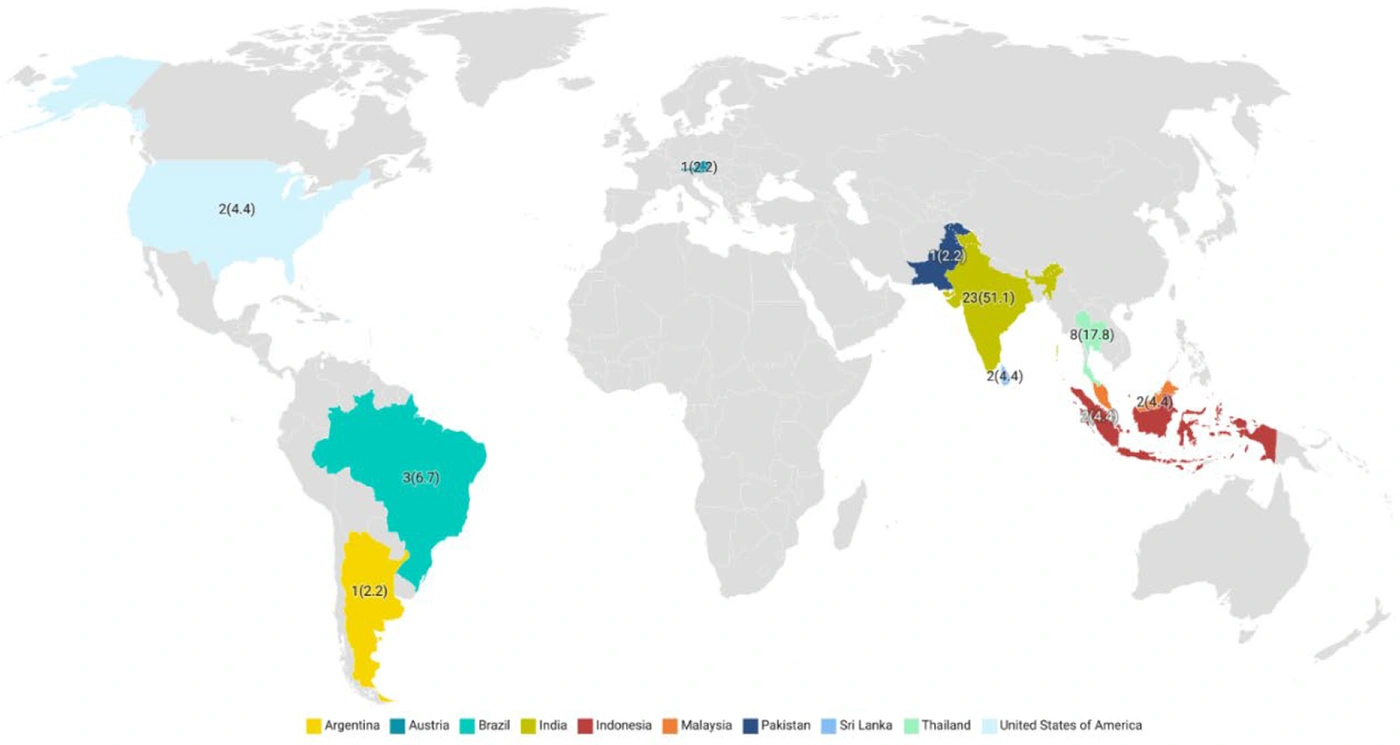
Figure 5.
Distribution of respiratory findings in dengue cases
Among 12 clinical respiratory manifestations, ARDS (24.4%) was a prominent feature of expanded dengue syndrome. Although pleural effusion (48.9%) was frequently reported, it is generally considered a secondary complication rather than a primary manifestation of the syndrome. Radiologically, ground-glass opacity (24.4%) was the predominant finding.
Concurrent infections included malaria (6.7%), H1N1, COVID-19, and Legionella pneumophila, while hypertension (4.4%) was the most common comorbidity. The dengue virus was mainly identified by serological tests (66.7%), and among studies reporting serotypes, DENV-2 predominated. Supportive therapy, including ventilatory support or fluid management, was the mainstay of treatment.
The predominance of pleural effusion and ARDS strongly indicates capillary leak syndrome as a key pathophysiological mechanism in severe dengue. Co-infections and comorbidities appear to exacerbate pulmonary outcomes, underscoring the need for early recognition and supportive respiratory care in these patients.
4. Discussion
This systematic review highlights the wide spectrum of atypical clinical features observed in patients with expanded dengue syndrome, emphasizing the importance of recognizing non-classical manifestations beyond the traditional febrile presentation of dengue virus infection. Diagnosing expanded dengue syndrome remains challenging because it can mimic numerous infectious, hematologic, rheumatologic, and gastrointestinal conditions. Coinfections and cross-reactivity in serological tests further complicate diagnostic accuracy, often delaying management. Clinicians should maintain a high index of suspicion and rely on a combination of careful clinical assessment, laboratory investigations, and molecular testing to differentiate expanded dengue syndrome from mimicking conditions and to prevent misdiagnosis and unnecessary interventions (17).
In our findings, gastrointestinal symptoms were among the most frequently reported atypical presentations in patients with dengue. Pancreatitis, vomiting, and diarrhea were predominant, supporting earlier studies showing that gastrointestinal involvement is a notable feature of severe dengue infections (18, 19). The most frequent laboratory findings associated with gastrointestinal complications included elevated liver enzymes, such as AST and ALT, and changes in platelet levels, which were observed in 46.2% of cases with gastrointestinal manifestations. Although these manifestations were not often fatal, they correlated with significant laboratory abnormalities, emphasizing the need for early laboratory evaluation and monitoring. Notably, diabetes emerged as the most common underlying condition in these patients, suggesting a possible link between metabolic dysfunction and increased vulnerability to gastrointestinal involvement.
Similarly, a cross-sectional study by Dinkar et al. (20) in India reported that among 220 dengue patients, severe hepatic dysfunction was significantly more frequent in secondary dengue (45.4%) than in primary dengue (11.1%). Pancytopenia was observed in 19.1% of cases, with a higher proportion in secondary dengue (36.6%). These findings are largely consistent with our results, indicating that gastrointestinal involvement, hematologic abnormalities, and multisystem derangements are more pronounced in secondary or severe dengue cases. This study also underscores the importance of monitoring liver enzymes, platelet counts, and blood cell indices to anticipate complications and guide clinical management (20).
Furthermore, rare but critical complications such as dengue-associated hemophagocytic lymphohistiocytosis (HLH) have been reported. For example, an 18-year-old female developed severe pancytopenia and progressive liver dysfunction secondary to dengue, ultimately meeting diagnostic criteria for HLH. She responded favorably to pulse methylprednisolone therapy, highlighting the importance of early recognition and prompt immunomodulatory intervention in such cases (21).
Cardiovascular manifestations, although less frequent than gastrointestinal findings, were associated with the highest mortality in our study. Myocarditis, arrhythmias, and pericardial diseases such as effusions were common in this group. Myocarditis has emerged as a significant predictor of morbidity and mortality in dengue patients; however, sufficient data on the incidence and comprehensive characterization of myocarditis in dengue infection are lacking. Our findings showed a mortality rate of 15.4% among patients with cardiovascular involvement, underscoring the need for close cardiac surveillance in dengue cases, particularly in those with severe manifestations. According to a study conducted by Baqi et al., the in-hospital mortality rate among patients with dengue-related myocarditis was 21.4% (22). Patients with this complication were nearly five times more likely to experience prolonged hospital stays. The study further reported that all patients with dengue myocarditis had elevated cardiac troponin I (cTn-I), 59.5% had at least one abnormal ECG finding, and 24% had reduced ejection fraction (EF < 55%). Additionally, there was a noticeable trend toward increased rates of shock and liver enzyme abnormalities among myocarditis patients (23).
Ocular manifestations in dengue were generally non-lethal but have the potential for long-term morbidity due to visual impairment. Although uncommon, ocular complications in dengue can affect both the anterior and posterior segments of the eye and are often linked to thrombocytopenia (24). Hemorrhage is the most frequently observed finding; however, conditions such as anterior uveitis and vasculitis may reflect underlying immune-driven mechanisms (25). Our findings indicate that, although no fatalities were reported among patients with ocular involvement, dengue infection can manifest with a diverse range of retinal, uveal, conjunctival, eyelid, and optic neuropathic complications. This broad spectrum of ocular findings may facilitate early recognition and clinical management of dengue-associated visual disturbances. Maculopathy was the most common retinal disease, highlighting its potential as a key indicator of dengue-associated ocular involvement. Dengue-related maculopathy encompasses a broad clinical presentation, most often occurring about a week after fever onset. Common features include intraretinal hemorrhages, macular swelling, optic disc edema, and foveolitis (26, 27).
Similarly, Dinkar et al. (28) reported a case of dengue-associated reversible blindness characterized by bilateral retinal hemorrhage and severe thrombocytopenia. This observation aligns with our findings, reinforcing that retinal involvement and hemorrhagic changes likely reflect immune-mediated and vascular injury mechanisms (28).
Respiratory manifestations of dengue infection were associated with a mortality rate of approximately 9% among patients with documented outcomes, underscoring the need for prompt respiratory evaluation and preparedness for intensive care, particularly in patients with signs of respiratory compromise. Radiologic findings, including ground-glass opacities, were frequently observed and may suggest secondary immune-mediated lung injury or viral pneumonitis (29). Co-infections, such as malaria, further complicate clinical management in endemic regions because of overlapping symptoms such as fever and hemolysis, which may obscure timely diagnosis and treatment. Although ARDS is relatively uncommon in dengue overall, it is more frequently reported in severe or fatal cases (30). However, given the limited data in the reviewed studies, the precise impact of co-infections on disease severity and patient outcomes remains unclear and warrants further investigation.
This aligns with a recent case report of a 27-year-old female with severe dengue who developed bilateral lung infiltrates, required prolonged ventilatory support, and had complications including severe hepatitis and pontine infarct (31).
Similarly, a 30-year-old female presented with ARDS, bilateral pulmonary infiltrates, and subdural and subarachnoid hemorrhage and was managed successfully with supportive care and ventilatory assistance (32). These cases highlight the importance of early recognition of respiratory compromise and readiness for intensive care interventions. Pleural effusion, noted in nearly half of the cases, further emphasizes the need for careful monitoring and timely interventions such as ventilatory support and intravenous fluids.
In summary, gastrointestinal, cardiovascular, ocular, and respiratory symptoms are critical to the clinical diagnosis and management of dengue, particularly when multiple organ systems are affected simultaneously. These manifestations often correlate with more severe disease outcomes, especially in patients with underlying comorbidities, and emphasize the need for multidisciplinary management.
4.1. Study Limitations
Several limitations should be acknowledged. First, the included studies were predominantly case reports and small case series, which may limit the generalizability of the findings. Second, systematic reporting of dengue virus serotypes, patient comorbidities, and long-term outcomes was largely absent. Third, most studies lacked standardized diagnostic criteria for atypical presentations, making cross-study comparisons difficult. Fourth, certain atypical manifestations, such as neurological symptoms, were not reported in the included studies, representing another limitation. Fifth, geographical bias is an important concern, as most reports originated from South and Southeast Asia, with limited representation from other endemic regions such as Africa and Oceania. Sixth, because the evidence was mainly case-based, methodological quality and data completeness varied considerably across studies.
Seventh, because of the exclusively descriptive nature of data derived from case reports and case series, together with the lack of access to individual-level data and control variables, advanced statistical analyses and causal inference were not feasible. This limitation has been duly acknowledged and highlighted in the study.
Eighth, an additional limitation is that this review was restricted to English-language publications. This approach was used to ensure accuracy in data extraction and minimize potential translation-related errors; however, it may have led to the exclusion of relevant evidence published in other languages.
4.2. Future Research
Future research should focus on developing standardized diagnostic criteria for expanded dengue syndrome to ensure consistency across clinical and epidemiological investigations. Larger multicenter cohort studies and prospective surveillance systems are needed to better define the true burden, spectrum, and pathophysiology of atypical manifestations. It is also essential to explore whether specific dengue virus serotypes are more strongly associated with particular complications, such as myocarditis or pancreatitis. Given the absence of effective antiviral therapies, the development of targeted treatment protocols for patients with severe organ involvement should be prioritized, along with investigations into host-related factors such as genetic susceptibility and pre-existing comorbidities.
Notably, no included study reported patients with simultaneous involvement of multiple organ systems (e.g., concurrent cardiac and gastrointestinal or respiratory manifestations), which likely reflects a lack of integrated clinical reporting rather than a true absence of multisystem disease.
Overall, this review underscores the pressing need for heightened clinical awareness of atypical dengue presentations, which often mimic other systemic illnesses and may lead to delayed or inappropriate treatment. The data suggest that organ-specific complications such as maculopathy and myocarditis may serve as early indicators of disease severity, meriting focused diagnostic and monitoring strategies. Moreover, the potential pathophysiological role of comorbidities, particularly diabetes and cardiovascular disease, in modulating the host immune response and disease course warrants further investigation. Future research should not only aim to clarify these mechanisms but also identify early biomarkers and develop risk stratification tools to guide management in resource-limited settings. Importantly, the absence of integrated reporting on multi-organ involvement highlights the need for comprehensive clinical documentation in future studies.
4.3. Conclusions
This systematic review highlights the distinctive clinical picture of expanded dengue syndrome, involving the gastrointestinal, cardiovascular, ocular, and respiratory systems. The most frequent complications were pancreatitis, myocarditis, maculopathy, and pleural effusion, with cardiovascular involvement associated with the highest mortality. Most reported cases originated from Asia, particularly the Indian subcontinent, Sri Lanka, and Pakistan, whereas cases from Europe, Africa, and Oceania were reported only occasionally.
These findings highlight the need for heightened clinical suspicion of atypical dengue presentations and call for standardization of case definitions and improved surveillance in endemic and non-endemic regions. Statements regarding organ-specific tropism or pathophysiology should be regarded as biologically plausible hypotheses rather than proven mechanisms.
Overall, this review emphasizes that the descriptive nature of case reports and case series limits the ability to draw causal inferences or estimate incidence. Future research should aim to confirm these observations in larger cohort studies and explore potential mechanistic pathways under controlled study designs.