
1. Background
Psittacosis is a zoonotic infection caused by Chlamydia psittaci and is primarily transmitted through exposure to infected birds or poultry. As an uncommon cause of community-acquired pneumonia (CAP), Chlamydia psittaci accounts for approximately 1.03% of CAP pathogens (1). However, because of its nonspecific clinical manifestations and the limited sensitivity of traditional detection methods (such as serology and culture), psittacosis pneumonia has often been missed or misdiagnosed, resulting in delayed treatment (2).
In recent years, the clinical application of metagenomic next-generation sequencing (mNGS) and targeted next-generation sequencing (tNGS) has significantly improved the detection rate of Chlamydia psittaci. Multiple studies have shown that Chlamydia psittaci infection has a high propensity to progress to severe disease, accounting for up to 8.0% of pathogens identified in severe pneumonia (3, 4), thereby posing a serious threat to patient health. Currently, tetracycline antibiotics are the primary treatment for psittacosis pneumonia, with quinolones serving as alternative options (5). Early definitive diagnosis and timely targeted antimicrobial therapy are crucial for improving respiratory symptoms and clinical outcomes. However, few systematic comparative studies have evaluated the clinical characteristics, laboratory parameters, and treatment outcomes in severe versus non-severe psittacosis pneumonia. This study retrospectively analyzes the clinical data of patients diagnosed with psittacosis pneumonia by mNGS or tNGS, compares clinical manifestations, laboratory findings, imaging features, antimicrobial treatment, and treatment outcomes between severe and non-severe psittacosis pneumonia, and aims to provide a reference for clinical practice.
2. Objectives
This study aimed to investigate differences in clinical presentations, radiologic and laboratory profiles, antimicrobial management, and patient outcomes between severe and non-severe psittacosis pneumonia and to provide practical insights for clinical management.
3. Methods
3.1. Patients and Setting
This multicenter retrospective study was conducted at three hospitals in Hunan Province, China: The First Affiliated Hospital of Hunan Traditional Chinese Medical College, The People's Hospital of Liuyang, and The People’s Hospital of Youxian. All three institutions are regional tertiary hospitals with comparable diagnostic and treatment protocols for CAP. No significant referral bias was identified because each hospital serves a local population without systematic inter-hospital transfer for psittacosis pneumonia.
Patients were identified by screening electronic medical records for cases with positive mNGS or tNGS results for Chlamydia psittaci in bronchoalveolar lavage fluid (BALF) or sputum samples between January 2023 and December 2025. All consecutive cases meeting the inclusion criteria during the study period were considered for enrollment. A total of 59 potentially eligible patients were initially identified; six were excluded because of severely incomplete clinical data (e.g., missing laboratory results or imaging reports), leaving 53 patients for the final analysis. Given the retrospective design and the exclusive use of de-identified medical records, the requirement for informed consent was waived.
3.2. mNGS and tNGS Workflows
The mNGS workflow was performed as follows. Within 8 hours of collection, 2 - 3 mL of BALF or sputum was obtained and centrifuged at 1,600 × g for 10 minutes at 4 °C. The resulting supernatants were transferred into 2-mL sterile centrifuge tubes and transported to the laboratory in a container with solid carbon dioxide for subsequent mNGS analysis. Nucleic acids were extracted using the TIANamp Micro DNA Kit (Tiangen Biotech, China). DNA sequencing libraries were constructed through fragmentation, end repair, adapter ligation, and PCR amplification. Library quality was assessed using the Agilent 2100 Bioanalyzer (Agilent Technologies, CA), and sequencing was performed on the BGISEQ-500 platform (Beijing Genomics Institute, China). Raw sequencing reads were aligned to the human reference genome (hg19) using the Burrows–Wheeler Alignment (BWA) tool to remove host sequences; the remaining non-human reads were retained for further analysis. These valid sequences were then compared against a microbial genome database (ftp://ftp.ncbi.nlm.nih.gov/genomes/) comprising 11,910 bacteria, 7,103 viruses, 1,046 fungi, and 305 parasite species associated with human infectious diseases. Finally, the aligned data were processed and analyzed, and potential pathogens were identified and summarized based on the number of strictly mapped reads, genome coverage, and sequencing depth.
The tNGS procedure included nucleic acid quantification, cDNA synthesis, targeted amplification, product purification, and library construction and purification. Total pathogenic DNA and RNA were extracted from clinical specimens and simulated mixed samples using the TIANamp Yeast DNA Kit (Tiangen, China) via a centrifugal column method. Samples were inactivated in a high-temperature water bath, and nucleic acid concentrations were determined using the Invitrogen high-sensitivity double-stranded DNA quantification kit (Invitrogen, USA). To detect RNA pathogens, the extracted nucleic acids were reverse-transcribed into cDNA. Multiplex PCR targeted amplification was then performed using specific primers, an amplification enzyme mix, and internal reference reagents, followed by magnetic bead purification. Sequencing libraries were constructed according to the protocols of the Sansure 296-pathogen tNGS library construction kit (Sansure, China). After passing quality control, the qualified libraries were sequenced on the Sansure FASTASeq 300 flow cell (Sansure, China). Bioinformatics analysis was performed on the CentOS 7.9 system. Low-quality sequences (raw reads > 50 bp) were filtered out, and only high-quality data with Q30 values above 85% were retained. After single-end sequence alignment, the read counts of targeted amplification sequences were calculated based on an in-house clinical pathogen database, with reference sequences derived from the GenBank, RefSeq, and Nucleotide databases of the National Center for Biotechnology Information (https://www.ncbi.nlm.nih.gov/).
3.3. Diagnostic Criteria and Grouping
The inclusion criteria were as follows: (1) age > 18 years; (2) pneumonia diagnosis consistent with the Guidelines for the diagnosis and treatment of adult community-acquired pneumonia in China (2016 edition) (6); and (3) detection of Chlamydia psittaci sequences by mNGS or tNGS in BALF or sputum samples. Cases were excluded if clinical data were severely incomplete, defined as missing information that precluded calculation of the primary outcome (time to clinical stability) or determination of severity group classification.
Severe pneumonia was diagnosed if patients met any one of the following major criteria or at least three minor criteria. Major criteria: (1) requiring invasive mechanical ventilation; (2) septic shock requiring vasopressors despite adequate fluid resuscitation. Minor criteria: (1) respiratory rate ≥ 30 breaths/min; (2) oxygenation index ≤ 250 mmHg (1 mmHg = 0.133 kPa); (3) multilobar infiltrates; (4) impaired consciousness and/or disorientation; (5) blood urea nitrogen ≥ 7.14 mmol/L; (6) systolic blood pressure < 90 mmHg requiring aggressive fluid resuscitation (6).
3.4. Outcome Assessment
The primary outcome measure of treatment efficacy was time to clinical stability, and the secondary outcome measure was length of stay. Clinical stability was defined as meeting all of the following five criteria (6): (1) body temperature ≤ 37.8°C; (2) heart rate ≤ 100 beats/min; (3) respiratory rate ≤ 24 breaths/min; (4) systolic blood pressure ≥ 90 mmHg; (5) oxygen saturation ≥ 90% (or arterial partial pressure of oxygen ≥ 60 mmHg while breathing room air).
3.5. Statistical Analysis
All statistical analyses were performed using MedCalc version 19.6.4 software. The Shapiro-Wilk test was used to assess the normality of continuous variables. Normally distributed data were expressed as mean ± standard deviation (SD), whereas non-normally distributed data were expressed as median with interquartile range (IQR). Categorical variables were presented as numbers and percentages (%). Comparisons between two groups were performed using the t-test for normally distributed continuous variables, the Mann-Whitney U test for non-normally distributed continuous variables, and the chi-square test or Fisher's exact test for categorical variables. All statistical tests were two-sided, and a P-value < 0.05 was considered statistically significant.
4. Results
4.1. Baseline Characteristics
A total of 53 patients with Chlamydia psittaci pneumonia were enrolled in this study, including 28 males (52.83%) and 25 females (47.17%), with a mean age of 58.57 ± 11.48 years. Underlying diseases were present in 36 patients (67.92%), and 19 patients (35.84%) had a clear history of exposure to parrots or poultry, such as pigeons, chickens, or ducks. Based on disease severity, patients were divided into the non-severe group (29 cases, 54.72%) and the severe group (24 cases, 45.28%).
Regarding clinical manifestations, fever was the most common symptom, occurring in 50 patients (94.34%), followed by cough (67.92%), myalgia (47.17%), fatigue (35.84%), dyspnea (33.96%), expectoration (26.41%), and headache (20.75%). Intergroup comparison showed that the incidence of dyspnea was significantly higher in the severe group than in the non-severe group (15 (62.50%) vs. 3 (10.34%), P < 0.01). No significant differences were observed in other symptoms between the two groups (P > 0.05). There were also no significant differences in age, gender, distribution of underlying diseases, or exposure history between the two groups (P > 0.05) (Table 1). These results indicate that these common demographic and baseline clinical factors did not confound the observed associations between disease severity and outcomes, thereby permitting a relatively unbiased comparison of severity-related differences.
Table 1.
Demographics and Clinical Symptoms of Patients with Psittacosis Pneumonia a
| Variables | Total (n = 53) | Non-severe (n = 29) | Severe (n = 24) | P-Value |
|---|---|---|---|---|
| Age (y) | 58.57 ± 11.48 | 57.32 ± 11.34 | 59.96 ± 11.70 | 0.41 |
| Male | 28 (52.83) | 13 (44.83) | 15 (62.50) | 0.27 |
| Underlying diseases | 36 (67.92) | 20 (68.97) | 16 (66.67) | 0.86 |
| Hypertension | 14 (26.42) | 8 (27.59) | 6 (25.00) | 0.83 |
| Diabetes | 10 (18.87) | 3 (10.34) | 7 (29.17) | 0.08 |
| Coronary heart disease | 2 (3.77) | 2 (6.90) | 0 (0.00) | 0.19 |
| Chronic liver disease | 2 (3.77) | 1 (3.45) | 1 (4.17) | 0.89 |
| Chronic pulmonary disease | 4 (7.55) | 3 (10.34) | 1 (4.17) | 0.40 |
| Anemia | 3 (5.66) | 2 (6.90) | 1 (4.17) | 0.67 |
| Renal insufficiency | 1 (1.89) | 1 (3.45) | 0 (0.00) | 0.36 |
| Exposure to parrots or poultry | 19 (35.84) | 11 (37.93) | 8 (33.33) | 0.78 |
| Symptoms | ||||
| Fever | 50 (94.34) | 28 (96.55) | 22 (91.67) | 0.45 |
| Cough | 36 (67.92) | 18 (62.07) | 18 (75.00) | 0.32 |
| Myalgia | 25 (47.17) | 16 (55.17) | 9 (37.50) | 0.20 |
| Fatigue | 19 (35.84) | 12 (41.38) | 7 (29.17) | 0.36 |
| Dyspnea | 18 (33.96) | 3 (10.34) | 15 (62.50) | < 0.01 |
| Expectoration | 14 (26.41) | 9 (31.03) | 5 (20.83) | 0.41 |
| Headache | 11 (20.75) | 8 (27.59) | 3 (12.50) | 0.18 |
| Vomiting | 8 (15.09) | 2 (6.90) | 6 (25.00) | 0.07 |
| Hemoptysis | 3 (5.66) | 2 (6.90) | 1 (4.17) | 0.67 |
| Abdominal pain | 2 (3.77) | 0 (0.00) | 2 (8.33) | 0.12 |
| Diarrhea | 2 (3.77) | 1 (3.45) | 1 (4.17) | 0.89 |
| Confirmed (d) | 11 (8.00 - 16.00) | 11 (7.00 - 13.25) | 11 (10.00 - 17.00) | 0.37 |
a Values are expressed as mean ± SD, No. (%) or median (IQR).
4.2. Laboratory and Imaging Findings
Compared with the non-severe group, the severe group had significantly higher white blood cell counts (9.19 × 10⁹/L (IQR, 8.02 - 12.55) vs. 6.69 × 10⁹/L (IQR, 6.03 - 8.28), P < 0.01), neutrophil counts (8.14 × 10⁹/L (IQR, 6.61 - 11.14) vs. 5.49 × 10⁹/L (IQR, 4.70 - 6.96), P < 0.01), C-reactive protein (174.13 ± 87.68 mg/L vs. 129.51 ± 61.17 mg/L, P = 0.04), procalcitonin (1.52 (IQR, 0.23 - 3.97) µg/L vs. 0.20 (IQR, 0.10 - 0.33) µg/L, P < 0.01), lactate dehydrogenase (360.0 U/L (IQR, 322.75 - 605.60) vs. 266.0 U/L (IQR, 197.43 - 387.22), P = 0.02), and creatine kinase levels (272.2 mmol/L (IQR, 113.75 - 861.92) vs. 47.9 mmol/L (IQR, 39.72 - 159.10), P = 0.04), whereas albumin levels (30.54 ± 6.34 g/L vs. 34.44 ± 4.23 g/L, P = 0.03) and the oxygenation index (217.05 ± 75.68 mmHg vs. 328.9 ± 56.95 mmHg, P < 0.01) were significantly lower, indicating more severe tissue damage and respiratory dysfunction in patients with severe disease. No significant differences were found in other laboratory parameters between the two groups (P > 0.05) (Table 2).
Table 2.
Laboratory Tests of Patients with Psittacosis Pneumonia a
| Variables | Total (n = 53) | Non-severe (n = 29) | Severe (n = 24) | P-Value |
|---|---|---|---|---|
| WBC, ×109/L | 7.94 (6.35 - 10.45) | 6.69 (6.03 - 8.28) | 9.19 (8.02 - 12.55) | < 0.01 |
| NEUT, ×109/L | 6.77 (5.07 - 8.55) | 5.49 (4.70 - 6.96) | 8.14 (6.61 - 11.14) | < 0.01 |
| LYM, ×109/L | 0.69 (0.56 - 1.13) | 0.70 (0.63 - 1.19) | 0.65 (0.40 - 0.96) | 0.23 |
| CRP, mg/L | 150.51 ± 77.38 | 129.51 ± 61.17 | 174.13 ± 87.68 | 0.04 |
| PCT, µg/L | 0.27 (0.12 - 1.60) | 0.20 (0.10 - 0.33) | 1.52 (0.23 - 3.97) | < 0.01 |
| ALT, U/L | 55.50 (38.35 - 92.45) | 71.00 (41.75 - 119.75) | 54.00 (35.65 - 75.10) | 0.27 |
| AST, U/L | 70.00 (43.53 - 125.05) | 80.50 (39.05 - 134.80) | 58.20 (48.08 - 109.83) | 0.71 |
| BUN, mmol/L | 4.72 (3.18 - 6.59) | 3.95 (2.95 - 5.87) | 5.74 (4.43 - 7.82) | 0.10 |
| Scr, mmol/L | 72.10 (52.20 - 92.29) | 72.20 (58.75 - 87.35) | 72.00 (50.40 - 94.28) | 0.72 |
| ALB, g/L | 32.79 ± 5.50 | 34.44 ± 4.23 | 30.54 ± 6.34 | 0.03 |
| LDH, U/L | 307.24 (237.00 - 517.15) | 266.00 (197.43 - 387.22) | 360.00 (322.75 - 605.60) | 0.02 |
| K, mmol/L | 3.40 ± 0.37 | 3.48 ± 0.35 | 3.26 ± 0.37 | 0.09 |
| Na, mmol/L | 133.74 ± 4.60 | 133.78 ± 4.09 | 133.63 ± 5.89 | 0.93 |
| D-dimer, mg/L | 545.00 (7.32 - 2184.50) | 520.50 (9.98 - 904.00) | 3235.50 (2.80 - 6005.00) | 0.26 |
| CK, mmol/L | 96.25 (41.00 - 373.02) | 47.90 (39.72 - 159.10) | 272.20 (113.75 - 861.92) | 0.04 |
| PO2/FiO2, mmHg | 263.65 ± 87.74 | 328.90 ± 56.95 | 217.05 ± 75.68 | < 0.01 |
| Reads | 475.00 (99.75 - 24530.75) | 294.00 (54.25 - 2805.75) | 996.00 (282.00 - 53324.00) | 0.17 |
a Values are expressed as mean ± SD or median (IQR). Abbreviations: WBC, white blood cell; NEUT, neutrophil count; LYM, lymphocyte count; CRP, C-reactive protein; PCT, procalcitonin; ALT, alanine aminotransferase; AST, aspartate aminotransferase; BUN, blood urea nitrogen; Scr, serum creatinine; ALB, albumin; LDH, lactate dehydrogenase; K, potassium; Na, sodium; CK, creatine kinase; PO2/FiO2, oxygenation index; SD, standard deviation; IQR, interquartile range.
All patients underwent a chest computed tomography (CT) examination upon admission. Regarding lesion distribution, unilateral lung involvement was observed in 36 cases (67.90%), and bilateral lung involvement in 17 cases (32.10%). The most common imaging finding was consolidation (38 cases, 71.70%), followed by patchy shadows (14 cases, 26.40%), ground-glass opacities (10 cases, 18.90%), and reverse halo sign (3 cases, 5.66%). Pleural effusion occurred in 24 cases (45.30%). Intergroup comparison revealed that the proportion of bilateral lung involvement was significantly higher in the severe group than in the non-severe group (12 (50.00%) vs. 5 (17.20%), P = 0.01), and the incidence of pleural effusion was also significantly higher (17 (70.80%) vs. 7 (24.10%), P < 0.01) (Table 3).
Table 3.
Radiographic Features a
| Variables | Total (n = 53) | Non-severe (n = 29) | Severe (n = 24) | P-Value |
|---|---|---|---|---|
| Distribution of lesions | ||||
| Bilateral involvement | 17 (32.08) | 5 (17.24) | 12 (50.00) | 0.01 |
| Unilateral, left lung | 15 (28.30) | 11 (37.93) | 4 (16.67) | 0.09 |
| Unilateral, right lung | 21 (39.62) | 13 (44.83) | 8 (33.33) | 0.40 |
| Patterns of lesions | ||||
| Consolidation | 38 (71.70) | 18 (62.07) | 20 (83.33) | 0.09 |
| Patchy shadow | 14 (26.4) | 8 (27.59) | 6 (25.00) | 0.83 |
| Ground-glass opacity | 10 (18.9) | 6 (20.69) | 4 (16.67) | 0.71 |
| Reverse halo sign | 3 (5.66) | 1 (3.45) | 2 (8.33) | 0.45 |
| Pleural effusion | 24 (45.28) | 7 (24.14) | 17 (70.83) | < 0.01 |
a Values are expressed as No. (%).
4.3. Antimicrobial Treatment
Before a definitive diagnosis of psittacosis pneumonia, most patients received empirical antimicrobial treatment. The most commonly used initial regimens included quinolones, penicillins, and carbapenems. Among these, 37 patients received combination therapy with two or more antibiotics; the main combination regimens included quinolones combined with penicillins (10 cases, 27.03%), quinolones combined with cephalosporins (9 cases, 24.20%), and quinolones combined with carbapenems (6 cases, 16.22%). Only 6 patients (11.32%) received tetracyclines as initial treatment (Figure 1).
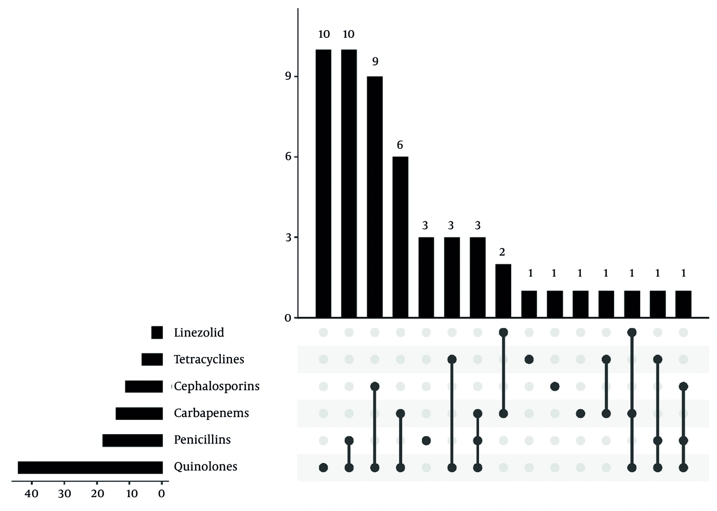
Figure 1.
Empirical antimicrobial therapy prior to etiological confirmation of psittacosis pneumonia. The UpSet plot displays the distribution of initial empirical antimicrobial regimens. Quinolones, penicillins, and carbapenems were the most frequent choices. Combination therapy was common, with quinolones plus penicillins as the most prevalent combination (n = 10). Tetracyclines were administered to only six patients (11.3%) before definitive diagnosis.
After confirmation of Chlamydia psittaci infection by mNGS or tNGS, 16 patients continued or switched to quinolone monotherapy, 13 patients continued or switched to tetracycline monotherapy, and 21 patients (39.62%) received combination therapy with quinolones and tetracyclines. Among those receiving combination therapy, 12 were in the severe group (50.00%) and 9 were in the non-severe group (31.03%), with a statistically significant difference between the groups (P < 0.05) (Figures 2 and 3).
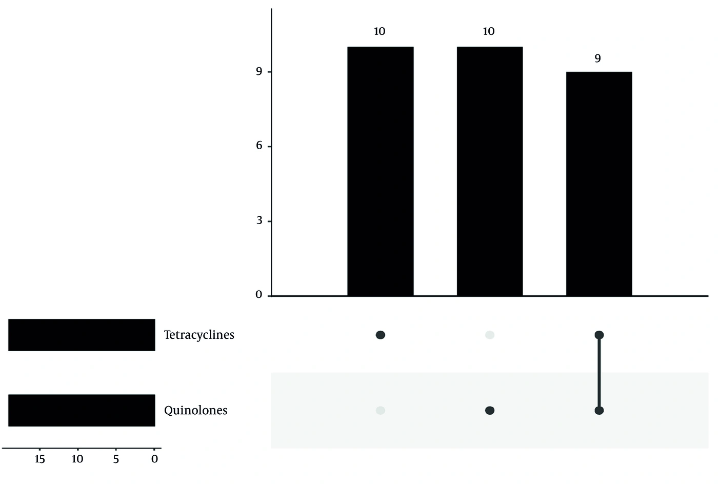
Figure 2.
Post-diagnosis antimicrobial therapy in non-severe psittacosis pneumonia. The UpSet plot shows the distribution of targeted regimens among 29 patients with non-severe disease. Quinolone monotherapy and tetracycline monotherapy were used with equal frequency; nine patients (31.0%) received combination therapy with both agents.
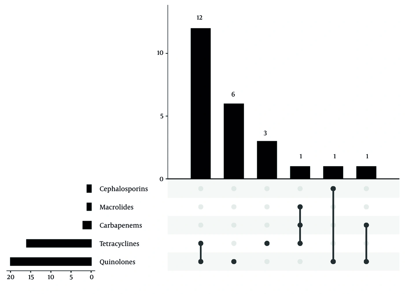
Figure 3.
Post-diagnosis antimicrobial therapy in severe psittacosis pneumonia. The UpSet plot shows targeted antibiotic regimens in 24 patients with severe disease. Combination therapy with a quinolone and a tetracycline predominated (n = 12, 50.0%).
4.4. Treatment Outcomes
All patients improved and were discharged after treatment. Overall, the median time to clinical stability was 8.00 (7.00 - 12.25) days, and the median length of stay was 12.50 (10.00 - 17.50) days. Compared with the non-severe group, the severe group had a significantly longer time to clinical stability (10.00 (8.00 - 14.25) vs. 7.00 (6.00 - 9.00), P < 0.01) and a longer hospital stay (14.00 (11.25 - 23.00) vs. 11.00 (9.00 - 15.00), P < 0.01) (Table 4).
Table 4.
Treatment Outcomes a
| Variables | Total (n = 53) | Non-severe (n = 29) | Severe (n = 24) | P-Value |
|---|---|---|---|---|
| Time to clinical stability (d) | 8.00 (7.00 - 12.25) | 7.00 (6.00 - 9.00) | 10.00 (8.00 - 14.25) | < 0.01 |
| Length of stay (d) | 12.50 (10.00 - 17.50) | 11.00 (9.00 - 15.00) | 14.00 (11.25 - 23.00) | < 0.01 |
a Values are expressed as median (IQR). Abbreviation: IQR, interquartile range.
5. Discussion
This study performed a comparative analysis of clinical features, laboratory parameters, imaging characteristics, antimicrobial treatment strategies, and clinical outcomes between severe and non-severe psittacosis pneumonia in a cohort of 53 patients diagnosed by mNGS or tNGS. Our findings indicate that severe psittacosis pneumonia is associated with a higher incidence of dyspnea, more pronounced inflammatory responses, greater tissue damage, more extensive radiological manifestations, a higher likelihood of combination antibiotic therapy, and significantly longer times to clinical stability and hospital stay. These results provide valuable insights for the early identification of severe psittacosis pneumonia and the optimization of clinical management.
In our study, the predominant clinical manifestations were fever, cough, and myalgia, consistent with previous reports on psittacosis pneumonia (7, 8). Dyspnea was markedly more prevalent in the severe group, occurring in 62.5% of patients, compared with 10.3% in the non-severe group. This finding is consistent with a large multicenter study in which shortness of breath was reported in 69.2% of the severe group versus 35.7% of the non-severe group (7). This underscores the critical importance of assessing respiratory symptoms when identifying patients at risk of disease progression. Although other symptoms, including cough, fatigue, and headache, did not differ significantly between groups, the prominence of dyspnea as a distinguishing feature is consistent with the pathophysiology of severe pneumonia, in which impaired gas exchange and increased respiratory effort often indicate clinical deterioration.
Laboratory findings further differentiated the two groups. Patients with severe disease had significantly higher white blood cell, neutrophil, C-reactive protein, and procalcitonin levels, indicating an intensified systemic inflammatory response. These elevations are consistent with cytokine-mediated lung injury in severe pneumonia (9). Beyond these conventional parameters, composite inflammatory indices have emerged as useful markers of systemic inflammation across various infections (10). Further research is needed to assess their role as adjunctive biomarkers for early risk stratification in psittacosis pneumonia, particularly in settings where mNGS/tNGS is unavailable. In addition, lactate dehydrogenase and creatine kinase levels were markedly higher in the severe group, suggesting increased tissue damage, including possible myositis, as reported in Chlamydia psittaci infections (11). By contrast, albumin levels and the oxygenation index were significantly lower in severe patients, reflecting poorer nutritional status and more severe respiratory dysfunction. Because the oxygenation index is one of the minor criteria for severe pneumonia classification (6), this difference is expected and reflects the validity of the grouping rather than an independent association. Although these biomarkers were significantly associated with disease severity in our study, the relatively small sample size precluded further analyses to evaluate their predictive value. Therefore, these findings should be interpreted as descriptive only and do not support the use of these biomarkers for early risk stratification.
Radiological analysis showed that bilateral lung involvement and pleural effusion were significantly more prevalent in the severe group. Notably, bilateral (multilobar) involvement is itself one of the minor criteria used to define severe pneumonia in the guidelines (6); therefore, its higher frequency in the severe group is an expected result of the classification algorithm rather than an independent novel finding. By contrast, pleural effusion, which is not part of the severity classification criteria, was also significantly more common in the severe group, suggesting that it may serve as an additional indicator of disease severity. Pleural effusion, present in 70.8% of the severe group, may reflect increased vascular permeability due to intense inflammation. Although consolidation was the predominant imaging pattern in both groups, its frequency did not differ significantly, suggesting that the extent of parenchymal involvement, rather than the pattern, is more important in determining disease severity.
Regarding antimicrobial therapy, tetracyclines and quinolones are the primary treatment options for psittacosis pneumonia. The Chinese Expert Consensus on Diagnosis and Treatment of Psittacosis recommends tetracyclines, particularly doxycycline and minocycline, as first-line agents, with quinolones and macrolides as alternatives (12). In our study, combination therapy with quinolones and tetracyclines was more frequent among patients with severe disease after etiological confirmation. However, because treatment allocation was clinician-driven and comparisons between regimens were not adjusted for disease severity, this observation should be reported descriptively rather than as evidence of therapeutic superiority. Accordingly, whether combination therapy provides better outcomes than monotherapy in severe psittacosis pneumonia remains to be established in prospective studies. The relatively low proportion of patients receiving tetracyclines as initial empirical therapy highlights the diagnostic challenges posed by this pathogen and underscores the importance of early molecular testing.
Clinically, patients with severe disease required a longer time to achieve stability and had longer hospital stays, reflecting a slower recovery trajectory. However, treatment decisions were not standardized and were at the discretion of attending physicians. Patients with more severe disease were also more likely to receive combination antibiotic therapy. Consequently, the observed differences in time to clinical stability and length of stay between severity groups should not be interpreted as direct effects of any specific treatment regimen, because they may be confounded by indication and baseline disease severity. These outcome measures are consistent with the greater disease burden and more intensive treatment requirements in the severe group. Long-term radiological follow-up in previous studies has shown that complete lesion absorption may take approximately 39 days on average, with an even longer course in severe cases (7), suggesting that recovery may extend beyond hospitalization.
Several limitations should be acknowledged. First, this retrospective study had a relatively small sample size, which may limit the generalizability of the findings. Second, the cohort was derived from three hospitals in China, and diagnosis relied on mNGS or tNGS, which may not be available in all clinical settings. Therefore, the findings may not be directly transferable to regions with different case mix, diagnostic access, referral pathways, or antimicrobial practices. Third, important unmeasured and treatment-related confounders should be considered. Most notably, nonstandardized, clinician-driven treatment decisions introduced indication bias: Patients who were more severely ill were more likely to receive combination antibiotic therapy, reflecting therapeutic escalation in response to clinical deterioration rather than protocol-based allocation. Consequently, the longer time to clinical stability and prolonged hospital stay in the severe group are largely confounded by baseline severity and cannot be attributed to any specific regimen. Other potential unmeasured confounders include time from symptom onset to diagnosis, pre-admission antibiotic use, detailed severity of pulmonary comorbidities, and nutritional status. Furthermore, although the three hospitals followed comparable CAP guidelines, site-specific variation in ICU admission thresholds, diagnostic test turnaround time, and documentation practices may have introduced residual confounding. In addition, information bias may arise from retrospective data extraction, including incomplete or inconsistent symptom recording. Fourth, given the modest sample size, we did not perform multivariable analyses to adjust for potential confounders. Thus, the associations identified between severity and various clinical features may be influenced by unmeasured factors and should be interpreted with caution.
Severe psittacosis pneumonia is associated with a higher incidence of dyspnea, more pronounced inflammatory responses, greater tissue damage, and more extensive radiological changes than non-severe disease. These patients more frequently require combination antibiotic therapy and experience prolonged clinical recovery and hospitalization. Early recognition of risk factors and timely initiation of appropriate antimicrobial therapy are essential to improve outcomes in severe psittacosis pneumonia. Future prospective studies with larger cohorts are warranted to validate these findings and optimize treatment strategies.