1. Background
Hepatitis C virus (HCV) infection remains a major cause of chronic liver disease, cirrhosis, and hepatocellular carcinoma worldwide (1-4). Anti-HCV antibodies indicate previous or current exposure, whereas detection of HCV RNA is required to establish current viremia; HCV core antigen has also been evaluated as an alternative marker of active infection when nucleic acid testing is less accessible (1, 5). Although direct-acting antiviral (DAA) therapy is highly effective, incomplete confirmatory testing and linkage to care remain barriers to HCV elimination. Disruption of attention to hepatitis and related services during the COVID-19 era further highlighted the vulnerability of testing and care pathways (6). The HCV burden may also be concentrated among high-risk populations, particularly people who use or inject drugs, underscoring the importance of behavioral context when interpreting clinical cohorts (7).
Global HCV genotype distribution is geographically heterogeneous: genotypes 1 and 3 predominate worldwide, genotype 4 is common in parts of the Middle East and Africa, and genotypes 5 and 6 have more restricted regional distributions (8-11). Xinjiang is geographically and demographically diverse, and HCV genotype distributions vary within the region (12-15). Aksu Prefecture comprises nine county-level administrative areas. Hospital-based data can characterize patients who undergo confirmatory HCV-RNA testing; however, such data cannot be used to estimate population prevalence. Local evidence from Aksu remains limited.
2. Objectives
This study aimed to describe HCV-RNA positivity among anti-HCV-positive patients tested at the Third People’s Hospital of Aksu Prefecture in 2024, evaluate the associations between sex, age, and region and HCV-RNA positivity, and describe the genotype distribution among HCV-RNA-positive patients.
3. Methods
3.1. Study Design and Data Collection
This retrospective, hospital-based study included 4,735 independent anti-HCV-positive patients with valid HCV-RNA results at the Third People’s Hospital of Aksu Prefecture during 2024. Each patient contributed one record, and repeated HCV-RNA tests were excluded. Individual-level data on sex, age, region, and HCV-RNA results were available for 3,842 patients from eight regions. For Wushi County, aggregate results were available for 893 patients (78 HCV-RNA positive and 815 negative), but individual sex and age data were unavailable for the HCV-RNA-negative patients. Wushi was therefore included in full-cohort regional estimates but excluded from sex-, age-, and multivariable analyses. By design, the source extract was restricted to patients already known to be anti-HCV positive; the total hospital anti-HCV screening denominator was not retrieved.
The study was approved by the Ethics Committee of the Third People’s Hospital of Aksu Prefecture (approval No. 2025KY004), and the requirement for informed consent was waived because existing clinical records were analyzed retrospectively.
3.2. Anti-HCV Detection and Genotype Identification
Anti-HCV was detected using the AiD™ anti-HCV ELISA Plus kit (double-antigen sandwich ELISA with biotin amplification; Beijing Wantai Biological Pharmacy Enterprise Co., Ltd., Beijing, China; catalog no. WC-31S96). In the available laboratory information system export, an HCV-RNA value < 50.00 IU/mL was classified as negative. HCV-RNA-positive samples were genotyped using the Hepatitis C Virus Genotyping Detection Kit (PCR–reverse dot blot hybridization; DAAN Gene, Guangzhou, China; product DA0450), which detects subtypes 1b, 2a, 3a, 3b, and 6a and has a stated detection limit of 5 × 10³ IU/mL.
3.3. Statistical Analysis
Analyses were performed using R version 4.4.3. Categorical variables were summarized as counts and percentages, and primary proportions were reported with Wilson 95% confidence intervals (CIs). Age was grouped as < 18, 18 - 30, 31 - 40, 41 - 50, 51 - 60, and ≥ 61 years; patients aged 60 years were included in the 51 - 60-year group. Pearson chi-square tests were used for global comparisons across regions and age groups; Yates’ continuity correction was used for the 2 × 2 sex comparison. County-specific sex and age estimates in Tables 1 and 2 were considered descriptive, and no county-specific hypothesis tests were performed. A multivariable logistic regression model was fitted to the eight-region subset, with HCV-RNA positivity as the outcome and sex, age group, and region as prespecified covariates. Adjusted odds ratios (aORs), 95% CIs, category-specific P values, and global likelihood-ratio tests were reported. All county-level genotype comparisons in Figure 1 were treated as secondary exploratory analyses. The nominal P values shown in Figure 1 were not adjusted for multiple comparisons and were not used for confirmatory inference. Correlations between genotype proportions and regional HCV-RNA positivity were not performed because they would constitute a part–whole ecological comparison. All primary tests were 2-sided, with P < 0.05 considered statistically significant.
Table 1.
HCV-RNA Positivity by Sex and Region in the Eight-Region Individual-Level Analytical Subset
| Area and Sex | Total | Negative | Positive | Positivity, % (95% CI) |
|---|---|---|---|---|
| Aksu City | ||||
| Male | 270 | 150 | 120 | 44.44 (38.64 - 50.41) |
| Female | 246 | 180 | 66 | 26.83 (21.68 - 32.69) |
| Awati County | ||||
| Male | 180 | 95 | 85 | 47.22 (40.06 - 54.50) |
| Female | 74 | 52 | 22 | 29.73 (20.53 - 40.93) |
| Baicheng County | ||||
| Male | 145 | 99 | 46 | 31.72 (24.70 - 39.69) |
| Female | 121 | 99 | 22 | 18.18 (12.32 - 26.00) |
| Kuqa City | ||||
| Male | 777 | 416 | 361 | 46.46 (42.98 - 49.98) |
| Female | 450 | 323 | 127 | 28.22 (24.26 - 32.55) |
| Keping County | ||||
| Male | 26 | 12 | 14 | 53.85 (35.46 - 71.24) |
| Female | 7 | 7 | 0 | 0.00 (0.00 - 35.43) |
| Shaya County | ||||
| Male | 282 | 186 | 96 | 34.04 (28.76 - 39.75) |
| Female | 314 | 250 | 64 | 20.38 (16.30 - 25.18) |
| Wensu County | ||||
| Male | 185 | 134 | 51 | 27.57 (21.63 - 34.41) |
| Female | 241 | 215 | 26 | 10.79 (7.47 - 15.34) |
| Xinhe County | ||||
| Male | 288 | 157 | 131 | 45.49 (39.83 - 51.26) |
| Female | 236 | 184 | 52 | 22.03 (17.22 - 27.75) |
Table 2.
HCV-RNA Positivity by Age Group and Region in the Eight-Region Individual-Level Analytical Subset a
| Area and Age Group (y) | Total | Negative | Positive | Positivity, % (95% CI) |
|---|---|---|---|---|
| Aksu City | ||||
| < 18 | 5 | 4 | 1 | 20.00 (3.62 - 62.45) |
| 18 - 30 | 24 | 18 | 6 | 25.00 (12.00 - 44.90) |
| 31 - 40 | 134 | 82 | 52 | 38.81 (30.98 - 47.26) |
| 41 - 50 | 128 | 70 | 58 | 45.31 (36.95 - 53.95) |
| 51 - 60 | 113 | 77 | 36 | 31.86 (23.99 - 40.92) |
| ≥ 61 | 112 | 79 | 33 | 29.46 (21.82 - 38.47) |
| Awati County | ||||
| < 18 | 1 | 1 | 0 | 0.00 (0.00 - 79.35) |
| 18 - 30 | 12 | 10 | 2 | 16.67 (4.70 - 44.80) |
| 31 - 40 | 82 | 42 | 40 | 48.78 (38.26 - 59.41) |
| 41 - 50 | 80 | 45 | 35 | 43.75 (33.41 - 54.66) |
| 51 - 60 | 41 | 27 | 14 | 34.15 (21.56 - 49.45) |
| ≥ 61 | 38 | 22 | 16 | 42.11 (27.85 - 57.81) |
| Baicheng County | ||||
| < 18 | 0 | 0 | 0 | NA |
| 18 - 30 | 6 | 5 | 1 | 16.67 (3.01 - 56.35) |
| 31 - 40 | 56 | 42 | 14 | 25.00 (15.52 - 37.69) |
| 41 - 50 | 73 | 49 | 24 | 32.88 (23.19 - 44.27) |
| 51 - 60 | 70 | 53 | 17 | 24.29 (15.75 - 35.50) |
| ≥ 61 | 61 | 49 | 12 | 19.67 (11.63 - 31.31) |
| Kuqa City | ||||
| < 18 | 9 | 6 | 3 | 33.33 (12.06 - 64.58) |
| 18 - 30 | 33 | 22 | 11 | 33.33 (19.75 - 50.39) |
| 31 - 40 | 268 | 129 | 139 | 51.87 (45.90 - 57.78) |
| 41 - 50 | 388 | 218 | 170 | 43.81 (38.96 - 48.79) |
| 51 - 60 | 291 | 190 | 101 | 34.71 (29.47 - 40.34) |
| ≥ 61 | 238 | 174 | 64 | 26.89 (21.66 - 32.86) |
| Keping County | ||||
| < 18 | 0 | 0 | 0 | NA |
| 18 - 30 | 1 | 1 | 0 | 0.00 (0.00 - 79.35) |
| 31 - 40 | 13 | 6 | 7 | 53.85 (29.14 - 76.79) |
| 41 - 50 | 11 | 7 | 4 | 36.36 (15.17 - 64.62) |
| 51 - 60 | 5 | 3 | 2 | 40.00 (11.76 - 76.93) |
| ≥ 61 | 3 | 2 | 1 | 33.33 (6.15 - 79.23) |
| Shaya County | ||||
| < 18 | 2 | 2 | 0 | 0.00 (0.00 - 65.76) |
| 18 - 30 | 39 | 30 | 9 | 23.08 (12.65 - 38.34) |
| 31 - 40 | 151 | 109 | 42 | 27.81 (21.29 - 35.44) |
| 41 - 50 | 154 | 115 | 39 | 25.32 (19.11 - 32.74) |
| 51 - 60 | 132 | 97 | 35 | 26.52 (19.73 - 34.63) |
| ≥ 61 | 118 | 83 | 35 | 29.66 (22.17 - 38.44) |
| Wensu County | ||||
| < 18 | 3 | 2 | 1 | 33.33 (6.15 - 79.23) |
| 18 - 30 | 11 | 8 | 3 | 27.27 (9.75 - 56.56) |
| 31 - 40 | 72 | 48 | 24 | 33.33 (23.53 - 44.82) |
| 41 - 50 | 93 | 79 | 14 | 15.05 (9.18 - 23.70) |
| 51 - 60 | 135 | 117 | 18 | 13.33 (8.60 - 20.09) |
| ≥ 61 | 112 | 95 | 17 | 15.18 (9.70 - 22.97) |
| Xinhe County | ||||
| < 18 | 2 | 1 | 1 | 50.00 (9.45 - 90.55) |
| 18 - 30 | 18 | 15 | 3 | 16.67 (5.84 - 39.22) |
| 31 - 40 | 148 | 82 | 66 | 44.59 (36.82 - 52.64) |
| 41 - 50 | 141 | 85 | 56 | 39.72 (32.02 - 47.96) |
| 51 - 60 | 112 | 79 | 33 | 29.46 (21.82 - 38.47) |
| ≥ 61 | 103 | 79 | 24 | 23.30 (16.19 - 32.33) |
a Values are counts and Wilson 95% CIs. Cells with fewer than five observations should be interpreted with caution; zero denominators are reported as NA. Wushi is not shown because individual-level age data were unavailable for its HCV-RNA-negative patients. County-specific hypothesis tests were not performed. Abbreviations: CI, confidence interval; HCV, hepatitis C virus.
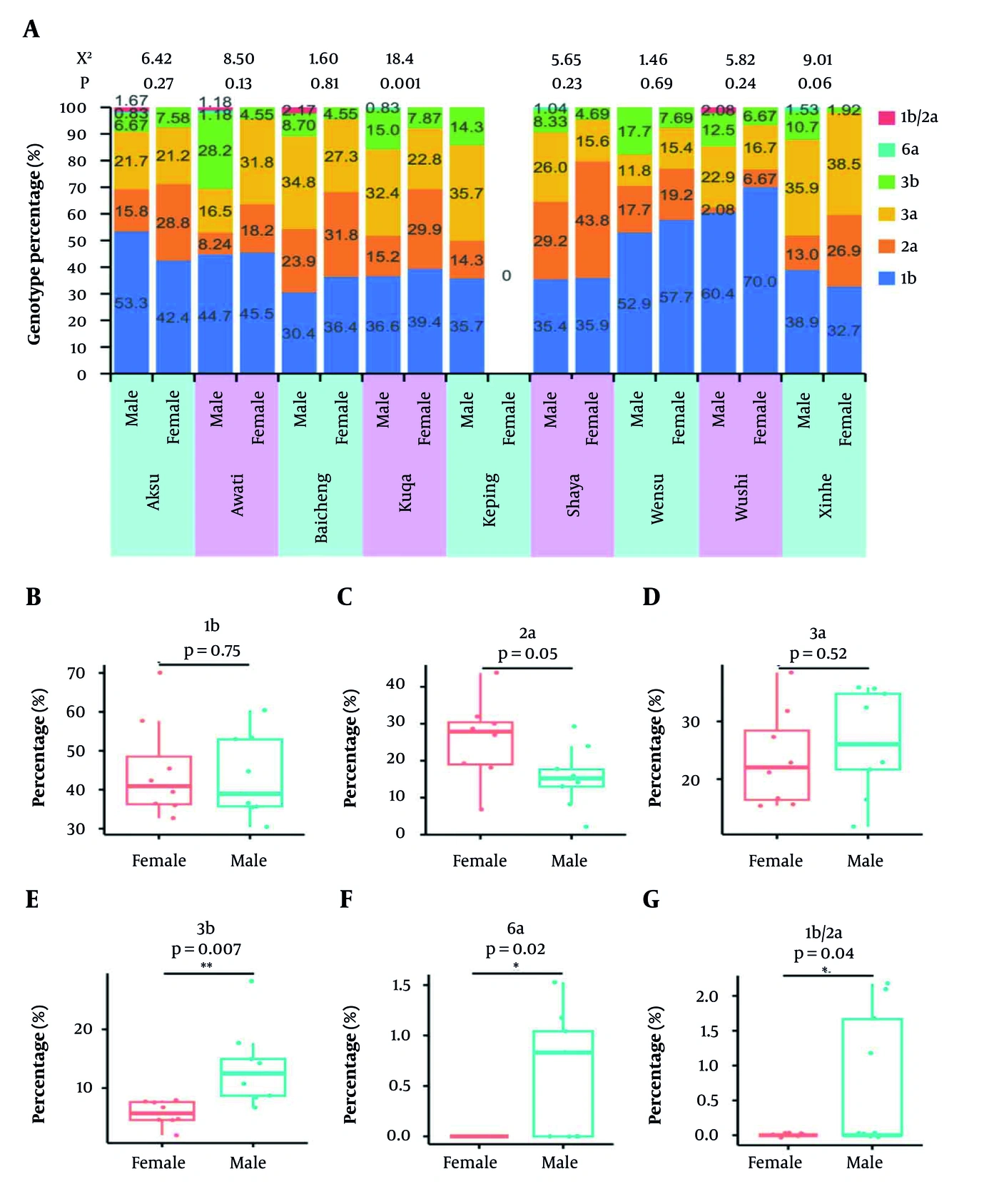
Figure 1.
Hepatitis C virus (HCV) genotype composition by sex and region among HCV-RNA-positive patients. (A) Genotype percentages by sex within each region. In Panel A, the red category labeled “1b/2a” represents mixed-genotype results, comprising mixed 1b/2a (n = 4) and mixed 1b/3b (n = 1). (B–G) Exploratory comparisons of county-level percentages between female and male patients for genotypes 1b, 2a, 3a, 3b, 6a, and the mixed category, respectively. Nominal P values are shown in the figure for exploratory purposes and were not adjusted for multiple comparisons. Detailed counts are reported in Table S2 in the Supplementary File (Parts A–C).
Values are counts and Wilson 95% CIs. Cells with fewer than five observations should be interpreted with caution. Wushi is not shown because individual-level sex data were unavailable for its HCV-RNA-negative patients. No county-specific hypothesis tests were performed. Abbreviations: CI, confidence interval; HCV, hepatitis C virus.
4. Results
4.1. Regional Differences in HCV-RNA Positivity Among Anti-HCV-Positive Patients
The study included 4,735 independent anti-HCV-positive patients with valid HCV-RNA results. HCV RNA was detected in 1,361 patients (28.74%; 95% CI, 27.47% - 30.05%). This proportion reflects HCV-RNA positivity within a hospital-tested anti-HCV-positive cohort, rather than population prevalence. Regional positivity ranged from 8.73% in Wushi County (78/893; 95% CI, 7.06% - 10.77%) to 42.42% in Keping County (14/33; 95% CI, 27.24% - 59.19%) and differed across the nine regions (χ2 = 321.88, df = 8, P < 0.001). Regional counts and CIs are provided in Table S1 in the Supplementary File and shown in Figure 2.
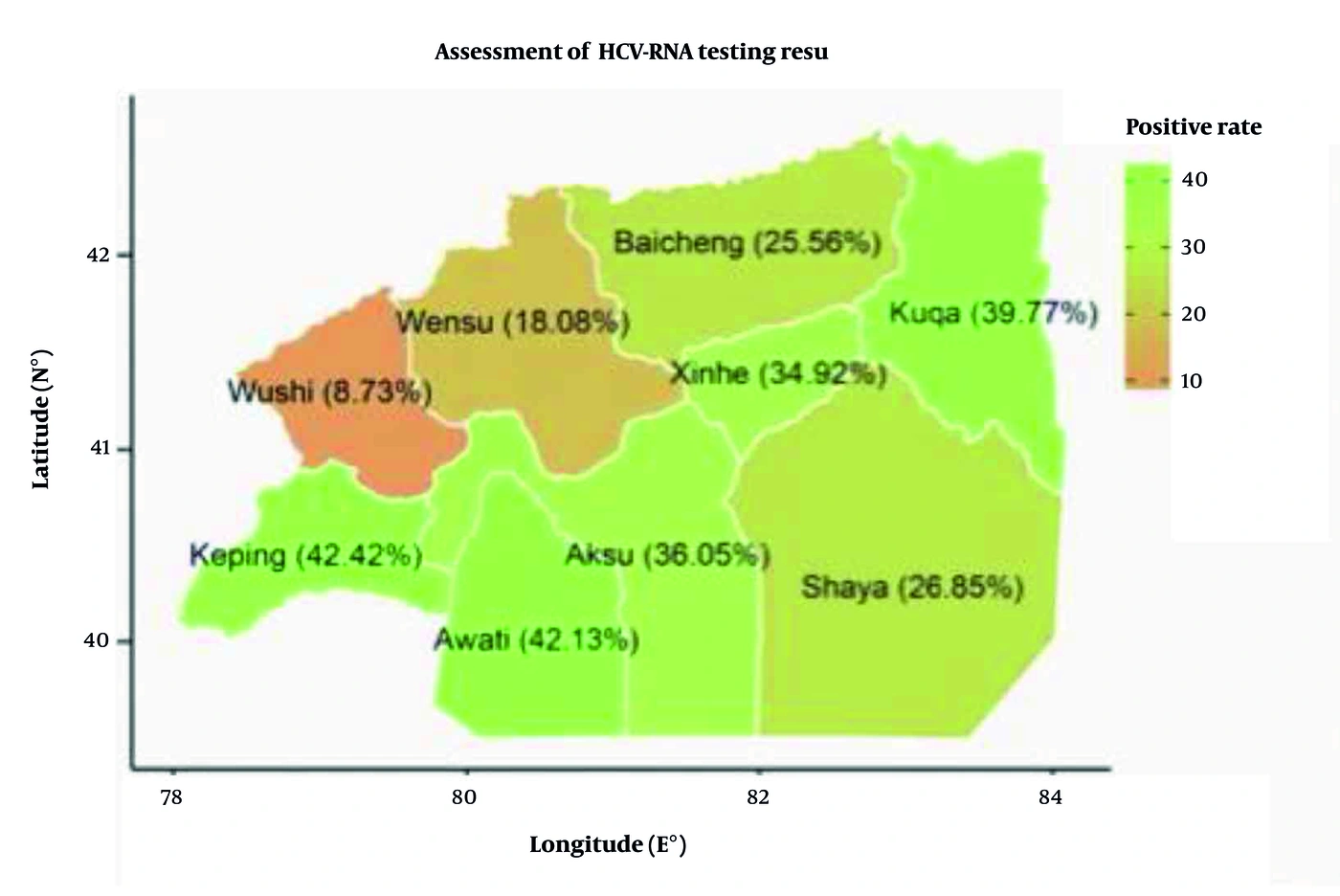
Figure 2.
Regional hepatitis C virus (HCV) RNA positivity among anti-HCV-positive patients in Aksu Prefecture. Wushi County had 78 HCV-RNA-positive results among 893 patients (8.73%); confidence intervals and denominators are reported in Table S1 in the Supplementary File.
4.2. Sex and Age Differences in HCV-RNA Positivity in the Eight-Region Analytical Subset
Among the 3,842 patients with individual-level covariates, 1,283 were HCV-RNA positive (33.39%; 95% CI, 31.92% - 34.90%). Positivity was higher in men (904/2,153; 41.99%; 95% CI, 39.92% - 44.09%) than in women (379/1,689; 22.44%; 95% CI, 20.51% - 24.49%; Yates-corrected χ2 = 161.74, P < 0.001). Positivity also differed across age groups (χ2 = 71.81, P < 0.001) and was highest among patients aged 31 - 40 years (384/924; 41.56%; 95% CI, 38.42% - 44.76%). Figure 3 shows the sex- and age-specific estimates; detailed regional strata and 95% CIs are presented in Table 1 and 2.
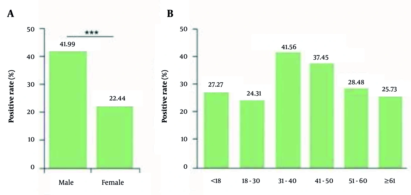
Figure 3.
Hepatitis C virus (HCV) RNA positivity by sex (A) and age group (B) in the eight-region individual-level analytical subset. The age groups include 51 - 60 and ≥ 61 years. Wushi was excluded because individual-level covariates were unavailable for its HCV-RNA-negative patients.
4.3. Descriptive Sex- and Age-Specific HCV-RNA Positivity by Region
Table 1 and 2 present county-by-sex and county-by-age estimates with Wilson 95% CIs and cautions regarding sparse cells. County-specific hypothesis tests were not performed to avoid interpreting numerous unadjusted comparisons as confirmatory.
Regional patterns were heterogeneous. Higher positivity among men was observed in several regions, whereas age-specific patterns varied. Estimates for Keping and several younger age strata were imprecise because of small denominators. Detailed values are provided in Tables 1 and 2 and are not repeated for each county in the text.
4.4. Multivariable Analysis of HCV-RNA Positivity
In the multivariable model including 3,842 patients, sex, age group, and region were each associated with HCV-RNA positivity in global likelihood-ratio tests (all P < 0.001). Men had higher adjusted odds than women (aOR, 2.22; 95% CI, 1.91 - 2.57), and patients aged 31 - 40 years had higher adjusted odds than those aged ≥ 61 years (aOR, 1.75; 95% CI, 1.41 - 2.16). The complete model is shown in Table 3.
Table 3.
Multivariable Logistic Regression of HCV-RNA Positivity in the Eight-Region Analytical Subset a
| Variable and Comparison | aOR | 95% CI | P Value |
|---|---|---|---|
| Sex | |||
| Male vs female | 2.22 | 1.91 - 2.57 | < 0.001 |
| Age group (y) | |||
| 51 - 60 vs ≥ 61 | 1.13 | 0.90 - 1.41 | 0.283 |
| 41 - 50 vs ≥ 61 | 1.41 | 1.15 - 1.74 | 0.001 |
| 31 - 40 vs ≥ 61 | 1.75 | 1.41 - 2.16 | < 0.001 |
| 18 - 30 vs ≥ 61 | 0.85 | 0.55 - 1.29 | 0.443 |
| < 18 vs ≥ 61 | 0.91 | 0.34 - 2.43 | 0.853 |
| Region | |||
| Awati vs Aksu | 1.07 | 0.78 - 1.47 | 0.672 |
| Baicheng vs Aksu | 0.59 | 0.42 - 0.83 | 0.002 |
| Keping vs Aksu | 0.97 | 0.47 - 2.01 | 0.936 |
| Kuqa vs Aksu | 1.08 | 0.86 - 1.34 | 0.516 |
| Shaya vs Aksu | 0.67 | 0.51 - 0.87 | 0.002 |
| Wensu vs Aksu | 0.42 | 0.31 - 0.58 | < 0.001 |
| Xinhe vs Aksu | 0.91 | 0.70 - 1.18 | 0.484 |
a Model n = 3,842 with 1,283 HCV-RNA-positive outcomes. Sex, age group, and region were included simultaneously. Reference categories were female sex, age ≥ 61 years, and Aksu City. Wushi was excluded because individual-level sex and age data were unavailable for its HCV-RNA-negative patients. Global likelihood-ratio tests for sex, age group, and region were all P < 0.001. Abbreviations: aOR, adjusted odds ratio; CI, confidence interval; HCV, hepatitis C virus.
4.5. Analysis of HCV Genotypes in Aksu Prefecture
All 1,361 HCV-RNA-positive patients had a recorded genotype result. Genotype 1b was the most frequent (566/1,361; 41.59%; 95% CI, 39.00% - 44.23%), followed by 3a (362/1,361; 26.60%; 95% CI, 24.32% - 29.01%), 2a (266/1,361; 19.54%; 95% CI, 17.52% - 21.74%), and 3b (154/1,361; 11.32%; 95% CI, 9.74% - 13.11%). Rare genotypes and mixed patterns are reported in Table S2 in the Supplementary File (Parts A–C).
Figure 1 presents genotype composition by sex and region. Panel A shows genotype percentages by sex within each region, and Panels B–G show secondary exploratory comparisons of county-level percentages. These comparisons were considered exploratory and were not used for confirmatory inference.
5. Discussion
In this retrospective hospital-based study, HCV RNA was detected in 28.74% of 4,735 anti-HCV-positive patients tested during 2024. The estimate of 33.39% reflects the eight-region individual-level analytical subset, not the complete cohort. Sex, age group, and region remained associated with HCV-RNA positivity in the adjusted eight-region analysis. These estimates describe a clinical convenience sample and should not be interpreted as population prevalence in Aksu Prefecture.
Genotype 1b remained the most frequent subtype, followed by 3a, 2a, and 3b. This ordering is broadly consistent with reports from China, although regional genotype composition is heterogeneous (13-15). Figure 1 presents these distributions descriptively; the data do not establish transmission routes or causal explanations for regional differences.
The clinical role of genotype has changed in the DAA era. Pan-genotypic regimens achieve high cure rates and have reduced routine reliance on genotype-guided treatment. However, genotype may remain relevant in selected patients, including those with genotype 3 infection, cirrhosis, previous DAA failure, or resistance-associated substitutions (16-19). A recent hospital-based study from Kashi also reported gaps in confirmatory HCV-RNA testing and access to DAA treatment, illustrating how healthcare pathways can shape regional hospital data (14). A study of Iranian blood donors comparing RT-qPCR with HCV core antigen likewise emphasizes the practical role of confirmatory testing for active infection (5), while COVID-19-era evidence underscores the need to sustain hepatitis services during broader health-system disruptions (6). Because treatment regimens and sustained virological response were not available in this study, treatment outcomes cannot be inferred from the observed genotype distribution.
County-level correlations between genotype proportions and HCV-RNA positivity were not performed because genotype proportions were derived from HCV-RNA-positive cases and therefore could not establish individual-level associations. Such aggregate comparisons are also vulnerable to ecological bias. Future studies examining genotype, transmission, or clinical outcomes should integrate individual epidemiological data with viral whole-genome sequencing and longitudinal treatment follow-up.
This study has several limitations. It was a single-center clinical convenience sample; the total number screened for anti-HCV was not available; and Wushi covariates were unavailable for HCV-RNA-negative patients. Socioeconomic status, healthcare access, migration, transfusion history, injection drug use, hemodialysis, previous DAA treatment, and other clinical or behavioral factors were not consistently recorded, leaving residual confounding. This limitation is important because HCV burden can be concentrated in high-risk populations, including people who use or inject drugs (7, 20). The commercial genotyping assay covered a limited set of subtypes, and no sequencing was performed. Sparse strata yielded imprecise estimates, and the observational design supports associations rather than causation.
In conclusion, among 4,735 anti-HCV-positive patients tested at a regional hospital in Aksu Prefecture during 2024, 28.74% had detectable HCV RNA. HCV-RNA positivity varied across demographic and regional strata, and genotype 1b was the most frequent. These findings may support local confirmatory-testing and molecular-surveillance activities but should not be generalized to the Aksu population.