
1. Background
Colorectal cancer is among the most commonly diagnosed malignancies and a leading cause of cancer-related death worldwide. GLOBOCAN 2022 estimated approximately 1.93 million new cases and 0.90 million deaths globally, including 12 899 incident cases in Iran, underscoring the importance of regional molecular epidemiology (1, 2).
Colorectal cancer is molecularly heterogeneous, and defects in the DNA mismatch repair (MMR) system represent one of its most clinically relevant molecular pathways. MMR deficiency can be assessed by the immunohistochemical loss of MMR protein expression, whereas microsatellite instability is evaluated using molecular methods such as polymerase chain reaction (PCR) or validated next-generation sequencing. Although deficient MMR (dMMR) and high microsatellite instability (MSI-H) status are strongly associated, they reflect results from different testing approaches and are not completely interchangeable (3, 4). In routine pathology practice, dMMR can be identified by immunohistochemistry (IHC) for MLH1, PMS2, MSH2, and MSH6, with loss of nuclear tumor staining interpreted only in the presence of appropriate internal positive controls. Because these proteins function as heterodimeric partners (MLH1 with PMS2 and MSH2 with MSH6), characteristic paired-loss patterns are expected, whereas isolated losses have specific clinical implications and recognized diagnostic pitfalls (5).
Assessment of MMR status has become an essential component of colorectal cancer evaluation. Guidance from the National Institute for Health and Care Excellence (NICE) recommends offering MMR/microsatellite instability testing to all patients with colorectal cancer at initial diagnosis, with defined downstream steps to distinguish sporadic MLH1-altered tumors from Lynch-associated cancers and to direct germline evaluation when appropriate (6). Professional guidance from multidisciplinary organizations similarly recommends MMR testing in colorectal cancer for Lynch syndrome risk assessment and prognostic stratification (7). In addition, dMMR/MSI-H status has major therapeutic implications, given the established efficacy of immune checkpoint inhibitors in advanced colorectal cancer and the emerging role of immunotherapy in localized dMMR rectal and colon cancers (8, 9).
Regional Iranian studies have examined MMR-related gene expression in colorectal cancer; however, population-level data on dMMR prevalence and immunophenotypic patterns remain limited (10). In addition, incomplete reporting of clinically relevant colorectal cancer pathology variables has been documented in national Iranian registry data (11).
2. Objectives
The primary aim of this study was to estimate the prevalence of dMMR among colorectal adenocarcinoma cases diagnosed and tested in Tabriz, Iran, over a 2-year period and to describe the distribution of MMR protein-loss patterns. Secondary aims were to explore associations between dMMR status and year of diagnosis, patient age, and sex, thereby providing foundational data for future outcomes studies and for planning biomarker-driven clinical trials.
3. Methods
3.1. Study Design and Case Selection
This retrospective cross-sectional study included all patients with histopathologically confirmed primary colorectal adenocarcinoma whose tissue specimens were evaluated at a specialized private pathology laboratory in Tabriz, Northwestern Iran. Cases were identified by retrospectively querying the laboratory's electronic database for pathology reports containing a diagnosis of colorectal adenocarcinoma. The study period began in May 2023, when the laboratory implemented routine immunohistochemical screening for MMR proteins, and ended in December 2024.
The cohort was assembled consecutively: every patient diagnosed after implementation of the IHC screening system who met the eligibility criteria was included in chronological order, without omission, to minimize selection bias.
The patient was the primary unit of analysis. To ensure independent observations, the analysis was restricted to 1 representative formalin-fixed, paraffin-embedded (FFPE) tissue block per patient. For surgical resections with multiple available blocks, or cases with both diagnostic biopsy and surgical resection specimens, the study pathologist evaluated and selected a single slide from the block with the highest viable tumor cellularity. The cohort was restricted to patients with solitary colorectal adenocarcinomas; patients with synchronous tumors were excluded.
A total of 902 consecutive patients were initially identified during this period. Patients were sequentially excluded for a history of neoadjuvant therapy (n = 15), prior treatment for colorectal cancer (n = 12), a borderline or uncertain pathological diagnosis (n = 11), or unavailable or exhausted FFPE blocks (n = 9), resulting in 47 exclusions. During the final evaluation, 2 additional cases were excluded because of incomplete or uninterpretable immunohistochemical assessment. The final analytic cohort therefore comprised 853 distinct patients (Figure 1).
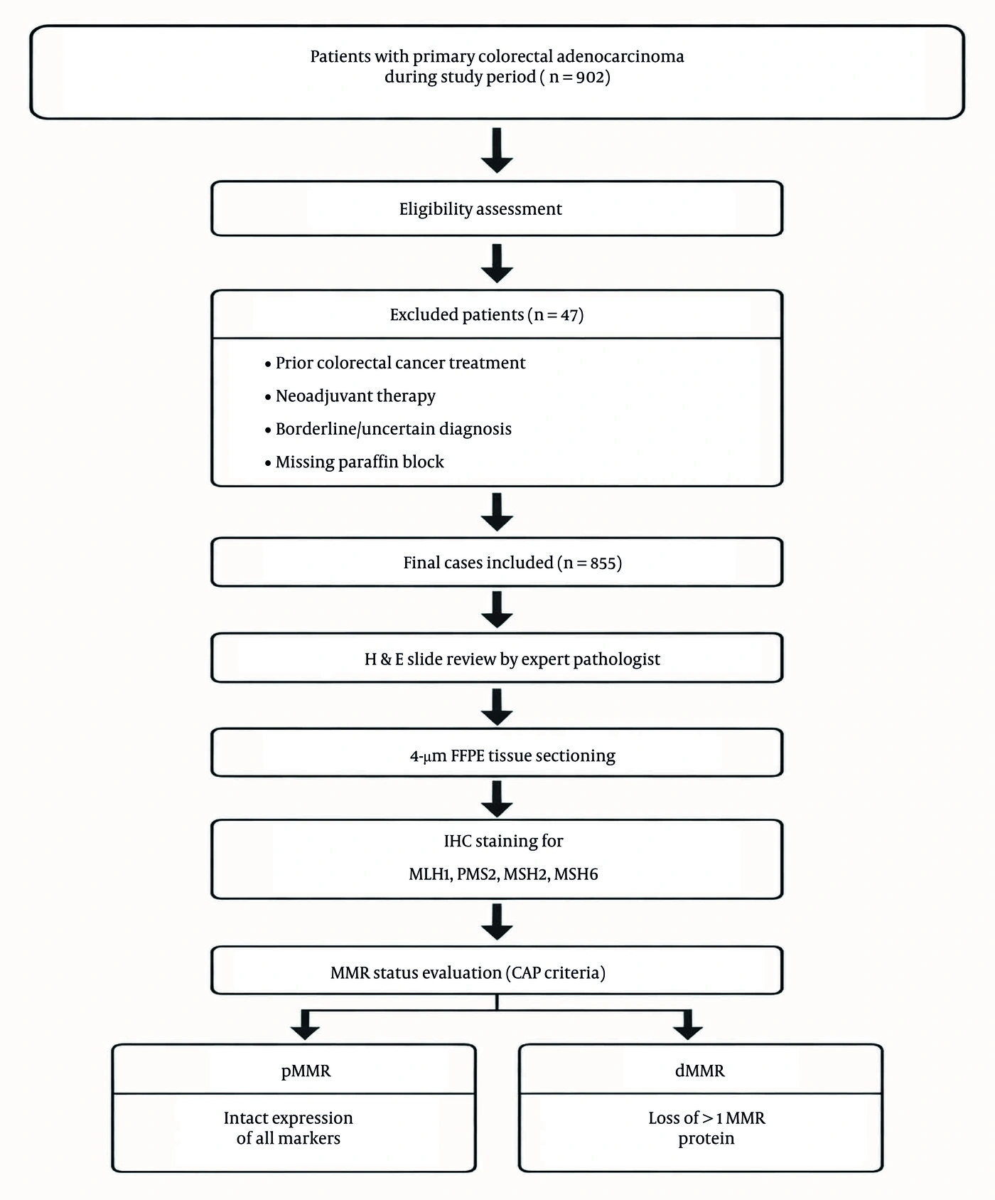
Figure 1.
Flowchart of the participant selection process. The diagram shows the derivation of the consecutive analytic cohort, the case-identification pathway, and the sequential exclusions.
3.2. Histopathology and Immunohistochemistry
FFPE blocks were sectioned at 4 µm using a SLEE CUT 5062+ microtome (SLEE medical GmbH, Germany). IHC staining was performed using a fully automated VitroStainer 42© platform (Vitro S.A., Spain). Ready-to-use primary monoclonal antibodies from Vitro Master Diagnóstica (Vitro S.A., Spain) included MLH1 (clone BS29), PMS2 (clone EP51), MSH2 (clone RBT-MSH2), and MSH6 (clone EP49). Heat-induced antigen retrieval was performed on the automated platform using Tris-EDTA buffer (High AR, pH 9.0). Detection was performed using the Master Polymer Plus Detection System with a diaminobenzidine (DAB) chromogen, followed by hematoxylin counterstaining.
Slides were interpreted by a single experienced gastrointestinal pathologist (A.V.), without an independent second reader or consensus verification. Because the study involved retrospective review of routinely processed specimens and finalized pathology records, the interpreting pathologist was not blinded to the corresponding diagnostic hematoxylin and eosin findings.
MMR protein status was evaluated by comparing nuclear staining in neoplastic cells with on-slide internal positive controls, including adjacent non-neoplastic colonic mucosa, stromal cells, and infiltrating lymphocytes. Proficient MMR (pMMR) was defined as retained nuclear expression of all 4 MMR proteins in tumor cells. Deficient MMR was defined as complete loss of nuclear expression of at least 1 MMR protein in tumor cells, with unequivocally retained nuclear staining in appropriate internal control cells. Cases in which internal controls did not demonstrate adequate nuclear staining were considered technically uninterpretable and excluded from analysis.
Because the study used retrospectively collected, routinely processed archival specimens, case-specific cold ischemia times and formalin fixation durations were not systematically recorded in the analytic database. Consequently, the uniformity of these preanalytical intervals across the study period could not be verified. The analytical IHC procedure was standardized using the same automated staining platform, antibody clones, antigen-retrieval conditions, detection system, and interpretation criteria. On-slide internal positive controls were evaluated for every interpretable case. However, dedicated case-level external positive/negative control slides, routine repeat immunostaining, alternative-block testing, and orthogonal molecular confirmation were not systematically performed specifically for isolated or discordant MMR-loss patterns. Unusual patterns, such as isolated PMS2 loss, were therefore classified strictly according to the observed immunohistochemical phenotype.
3.3. Statistical Analysis
Variables available for analysis included age, sex, diagnosis year, overall MMR status, and patterns of MLH1, PMS2, MSH2, and MSH6 loss. Tumor location, histologic grade, pathological stage, histologic subtype, and other detailed clinicopathological variables were not systematically captured in the analytic database and could not be reliably included in the statistical analyses. Continuous variables were summarized as mean ± standard deviation or median (interquartile range), and categorical variables as frequencies and percentages. The 95% confidence intervals (CIs) for prevalence estimates were calculated using the Wilson score interval method. Group comparisons used Welch's t-test or the chi-square test, as appropriate.
An exploratory multivariable logistic regression analysis assessed the association between diagnosis year and dMMR status. Age and sex were included as covariates because they were the only relevant patient-level variables systematically available for the entire cohort. The absence of tumor location, grade, stage, and other clinicopathological characteristics precluded adjustment for these potential confounders. To assess model validity, diagnostics included evaluation of linearity between age and the logit of the outcome using the Box-Tidwell procedure, goodness of fit using the Hosmer-Lemeshow test, and influential observations using Cook's distance. A 2-sided P value < 0.05 was considered statistically significant. Analyses were conducted using SPSS version 24 (IBM Corp., Armonk, NY, USA), and figures were generated using Python version 3. The final analytic dataset was complete for MMR protein results, age, sex, and diagnosis year. All analyses therefore used complete cases (n = 853), and no imputation was required.
4. Results
A total of 853 patients with colorectal adenocarcinoma were included in the final analysis. Of these, 103 were classified as dMMR, yielding an overall prevalence of 12.1% (95% CI, 10.1% - 14.4%). The remaining 750 were classified as pMMR. Clinicodemographic characteristics by MMR status are summarized in Table 1.
Table 1.
Clinicodemographic Characteristics of Patients According to MMR Status a
| Characteristic | Total Cohort (n = 853) | pMMR (n = 750) | dMMR (n = 103) | P Value |
|---|---|---|---|---|
| Age, y | ||||
| Mean ± SD | 59.35 ± 12.44 | 59.39 ± 12.48 | 59.02 ± 12.22 | 0.773 |
| Median (IQR) | 61 (51 - 68) | 61 (51 - 68) | 60 (52 - 67.5) | - |
| Gender | 0.538 | |||
| Male | 459 (53.8) | 407 (54.3) | 52 (50.5) | |
| Female | 394 (46.2) | 343 (45.7) | 51 (49.5) | |
| Diagnosis year | 0.003 | |||
| 2023 | 352 (41.3) | 295 (39.3) | 57 (55.3) | |
| 2024 | 501 (58.7) | 455 (60.7) | 46 (44.7) |
a Values are expressed No. (%) unless otherwise indicated. Abbreviations: MMR, mismatch repair; dMMR, deficient mismatch repair; IQR, interquartile range; pMMR, proficient mismatch repair; SD, standard deviation.
Age and sex were not significantly associated with dMMR (age, P = 0.773; sex, P = 0.538). The prevalence of dMMR also did not differ significantly across age groups (P = 0.935) but was higher in 2023 than in 2024 (16.2% vs. 9.2%; P = 0.003) (Table 1, Figure 2).
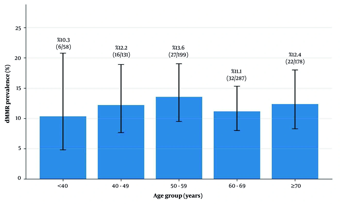
Figure 2.
Prevalence of deficient mismatch repair according to age group. Bars represent the percentage of dMMR cases within each age category, and error bars indicate Wilson 95% confidence intervals.
The reason for the difference in dMMR prevalence between the 2 study periods could not be determined from the available data. Referral source, indication for MMR testing, tumor location, histologic grade, stage, and other potential case-mix indicators were not systematically available for comparison between 2023 and 2024. This temporal difference should therefore be regarded as an unexplained observational finding rather than evidence of a true change in population-level dMMR prevalence.
Among dMMR tumors, combined MLH1/PMS2 loss (35.0%) and isolated PMS2 loss (30.1%) were the predominant patterns. Isolated MSH2 and MLH1 loss accounted for 11.7% and 9.7%, respectively, while combined MSH2/MSH6 loss accounted for 6.8%. The remaining patterns were uncommon (Table 2, Figure 3).
Table 2.
Descriptive Frequencies of Immunohistochemical MMR Protein-Loss Patterns Among dMMR Tumors (N = 103) a
| Loss Pattern | No. | Percentage of dMMR (n = 103) b |
|---|---|---|
| Isolated MLH1 | 10 | 9.7 |
| Isolated PMS2 | 31 | 30.1 |
| Isolated MSH2 | 12 | 11.7 |
| Isolated MSH6 | 2 | 1.9 |
| MLH1 + PMS2 | 36 | 35.0 |
| MLH1 + MSH6 | 1 | 1.0 |
| PMS2 + MSH2 | 2 | 1.9 |
| PMS2 + MSH6 | 1 | 1.0 |
| MSH2 + MSH6 | 7 | 6.8 |
| MLH1 + PMS2 + MSH2 | 1 | 1.0 |
a Abbreviations: dMMR, deficient mismatch repair; MMR, mismatch repair.
b Data are presented strictly descriptively. Because several atypical loss categories contain few cases, formal confidence intervals are not provided to avoid implying undue statistical precision.
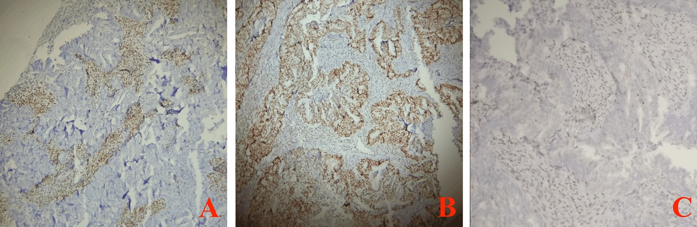
Figure 3.
Distribution of the five most frequent MMR protein-loss patterns and pooled less common patterns among dMMR colorectal adenocarcinomas.
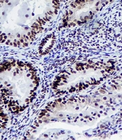
Figure 4 shows representative immunohistochemical examples of MMR protein expression patterns identified in the study cohort, including loss of tumor nuclear staining with preserved internal positive controls.
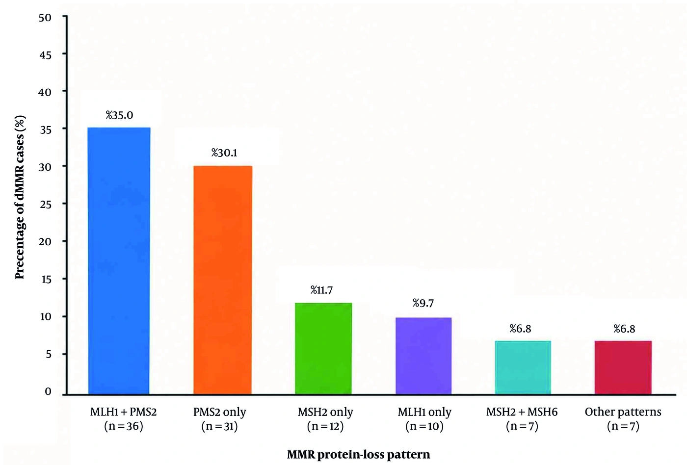
Figure 4.
Representative immunohistochemical staining patterns of mismatch repair proteins in colorectal adenocarcinoma. A, MLH1 showing loss of nuclear staining in tumor cells with preserved nuclear staining in internal control stromal/inflammatory cells; B, MSH6 showing retained nuclear staining in internal control cells with loss of staining in tumor cells, consistent with deficient mismatch repair; C, PMS2 showing loss of nuclear tumor staining with preserved internal positive controls. These panels illustrate representative abnormal MMR immunohistochemical patterns identified in the study cohort.
In multivariable analysis, diagnosis year remained associated with dMMR (2024 vs. 2023: odds ratio [OR], 0.52; 95% CI, 0.34 - 0.79; P = 0.002), whereas age and sex were not associated with dMMR (Table 3). Model diagnostics indicated adequate fit, linearity for age, and no highly influential observations.
Table 3.
Multivariable Logistic Regression Analysis of Factors Associated with dMMR Status
| Predictor | OR (95% CI) a | P Value |
|---|---|---|
| Diagnosis year (2024 vs. 2023) | 0.52 (0.34 - 0.79) | 0.002 |
| Age (Continuous, per 1-year increase) | 0.99 (0.98 - 1.01) | 0.778 |
| Sex (Reference: Female) | 0.85 (0.56 - 1.28) | 0.431 |
a The multivariable logistic regression model used complete-case analysis (n = 853).
5. Discussion
This retrospective cross-sectional study evaluated the prevalence and immunohistochemical patterns of mismatch repair deficiency in a large cohort of colorectal adenocarcinoma cases from Northwestern Iran. Overall, dMMR was identified in 12.1% of cases, indicating that approximately 1 in 8 colorectal adenocarcinomas in this cohort showed loss of at least 1 MMR protein by IHC.
The observed dMMR prevalence of 12.1% is consistent with global epidemiological estimates, which typically indicate dMMR in 10% - 15% of sporadic colorectal adenocarcinomas (12-14). Regionally, this rate is slightly lower than the 14% prevalence reported in an early Iranian cohort by Molaei et al. but remains consistent with the broader Middle Eastern literature (15, 16). Minor variations in these regional estimates are expected and likely reflect differences in institutional testing criteria, particularly the transition from targeted, age-restricted screening to universal screening, as well as differences in the sensitivity of immunohistochemical platforms used across different periods.
Our findings support the view that the prevalence of dMMR colorectal adenocarcinoma in Northwestern Iran is generally consistent with national and international reports. These regional data are clinically relevant because universal MMR testing requires appropriate laboratory capacity, standardized interpretation, and clear referral pathways for patients with abnormal findings. Combined MLH1/PMS2 loss was the most frequent abnormal immunohistochemical pattern, accounting for 35.0% of dMMR cases. This finding is biologically plausible and consistent with the heterodimeric relationship between MLH1 and PMS2, in which loss of MLH1 commonly leads to secondary loss of PMS2 expression. Previous studies have similarly identified MLH1/PMS2 co-loss as the dominant abnormal staining pattern in many colorectal cancer screening cohorts, particularly in sporadic dMMR tumors associated with MLH1 promoter hypermethylation (5, 17, 18). Combined MSH2/MSH6 loss was less frequent in our cohort, consistent with its stronger association with Lynch syndrome-related hereditary dMMR.
A notable finding was the unexpectedly high frequency of isolated PMS2 loss. This pattern occurred in 31 of 103 dMMR tumors (30.1%), corresponding to 31 of 853 tumors (3.6%) in the overall cohort. Isolated PMS2 loss is generally considered an uncommon MMR immunophenotype; a large multicenter series reported this pattern in approximately 4% of colorectal carcinomas with microsatellite instability (19). The comparatively high proportion among dMMR tumors in our cohort therefore requires cautious interpretation.
Both biological and technical mechanisms may contribute to atypical MMR immunophenotypes. Preanalytical conditions are particularly relevant because experimental evaluation of colorectal cancer specimens has shown that formalin hypofixation and hyperfixation can adversely affect MMR IHC, with MLH1 and PMS2 among the proteins most susceptible to reduced staining intensity and other staining abnormalities (20). In this retrospective cohort, case-specific cold ischemia and fixation times were not systematically recorded, and isolated-loss cases did not routinely undergo repeat staining, alternative-block assessment, or orthogonal molecular confirmation. Consequently, the unexpectedly high frequency of isolated PMS2 loss cannot be assumed to represent a molecularly confirmed PMS2-deficient subgroup. These results should be interpreted strictly as observed immunohistochemical phenotypes requiring further technical and molecular validation.
Because this study relied exclusively on immunohistochemical screening without orthogonal molecular validation, these data cannot be used to estimate the prevalence of Lynch syndrome in the cohort. Without accompanying PCR-based microsatellite instability testing, MLH1 promoter hypermethylation analysis, BRAF V600E mutational profiling, or germline sequencing, we cannot definitively classify any dMMR tumor as sporadic or hereditary.
The prevalence of dMMR was significantly higher in 2023 than in 2024 (16.2% vs. 9.2%), and the diagnosis year remained associated with dMMR status after adjustment for age and sex. However, the underlying reason for this temporal difference could not be determined from the available data. The study database did not systematically capture referral source, indication for MMR testing, tumor location, histologic grade, pathological stage, or other variables needed to assess changes in case mix between the 2 periods. The difference may reflect unmeasured sampling or referral characteristics, operational factors, or random temporal variation, but none of these explanations can be established from this study. The pooled dMMR prevalence estimate of 12.1% should therefore be interpreted in light of this temporal heterogeneity, and confirmation in longer-term multicenter cohorts would improve its generalizability.
Clinically, these findings reinforce the importance of routine MMR testing in colorectal cancer. Current guidelines recommend universal MMR or microsatellite instability assessment in newly diagnosed colorectal carcinoma because of its role in Lynch syndrome screening, prognostic stratification, and treatment selection (6, 7). Deficient MMR and molecularly defined MSI-H are clinically actionable biomarkers, given the efficacy of immune checkpoint inhibitors in advanced disease and their emerging role in localized colorectal cancer (8, 9). In this study, however, MMR status was determined exclusively by IHC, and no molecular microsatellite instability testing was performed. Identification of a substantial dMMR subgroup in this regional cohort supports the need for standardized pathology-based screening and appropriate downstream molecular or genetic evaluation when clinically indicated.
5.1. Study Limitations
This study has several limitations. First, its retrospective, single-center design may limit generalizability beyond the source population. Second, important clinicopathological variables, including tumor location, histologic grade, pathological stage, histologic subtype, specimen type, detailed treatment characteristics, family history, and survival outcomes, were not systematically available in the analytic database. Their absence precluded evaluation of established clinicopathological correlates of dMMR and limited adjustment for potential confounding. Third, case-specific cold ischemia times and formalin fixation durations were not systematically recorded, precluding direct assessment of the contribution of preanalytical variation to the observed staining patterns. This limitation is particularly relevant to PMS2 because MMR IHC, including PMS2 staining, can be affected by suboptimal fixation conditions. Fourth, IHC interpretation was performed by a single experienced gastrointestinal pathologist without independent second-reader or consensus verification. Interobserver reliability therefore could not be assessed, particularly for weak, focal, heterogeneous, or otherwise borderline staining patterns. Fifth, isolated and discordant MMR-loss patterns were not systematically reassessed using repeat immunostaining, alternative tumor blocks, or orthogonal molecular testing. MSI-PCR, MLH1 promoter methylation analysis, BRAF V600E testing, and germline sequencing were not performed. This limitation is particularly important given the unexpectedly high proportion of isolated PMS2 loss among dMMR tumors and means that the observed IHC phenotype cannot be assumed to represent molecular MSI-H status or a specific hereditary mechanism. The study therefore cannot determine the prevalence of Lynch syndrome or distinguish sporadic from hereditary causes of dMMR. Finally, the significant difference in dMMR prevalence between 2023 and 2024 remains unexplained because the referral source, indication for testing, tumor location, stage, grade, and other potential measures of case mix were unavailable. This temporal heterogeneity introduces further uncertainty regarding the generalizability of the pooled prevalence estimate.
5.2. Conclusions
In this retrospective cohort from Northwestern Iran, dMMR was identified by IHC in 12.1% of colorectal adenocarcinoma cases. Combined MLH1/PMS2 loss was the most frequent abnormal pattern, whereas isolated PMS2 loss occurred unexpectedly often among dMMR tumors. Because molecular microsatellite instability testing and systematic confirmatory assessment of isolated-loss patterns were not performed, these findings should be interpreted strictly as immunohistochemical phenotypes rather than as molecularly confirmed MSI-H or hereditary MMR abnormalities. The unexplained variation in dMMR prevalence between study years and the absence of several clinicopathological covariates support the need for multicenter studies incorporating standardized preanalytical protocols, independent pathological review, and orthogonal molecular validation.