Rhabdomyosarcoma of Cervix: A Case Report

Author(s):
Maryam Sadat HosseiniMaryam Sadat Hosseini1, Tahereh AshrafganjoeiTahereh Ashrafganjoei1, Ainaz SouratiAinaz Sourati2,*, Morteza TabatabeifarMorteza Tabatabeifar2, Mahdiss MohamadianamiriMahdiss Mohamadianamiri1
1Department of Obstetrics and Gynecology, Preventative Gynecology Research Center, Imam Hossein Hospital, Shahid Beheshti University of Medical Sciences, Tehran, IR Iran
2Department of Radiotherapy and Oncology, Imam Hossein Hospital, Shahid Beheshti University of Medical Sciences, Tehran, IR Iran

International Journal of Cancer Management:Vol. 9, issue 3; e4383
Published online:Jun 18, 2016
Article type:Case Report
Received:Oct 20, 2015
Accepted:May 30, 2016
How to Cite:Hosseini MS, Ashrafganjoei T, Sourati A, Tabatabeifar M, Mohamadianamiri M. Rhabdomyosarcoma of Cervix: A Case Report. Int J Cancer Manag. 2016;9(3):e4383. doi: https://doi.org/10.17795/ijcp-4383

Abstract

Introduction:

Rhabdomyosarcoma has known as a highly malignant soft tissue sarcoma. It has been the most common soft tissue sarcoma in childhood, accounting for about 3 to 4 % of all cases of childhood cancer. Rhabdomyosarcoma was rare in adults, accounting for 3% of all soft-tissue sarcomas. embryonal rhabdomyosarcoma of female genital tract including uterine cervix in an adult was rare.

Case Presentation:

This study has reported a 33-year-old woman presented with abnormal vaginal discharge. Gynecologic examination revealed a cervical mass with grape- like feature protruding into vagina with posterior- superior vaginal wall involvement. Biopsy has performed and pathologic examination was consistent with embryonal botryoid type rhabdomyosarcoma. She has undergone the staging work up measurements including thoracic computed tomography (CT) scan, abdominopelvic magnetic resonance imaging (MRI), bone scan and bone marrow examination. In exception of abdominopelvic MRI, with 2 suspicious pelvic lymph nodes in addition of cervical mass, all others were normal. Radical hysterectomy with lymph node debulking and ovarian preservation has performed. Final results have shown embryonal botryoid type rhabdomyosarcoma of cervix. ovaries, endometrium, parametrium, and follopian tubes were unremarkable. Pelvic lymph nodes pathology and intraabdominal fluid cytology were negative for malignancy. Lymphovascular invasion was identified. She has advised for adjuvant chemotherapy.

Conclusions:

This case has reminded that embryonal rhabdomyosarcoma could occur in uncommon site and older female. Longer follow up of these cases has required due to lack of survival data for embryonal rhabdomyosarcoma of this site and age group.

1. Introduction

Rhabdomyosarcoma was a highly malignant soft tissue sarcoma which arised from embryonic muscle cells (1). It has known as the most common soft tissue sarcoma in childhood, accounting for about 3 to 4 % of all cases of childhood cancer. Approximately 350 new cases have diagnosed each year in patients younger than 20 years (2). Rhabdomyosarcoma was rare in adults, with soft-tissue sarcomas making up less than 1% of malignancies in adults and rhabdomyosarcoma accounting for 3% of all soft-tissue sarcomas (3). Male to female ratio was 1.5 to 1 and male had slightly better overall survival (4).
The head and neck were the most frequent (35 - 40%) sites of origin, followed by the genitourinary tract, extremities, trunk, retroperitoneum, and uncommon regions (e.g. intrathoracic, GI tract, perianal and anal regions) (5). One of the least common sites for rhabdomyosarcoma in the genitourinary tract is the uterine cervix (6).
Five major histologic subtypes of rhabdomyosarcoma have identified including embryonal, alveolar, botryoid embryonal, spindle cell embryonal, and anaplastic.
The embryonal subtype was the most common subtype in childhood, although there have been a few reports of this neoplasm in adult (7). Most were of the classic subtype, and the botryoid and spindle cell variants comprised 6 and 3 percent, respectively (8). Botryoid rhabdomyosarcoma is a subtype of embryonal rhabdomyosarcoma that could observe in the walls of hollow, mucosa lined structures such as vagina, bladder nasopharynx and rarely cervix and uterine fundus. It has resembled a bunch of grapes. Sarcoma botryoides normally has found in children under 8 years of age (9, 10). However cases of older women with this pathology have also reported (11).
Although adults with rhabdomyosarcoma had generally worse survival than children with similar tumors, cervical botyroid rhabdomyosarcoma, usually occurring in second decade has higher level of survival and better prognosis than vaginal lesions with peak of incidence in infants. The survival rate of vaginal and cervical lesions has reported to be of 96% and 60%, respectively (12).

2. Case Presentation

A 33-year-old woman, G2L2P2, presented with abnormal vaginal discharge. Gynecologic examination has revealed a cervical mass with grape-like feature protruding into vagina with posterior-superior vaginal wall involvement. She had normal menstrual cycles and her past medical history was unremarkable. Cervical mass biopsy has performed and pathologic examination was consistent with embryonal botryoid type rhabdomyosarcoma which has confirmed with pathologic review. Transvaginal sonography has shown a 38 × 27 × 60 mm cervical mass at inferior-posterior portion of cervix with superior vaginal wall infiltration. She has undergone the staging work up measurements including thoracic computed tomography (CT) scan, bone scan and bone marrow examination. In abdominopelvic magnetic resonance imaging (MRI), in addition of cervical mass, 2 suspicious pelvic lymph nodes have revealed. All other measurements were normal. Radical hysterectomy with lymph node debulking and ovarian preservation has performed. There was a grossly huge cervical mass protruding into vagina with surface irregularities measuring 10×10 cm. On sections, there was a gray tumor with spindle cell proliferation, measuring 8×5×2.5 cm. On microscopic examination, tumor has covered by attenuated epithelium with condensed neoplastic cells layer beneath epithelium (cambium layer) and consisted of the spindle shaped embryonal rhabdomyoblasts cells with eosinophilic cytoplasm and atypical nuclei. Final results have shown embryonal botryoid type rhabdomyosarcoma of cervix, consistent with the intergroup RMS study Group I. Ovaries, endometrium, parametrium, and follopian tubes were unremarkable. Pelvic lymph nodes pathology and intraabdominal fluid cytology were negative for malignancy. Lymphovascular invasion has identified. She has advised for adjuvant chemotherapy.
Based on medical ethics committee of Shahid Beheshti University of Medical Sciences, a written informed consent has obtained from the patient for publication of this manuscript and accompanying images.
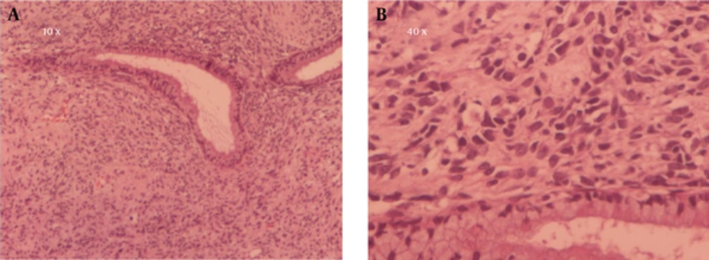
The Benign Edocervical Glands Surrounded By Pleomorphic Spindle to Round Cells with Dark Pyknotic Nuclei and Scant Cytoplasm in Abundant Loose Vascular Myxoid Stroma
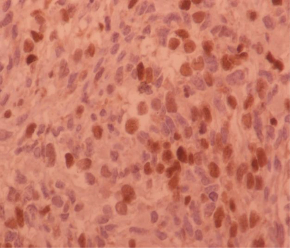
Figure 1.

The Benign Edocervical Glands Surrounded By Pleomorphic Spindle to Round Cells with Dark Pyknotic Nuclei and Scant Cytoplasm in Abundant Loose Vascular Myxoid Stroma

Myogen IHC Staining: Strong Nuclear Positivity Which is Highly Specific for Rhabdomyoblastic Differentiation. Microscopic Image at 40 X
Figure 2.

Myogen IHC Staining: Strong Nuclear Positivity Which is Highly Specific for Rhabdomyoblastic Differentiation. Microscopic Image at 40 X

3. Discussion

Genital tract has been the second most common site of embryonal rhabdomyosarcoma. It has happened in female genital tract in childhood period but rarely in female adults. cervical rhabdomyosarcoma usually has occurred in the second decade however few cases have reported in later ages. Shim et al. has reported a case of cervical embryonal rhabdomyosarcoma in a 52-year-old woman with vaginal bleeding and the feeling of a mass protruding from the introitus (3).
Kaushal et al. has reported a 44-year-old premenopausal female with embryonal rhabdomyosarcoma presented with complaints of bleeding per vagina and irregular menstruation (13).
Our case was a 33-year-old that has been older than the peak incidence age of cervical rhabdomyosarcoma. Presenting symptom was abnormal vaginal discharge.
Dehner et al. has examined 14 case of embryonal rhabdomyosarcoma, shown that the most of patients have presented with cervical mass in the range of 1.5 - 5 cm (14). Our case had a large mass measuring 6 cm.
The surgical treatment for cervical rhabdomyosarcoma could be conservative or radical surgery depending of patient parity and tumor extension. A 52-year-old case has reported with Shim et al., undergone radical abdominal hysterectomy with bilateral salpingo-oophorectomy, and bilateral pelvic lymph node (3). Most case has reported by Dehner et al., treated with local excision (14).
This case has reminded that embryonal rhabdomyosarcoma could occur in uncommon site and older female. Longer follow up of these cases has required due to lack of survival data for embryonal rhabdomyosarcoma of this site and age group.

Acknowledgments

Footnotes

References

  • 1.
    Behtash N, Mousavi A, Tehranian A, Khanafshar N, Hanjani P. Embryonal rhabdomyosarcoma of the uterine cervix: case report and review of the literature. Gynecol Oncol. 2003;91(2):452-5. [PubMed ID: 14599884].
  • 2.
    Jemal A, Siegel R, Ward E, Murray T, Xu J, Thun MJ. Cancer Statistics, 2007. Ca-Cancer J Clin. 2007;57(1):43-66. https://doi.org/10.3322/canjclin.57.1.43.
  • 3.
    Shim AR, Lee M, Paek JH, Kim MJ, Kim SW. A case of embryonal rhabdomyosarcoma of the uterine cervix in a middle-aged woman. Korean J Obstet Gynecol. 2011;54(11):707. https://doi.org/10.5468/kjog.2011.54.11.707.
  • 4.
    Ognjanovic S, Linabery AM, Charbonneau B, Ross JA. Trends in childhood rhabdomyosarcoma incidence and survival in the United States, 1975-2005. Cancer. 2009;115(18):4218-26. [PubMed ID: 19536876]. https://doi.org/10.1002/cncr.24465.
  • 5.
    Crist WM, Anderson JR, Meza JL, Fryer C, Raney RB, Ruymann FB, et al. Intergroup rhabdomyosarcoma study-IV: results for patients with nonmetastatic disease. J Clin Oncol. 2001;19(12):3091-102. [PubMed ID: 11408506].
  • 6.
    Daya DA, Scully RE. Sarcoma botryoides of the uterine cervix in young women: a clinicopathological study of 13 cases. Gynecol Oncol. 1988;29(3):290-304. [PubMed ID: 3278956].
  • 7.
    Lloyd RV, Hajdu SI, Knapper WH. Embryonal rhabdomyosarcoma in adults. Cancer. 1983;51(3):557-65. [PubMed ID: 6821833].
  • 8.
    Okcu MF, Hicks J, Horowitz M. Rhabdomyosarcoma and undifferentiated sarcoma in childhood and adolescence. Netherland: UpToDate; 2006.
  • 9.
    Hilgers RD. Pelvic exenteration for vaginal embryonal rhabdomyosarcoma: a review. Obstet Gynecol. 1975;45(2):175-80. [PubMed ID: 1090863].
  • 10.
    Atlante M, Dionisi B, Cioni M, Di Ruzza D, Sedati P, Mariani L. Sarcoma botryoides of the uterine cervix in a young woman: a case report. Eur J Gynaecol Oncol. 2000;21(5):504-6. [PubMed ID: 11198044].
  • 11.
    Reynolds EA, Logani S, Moller K, Horowitz IR. Embryonal rhabdomyosarcoma of the uterus in a postmenopausal woman. Case report and review of the literature. Gynecol Oncol. 2006;103(2):736-9. [PubMed ID: 16684558]. https://doi.org/10.1016/j.ygyno.2006.03.033.
  • 12.
    Sultan I, Qaddoumi I, Yaser S, Rodriguez-Galindo C, Ferrari A. Comparing adult and pediatric rhabdomyosarcoma in the surveillance, epidemiology and end results program, 1973 to 2005: an analysis of 2,600 patients. J Clin Oncol. 2009;27(20):3391-7. [PubMed ID: 19398574]. https://doi.org/10.1200/JCO.2008.19.7483.
  • 13.
    Kaushal A, Patel A, Shah S, Patel K, Trivedi P. Rhabdomyosarcoma of Uterine Cervix in a 44 year female: A rare presentation. Indian J Med Paediatr Oncol. 2006;27:35-7.
  • 14.
    Dehner LP, Jarzembowski JA, Hill DA. Embryonal rhabdomyosarcoma of the uterine cervix: a report of 14 cases and a discussion of its unusual clinicopathological associations. Mod Pathol. 2012;25(4):602-14. [PubMed ID: 22157934]. https://doi.org/10.1038/modpathol.2011.185.

Similar Articles

26
Sep
2017

Renal Rhabdomyosarcoma in a Young Woman with Gross Hematuria: A Case Report

Farzad Allameh,
Morteza Fallah Karkan,
Azadeh Rakhshan

Allameh F, Fallah Karkan M, Rakhshan A. Renal Rhabdomyosarcoma in a Young Woman with Gross Hematuria: A Case Report. Int J Cancer Manag. 2017;10(9):e10649. doi: https://doi.org/10.5812/ijcm.10649

19
Jun
2002

Extranodal Non-Hodgkin’s Lymphoma of the Uterine Cervix (a Case Report)

N Amirifard

Amirifard N. Extranodal Non-Hodgkin’s Lymphoma of the Uterine Cervix (a Case Report). J Kermanshah Univ Med Sci. 2002;6(1):e80970. doi:

7
Dec
2022
Int J Cancer Manag

Primary Malignant Melanoma of Uterine Cervix with Disseminated Involvement of Lower Genitourinary Tract

Zahra Shiravani,
Minoo Robati,
Ali Ariafar,
Akbar Safaei,
Mojgan Hajisafari Tafti,
Shaghayegh Moradi Alamdarloo

Shiravani Z, Robati M, Ariafar A, Safaei A, Hajisafari Tafti M, et al. Primary Malignant Melanoma of Uterine Cervix with Disseminated Involvement of Lower Genitourinary Tract. Int J Cancer Manag. 2022;15(12):e114156. doi: https://doi.org/10.5812/ijcm-114156

4
Apr
2022
Int J Cancer Manag

Coincidence of Undifferentiated Pleomorphic Sarcoma and Epithelioid Leiomyosarcoma in a Patient; A Case Report

Masoud Mirkazemi,
Pedram Fadavi,
Kambiz Novin,
Mastane Saneii,
Ramyar Hariri,
Maryam Garousi

Mirkazemi M, Fadavi P, Novin K, Saneii M, Hariri R, et al. Coincidence of Undifferentiated Pleomorphic Sarcoma and Epithelioid Leiomyosarcoma in a Patient; A Case Report. Int J Cancer Manag. 2022;15(2):e121892. doi: https://doi.org/10.5812/ijcm.121892

18
Jan
2016

Rhabdomyosarcoma Metastasis to the Breast: Appearance on Diffusion-Weighted Imaging and Apparent Diffusion Coefficient Sequences

Chermaine Deepa Antony,
Kartini Rahmat,
Anushya Vijayananthan,
Jayalakshmi Pailoor

Antony CD, Rahmat K, Vijayananthan A, Pailoor J. Rhabdomyosarcoma Metastasis to the Breast: Appearance on Diffusion-Weighted Imaging and Apparent Diffusion Coefficient Sequences. I J Radiol. 2017;14(1):e13457. doi: https://doi.org/10.5812/iranjradiol.23273

Indexed in

Crossmark
Crossmark
Checking
Share on
Cited by
Metrics

Ordering Reprints

Articles are published under the Creative Commons license stated on each article. No permission or royalty fee is required for uses permitted by that license. CCC handles optional bulk and customized reprint orders. Any quotation covers production and delivery services only, not copyright permission. > Request Reprints from CCC 

Search Relations

Author(s):

Related Articles