
Fulltext
Full text is available in PDF.
International Journal of Cancer Management
The Official Journal of Cancer Research Center (CRC), Shahid Beheshti University of Medical Sciences
Authors
Background: Mammography is the primary imaging technique for detection and diagnosis of breast cancer; however, the contrast of a mammogram image is often poor, especially for dense and glandular tissues. In these cases the radiologist may miss some diagnostically important microcalcifications. In order to improve diagnosis of cancer correctly, image enhancement technology is often used to enhance the image and help radiologists.
Methods: This paper presents a comparative study in digital mammography image enhancement based on four different algorithms: wavelet-based enhancement (Asymmetric Daubechies of order 8), Contrast-Limited Adaptive Histogram Equalization (CLAHE), morphological operators and unsharp masking. These algorithms have been tested on 114 clinical digital mammography images. The comparison for all the proposed image enhancement techniques was carried out to find out the best technique in enhancement of the mammogram images to detect microcalcifications.
Results: For evaluation of performance of image enhancement algorithms, the Contrast Improvement Index (CII) and profile intensity surface area distribution curve quality assessment have been used after any enhancement. The results of this study have shown that the average of CII is about 2.61 for wavelet and for CLAHE, unsharp masking and morphology operation are about 2.047, 1.63 and 1.315 respectively.
Conclusion: Experimental results strongly suggest that the wavelet transformation can be more effective and improve significantly overall detection of the Computer-Aided Diagnosis (CAD) system especially for dense breast. Compare to other studies, our method achieved a higher CII.
© 2012, Author(s). This open-access article is available under the Creative Commons Attribution 4.0 (CC BY 4.0) International License (https://creativecommons.org/licenses/by/4.0/), which allows for unrestricted use, distribution, and reproduction in any medium, provided that the original work is properly cited.
Langarizadeh M, Mahmud R, Ramli A, Napis S, Beikzadeh M, et al. Effects of Enhancement Methods on Diagnostic Quality of Digital Mammogram Images. Int J Cancer Manag. 2010;3(1):e80655. doi:
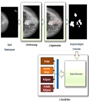
Niroomand Fam B, Nikravanshalmani A, Khalilian M. An Efficient Method for Automated Breast Mass Segmentation and Classification in Digital Mammograms. I J Radiol. 2021;18(3):e106717. doi: https://doi.org/10.5812/iranjradiol.106717
MokhtariDizaji M. Discriminate of microcalcification in breast tissue with digital mammography: the presentation a new method of processing. koomesh. 2001;2(3):e151935. doi:

Yousefibanaem H, Mehri Dehnavi A, Shahnazi M. Ensemble Supervised Classification Method Using the Regions of Interest and Grey Level Co-Occurrence Matrices Features for Mammograms Data. I J Radiol. 2015;12(3):e91069. doi: https://doi.org/10.5812/iranjradiol.11656
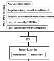
Torabi Jafroudi F, Kiani-Sarkaleh A, Nirouei M. Automatic Classification of Cancerous Masses in Digital Mammograms Using Curvelet Analysis and Hybrid Genetic Algorithm-Artificial Neural Network Model. I J Radiol. 2024;21(2):e146102. doi: https://doi.org/10.5812/iranjradiol-146102
Last Update: 2 weeks ago
Last Update: 2 weeks ago
Last Update: 2 weeks ago
Last Update: 7 hours ago
Last Update: 1 day ago
Last Update: 5 days ago