Fulltext
Full text is available in PDF.
International Journal of Cancer Management
The Official Journal of Cancer Research Center (CRC), Shahid Beheshti University of Medical Sciences
Authors
Background: Breast cancer is one of the most important diseases in females. Malaysian women have not excluded. According to the Malaysian Oncology Society [1], about 4% of women (who are 40 years old and above) have involved by breast cancer. Masses and microcalcifications are two important signs for breast cancer diagnosis on mammography. According to our estimation, radiologists could diagnose breast cancer on mammogram screening program, with approximately 75% accuracy. About 25% of breast cancers have missed on mammograms. This study aimed to explore the effects of enhancement methods on digital mammograms.
Methods: SPSS software have used for data analysis. Wilcox on ranked test and ROC have used to compare the original and manipulated images. In this study, 60 digital mammogram images which include 20 normal and 40 confirmed diagnosed cases of breast cancer (masses), have selected and manipulated by using histogram equation, histogram stretching and median filter.
Results: The results have shown that the histogram stretching and median filter methods could improve image quality for detection of masses with increased sensitivity and specificity by 5%.
Conclusion: The sensitivity and specificity have improved by using histogram stretching and median filter. The results of this study have shown results as below ; the histogram equation have improved the sensitivity up to 97.5% ,while the median filter could improve sensitivity (97.5%) and specificity (85.5%). It means that the median filter could be more effective than the other enhancement methods
© 2010, Author(s). This open-access article is available under the Creative Commons Attribution 4.0 (CC BY 4.0) International License (https://creativecommons.org/licenses/by/4.0/), which allows for unrestricted use, distribution, and reproduction in any medium, provided that the original work is properly cited.

Moradmand H, Setayeshi S, Karimian AR, Sirous M, Akbari ME. Comparing the Performance of Image Enhancement Methods to Detect Microcalcification Clusters in Digital Mammography. Int J Cancer Manag. 2012;5(2):e80801. doi:
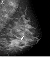
Arslan ZF, Altunkeser A, Aksoy N, Korez MK, Omeroglu E. Which Modality Should Be Integrated to Increase the Diagnostic Efficiency of BI-RADS 0, 3, and 4 Lesions? Ultrasonography or Digital Breast Tomosynthesis?. I J Radiol. 2021;18(4):e113845. doi: https://doi.org/10.5812/iranjradiol.113845

Altunkeser A, Arslan ZF, Eryilmaz MA, Korez MK, Bayramoğlu Z. Is Digital Breast Tomosynthesis Superior to Digital Mammography? A Preclinical Evaluation of Tumor Histopathological Markers Using Digital Breast Tomosynthesis. I J Radiol. 2021;18(4):e113846. doi: https://doi.org/10.5812/iranjradiol.113846
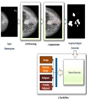
Niroomand Fam B, Nikravanshalmani A, Khalilian M. An Efficient Method for Automated Breast Mass Segmentation and Classification in Digital Mammograms. I J Radiol. 2021;18(3):e106717. doi: https://doi.org/10.5812/iranjradiol.106717
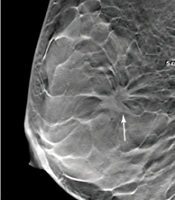
Xu H, Zeng W, Fu Z, Cui Q. Diagnostic Value of Digital Breast Tomosynthesis Combined with Magnetic Resonance Imaging for Breast Cancer in Dense Breast Tissues. I J Radiol. 2021;18(4):e114976. doi: https://doi.org/10.5812/iranjradiol.114976
Last Update: 3 weeks ago
Last Update: 3 weeks ago
Last Update: 3 weeks ago
Last Update: 9 hours ago
Last Update: 1 day ago
Last Update: 4 days ago