Fulltext
Full-text is available in PDF file.
Authors
An electrocardiogram from a patient implanted with a dual-chamber DDD pacemaker showed paced QRS complexes whose morphology and frontal-plane axis were consistent with biventricular stimulation with right ventricular lead located at the apex. However, some electrocardiographic findings were suggestive, rather, of univentricular right apical pacing and sustained ventricular fusion with competing native atrioventricular conduction in the presence of patient's spontaneous QRS showing right bundle branch block plus left anterior hemiblock. Shortening atrioventricular delay with magnet application advanced right ventricle stimulation and prevented the supraventricular impulse to contribute to ventricular depolarization, thereby making clear the mechanism of right v
© 2017, International Journal of Cardiovascular Practice. This open-access article is available under the Creative Commons Attribution-NonCommercial 4.0 (CC BY-NC 4.0) International License (https://creativecommons.org/licenses/by-nc/4.0/), which allows for the copying and redistribution of the material only for noncommercial purposes, provided that the original work is properly cited.
Eysenck W, Sulke N, Gallagher A, Jouhra F, Patel N, et al. Right Ventricular Septal Pacing vs. Right Ventricular Apical Pacing Following Atrioventricular Node Ablation: A 10-Year Follow-up. Int Cardiovasc Res J. 2018;12(3):e69785. doi:

Moaref A, Fallahzadeh E, Zamani J, Najib K. The influence of right ventricular apical pacing on left atrial volume in patients with normal left ventricular function. Int Cardiovasc Res J. 2018;1(3):e79410. doi:
Jorat MV, Aslani A, Nikoo MH. Implanting CRT via Endo-Cardiac Route When Tricuspid Valve is Metallic. Int Cardiovasc Res J. 2017;8(1):e12059. doi:
Rastravan R, Azarfarin R, Babaei T, Tirgarfakheri K, Ghanbari M. Life Threatening Right Ventricle and Atrium Perforation and Tricuspid Valve Injury Due to Pacemaker Lead Dislodgment- A Case Report. Multidiscip Cardio Annal. 2022;13(1):e123135. doi: https://doi.org/10.5812/mca-123135
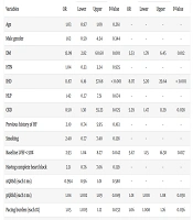
Payami B, Zakerinia M, Moradi A, Easapour Moghadam M, Akiash N. Pacing-Induced Cardiomyopathy in Right Ventricular Pacemaker Patients: Prevalence and Risk Factors in an Iranian Cohort. Int Cardiovasc Res J. 2025;19(1):e164447. doi: https://doi.org/10.5812/icrj-164447
Last Update: 3 days ago
Last Update: 3 days ago
Last Update: 3 days ago
Last Update: 6 days ago
Last Update: 5 hours ago
Last Update: 4 days ago