
1. Background
Cardiovascular diseases (CVDs) remain the leading cause of morbidity and mortality worldwide. Coronary artery disease (CAD) is the most prevalent manifestation of CVDs. Despite advances in preventive strategies and medical therapy, the global burden of CAD continues to increase because of population aging and the rising prevalence of modifiable cardiovascular risk factors, including smoking, obesity, physical inactivity, and unhealthy dietary habits (1-3). Progressive atherosclerotic plaque formation within the coronary arteries results in luminal narrowing and impaired myocardial perfusion, whereas plaque destabilization and rupture may precipitate thrombus formation, acute coronary occlusion, and myocardial ischemia requiring coronary revascularization (4, 5).
Coronary artery bypass grafting (CABG) has long been the standard revascularization strategy for patients with complex multivessel or left main coronary artery disease. However, percutaneous coronary intervention (PCI) has become the preferred treatment for many patients because of its minimally invasive nature and favorable short-term outcomes. Current guidelines recommend CABG for patients with left main disease or complex multivessel CAD, whereas PCI is considered appropriate in many other clinical settings (6, 7). Nevertheless, a considerable proportion of patients who undergo PCI ultimately require CABG because of restenosis, progression of atherosclerosis, or recurrent ischemic events (8).
Histopathological studies of coronary arteries have primarily focused on the morphology of atherosclerotic plaques and endothelial alterations associated with plaque instability. Vulnerable plaques are typically characterized by a lipid-rich necrotic core covered by a thin fibrous cap and are frequently accompanied by neovascularization, inflammatory cell infiltration, intraplaque hemorrhage, and spotty calcification, all of which contribute to plaque rupture and subsequent thrombotic events (9-11). In addition to plaque characteristics, local hemodynamic forces such as shear stress play a critical role in vascular remodeling and plaque destabilization.
Accumulating evidence indicates that PCI itself may induce biological changes within the coronary arterial wall beyond its mechanical revascularization effect. Coronary stent implantation has been associated with altered local shear stress, endothelial injury, activation of inflammatory pathways, increased cytokine production, inflammatory cell infiltration, vascular wall edema, and vascular smooth muscle cell activation, all of which may contribute to restenosis and disease progression (12-14). These findings suggest that PCI may trigger persistent inflammatory and remodeling responses within the vessel wall that extend beyond the immediate postprocedural period.
2. Objectives
Despite these observations, little is known about the histopathological characteristics of coronary arteries in patients who subsequently undergo CABG after PCI. In particular, comparative data on inflammatory and proliferative changes in the coronary arterial wall between patients with and without previous PCI are limited. Therefore, this study aimed to evaluate histopathological alterations and the immunohistochemical expression of inflammatory and proliferative markers in coronary artery specimens obtained during CABG and to compare these findings between patients with and without a history of PCI.
3. Methods
3.1. Study Population
This pilot case-control study was conducted over a six-month period, from March to September 2024. Consecutive patients with angiographically confirmed CAD who were scheduled to undergo CABG and met the eligibility criteria were enrolled after providing written informed consent.
Participants were assigned to one of two groups according to their history of PCI. The PCI group consisted of patients with a prior history of coronary stent implantation, whereas the control group included patients with CAD who had undergone diagnostic coronary angiography only and had no history of PCI.
Patients were excluded if they had active systemic inflammatory or autoimmune diseases, such as systemic lupus erythematosus or rheumatoid arthritis, because of their potential effects on inflammatory cell infiltration. Additional exclusion criteria included active malignancy or ongoing chemotherapy, end-stage renal disease requiring dialysis or severe renal failure associated with chronic systemic inflammation, and inadequate tissue specimens with substantial processing artifacts or insufficient tissue for reliable histopathological evaluation of the endothelial and neointimal layers.
This study was conducted in accordance with the Declaration of Helsinki and was approved by the Ethics Committee of Shahid Beheshti University of Medical Sciences (IR.SBMU.MSP.REC.1402.3). All patient information was kept confidential, and the results were reported anonymously.
3.2. Clinical Evaluation
Demographic and clinical data, including age, sex, comorbidities, previous coronary angiography, and history of PCI, were recorded for all participants. During CABG, coronary artery tissue specimens were obtained from the edge of the coronary arteriotomy at the distal anastomotic site. The harvested specimens were processed for histopathological examination.
All histopathological assessments were performed by an experienced pathologist who was blinded to the patients’ clinical characteristics. Tissue sections were evaluated for atherosclerotic plaques, inflammatory changes, inflammatory cell infiltration, endothelial injury, and extracellular matrix damage within the coronary arterial wall. Immunohistochemical staining was performed to assess macrophage infiltration using CD68, T-lymphocyte infiltration using CD45RO, and vascular smooth muscle cell proliferation using α-smooth muscle actin (α-SMA). Collagen deposition was also evaluated. Each histopathological parameter was recorded as positive or negative according to predefined pathological criteria. The frequencies of these histopathological findings were subsequently compared between patients with and without a history of PCI.
3.3. Statistical Analysis
Statistical analyses were performed using IBM SPSS Statistics version 22. Continuous variables were summarized as mean ± standard deviation, along with minimum and maximum values, whereas categorical variables were expressed as frequencies and percentages. The normality of continuous variables was assessed using the Kolmogorov-Smirnov test. Between-group comparisons were performed using the independent-samples t-test for normally distributed variables and the Mann-Whitney U test for nonnormally distributed variables. Categorical variables were compared using the chi-square test or Fisher exact test, as appropriate.
To evaluate the effect of the time interval between PCI and CABG, patients in the PCI group were stratified into two subgroups: the urgent group (CABG performed within one week of PCI) and the emergent group (immediate CABG following PCI). Histopathological parameters were then compared between these two subgroups using the chi-square test. A two-sided P value ≤ 0.05 was considered statistically significant.
4. Results
4.1. Baseline Demographic and Clinical Characteristics
A total of 22 patients undergoing CABG were included, comprising 11 patients with a history of PCI and 11 patients without prior PCI. The demographic characteristics and baseline clinical features of the study population are summarized in Table 1.
Table 1.
Baseline Demographic and Clinical Characteristics of the Study Population a
| Variables | Total (n = 22) | PCI (n = 11) | Non-PCI (n = 11) | P-Value |
|---|---|---|---|---|
| Male sex | 14 (63.6) | 7 (63.6) | 7 (63.6) | 0.990 |
| Age (y) | 64.27 ± 6.00 | 65.27 ± 5.13 | 63.27 ± 6.87 | 0.449 |
| Diabetes mellitus | 10 (45.5) | 6 (54.5) | 4 (36.4) | 0.670 |
| Hypertension | 13 (59.1) | 7 (63.6) | 6 (54.5) | 0.990 |
| Hyperlipidemia | 12 (54.5) | 7 (63.6) | 5 (45.5) | 0.660 |
| Current smoking | 11 (50.0) | 7 (63.6) | 4 (36.4) | 0.395 |
| Chronic kidney disease | 3 (13.6) | 1 (9.1) | 2 (18.2) | 0.990 |
a Variables are expressed as No. (%) or mean ± SD.
No significant differences were observed between the PCI and non-PCI groups in age, sex distribution, or the prevalence of major cardiovascular risk factors. The mean age of the study population was 64.27 ± 6.00 years, with comparable ages in the PCI and non-PCI groups (65.27 ± 5.13 vs. 63.27 ± 6.87 years, P = 0.449). Males constituted 63.6% of patients in both groups. Similarly, the prevalence of diabetes mellitus (54.5% vs. 36.4%, P = 0.670), hypertension (63.6% vs. 54.5%, P = 0.990), hyperlipidemia (63.6% vs. 45.5%, P = 0.660), smoking (63.6% vs. 36.4%, P = 0.395), and chronic kidney disease (9.1% vs. 18.2%, P = 0.990) did not differ significantly between the groups, indicating well-balanced baseline clinical characteristics.
4.2. Histopathological Changes in the Coronary Artery Wall According to Previous PCI
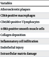
The histopathological findings in coronary artery specimens are presented in Table 2. Atherosclerotic plaques were identified in all patients, regardless of PCI history (100% in both groups, P = 0.990).
Table 2.
Comparison of Histopathological Findings Between Patients with and Without Previous PCI a
| Variables | Total (n = 22) | PCI (n = 11) | Non-PCI (n = 11) | P-Value |
|---|---|---|---|---|
| Atherosclerotic plaques | 22 (100.0) | 11 (100.0) | 11 (100.0) | 0.990 |
| CD68-positive macrophages | 9 (40.9) | 8 (72.7) | 1 (9.1) | 0.008 |
| CD45RO-positive T lymphocytes | 12 (54.5) | 9 (81.8) | 3 (27.3) | 0.030 |
| α-SMA-positive smooth muscle cells | 14 (63.6) | 10 (90.9) | 4 (36.4) | 0.024 |
| Collagen deposition | 10 (45.5) | 6 (54.5) | 4 (36.4) | 0.670 |
| Inflammatory cell infiltration | 14 (63.6) | 10 (90.9) | 4 (36.4) | 0.024 |
| Endothelial injury | 4 (18.2) | 3 (27.3) | 1 (9.1) | 0.586 |
| Extracellular matrix damage | 3 (13.6) | 2 (18.2) | 1 (9.1) | 0.990 |
a Values are expressed as No. (%).
Compared with patients without previous PCI, those with prior PCI showed significantly greater inflammatory and proliferative changes within the coronary artery wall. Positive CD68 staining, indicating macrophage infiltration, was observed in 72.7% of patients with previous PCI compared with 9.1% of patients without PCI (P = 0.008). Similarly, CD45RO-positive T-lymphocyte infiltration was more frequent in the PCI group than in the non-PCI group (81.8% vs. 27.3%, P = 0.030).
Expression of α-smooth muscle actin (α-SMA), a marker of vascular smooth muscle cell proliferation, was also significantly higher in patients with prior PCI (90.9% vs. 36.4%, P = 0.024). Inflammatory cell infiltration followed a similar pattern, occurring in 90.9% of the PCI group compared with 36.4% of the control group (P = 0.024).
In contrast, collagen deposition, endothelial injury, and extracellular matrix damage did not differ significantly between the groups. Positive collagen staining was detected in 54.5% of patients with previous PCI and 36.4% of controls (P = 0.670). Likewise, endothelial injury (27.3% vs. 9.1%, P = 0.586) and extracellular matrix damage (18.2% vs. 9.1%, P = 0.990) were comparable between the groups.
4.3. Effect of the Interval Between PCI and CABG on Histopathological Changes
Patients with previous PCI were further categorized, according to the interval between PCI and CABG, into the urgent group (CABG performed within one week after PCI; n = 7) and the emergent group (immediate CABG following PCI; n = 4). A comparison of histopathological findings between these subgroups is shown in Table 3.
Table 3.
Comparison of Histopathological Findings According to the Interval Between PCI and CABG a
| Variables | Urgent (n = 7) | Emergent (n = 4) | P-Value |
|---|---|---|---|
| Atherosclerotic plaques | 7 (100.0) | 4 (100.0) | 0.990 |
| CD68-positive macrophages | 5 (71.4) | 3 (75.0) | 0.990 |
| CD45RO-positive T lymphocytes | 5 (71.4) | 4 (100.0) | 0.491 |
| α-SMA-positive smooth muscle cells | 6 (85.7) | 4 (100.0) | 0.990 |
| Collagen deposition | 4 (57.1) | 4 (100.0) | 0.236 |
| Inflammatory cell infiltration | 7 (100.0) | 3 (75.0) | 0.364 |
| Endothelial injury | 2 (28.6) | 1 (25.0) | 0.990 |
| Extracellular matrix damage | 1 (14.3) | 1 (25.0) | 0.990 |
a Values are expressed as No. (%).
No statistically significant differences were identified between the urgent and emergent groups for any evaluated histopathological parameter. Although CD45RO-positive T-lymphocyte staining (100% vs. 71.4%), α-SMA expression (100% vs. 85.7%), and collagen deposition (100% vs. 57.1%) were numerically higher in the emergent group, these differences did not reach statistical significance. Similarly, the frequencies of CD68-positive macrophages, inflammatory cell infiltration, endothelial injury, and extracellular matrix damage were comparable between the two groups (all P > 0.05).
5. Discussion
The present study investigated and compared histopathological and immunological changes in the coronary artery wall of patients with and without a history of PCI before undergoing CABG. Although the two groups were well matched in demographic characteristics and baseline cardiovascular risk factors, patients with previous PCI showed significantly higher expression of the inflammatory markers CD68 (macrophages) and CD45RO (T lymphocytes), as well as significantly increased expression of α-smooth muscle actin (α-SMA), a marker of vascular smooth muscle cell proliferation. In contrast, no significant differences were observed between the groups in extracellular matrix damage, collagen deposition, or endothelial injury, and atherosclerotic plaques were present in all patients. Furthermore, the interval between PCI and CABG (immediate versus within one week) did not influence the histopathological findings. Collectively, these findings suggest that PCI promotes a persistent inflammatory and proliferative response within the coronary artery wall without substantial structural disruption of the extracellular matrix.
The lack of significant differences in demographic characteristics and baseline clinical risk factors between the PCI and non-PCI groups strengthens the validity of our findings by minimizing the influence of potential confounders. Consequently, the observed histopathological differences are more likely to reflect the biological consequences of PCI itself than differences in patient characteristics or underlying cardiovascular risk profiles.
One of the principal findings of this study was the significantly greater infiltration of CD68-positive macrophages and CD45RO-positive T lymphocytes in coronary artery specimens from patients with previous PCI. These observations are consistent with the current understanding of the vascular response to mechanical injury. Immediately after PCI, endothelial disruption triggers platelet activation and the expression of adhesion molecules such as P-selectin, promoting the initial recruitment of circulating leukocytes (15-17). Stable leukocyte adhesion and transmigration are subsequently mediated by integrins, particularly Mac-1 (CD11b/CD18), expressed on neutrophils and monocytes (18, 19). This inflammatory response is further amplified by chemokines, including monocyte chemoattractant protein-1 (MCP-1), which recruits monocytes and activated T lymphocytes, and interleukin-8 (IL-8), which attracts neutrophils to the injured vessel wall (20).
In addition to mechanical injury, the implanted stent itself may contribute to persistent vascular inflammation. Experimental studies have demonstrated that corrosion products, metal ions, and nanoparticles released from coronary stents can activate inflammatory pathways and promote neointimal remodeling, thereby contributing to in-stent restenosis (ISR) (21-24). Moreover, Shlofmitz et al. reported that local vascular inflammation, particularly in the presence of dyslipidemia, plays a central role in the progression of atherothrombosis and stimulates aggressive neointimal proliferation (25). Therefore, the increased expression of CD68 and CD45RO observed in our study likely reflects the combined effects of mechanical vascular injury, chemokine-mediated leukocyte recruitment, and chronic immune activation induced by stent-related materials.
Another important finding was the significantly increased expression of α-SMA in the PCI group, indicating enhanced vascular smooth muscle cell (VSMC) activation and neointimal hyperplasia (26). This observation highlights the distinction between native atherosclerosis and restenosis, which are biologically distinct processes. Although advanced atherosclerotic plaques were present in all patients, increased α-SMA expression was observed only in patients with prior PCI, suggesting that coronary intervention activates a specific proliferative pathway beyond the underlying atherosclerotic disease.
Several mechanisms may explain this proliferative response. First, vascular injury after PCI induces substantial production of reactive oxygen species (ROS), particularly through activation of NADPH oxidase (NOX), which directly stimulates VSMC migration and proliferation (27). Second, although drug-eluting stents (DES) effectively inhibit neointimal growth, they may also delay endothelial regeneration and reduce nitric oxide (NO) bioavailability through impaired endothelial nitric oxide synthase (eNOS) activity (28). Reduced NO signaling subsequently favors VSMC proliferation and vascular remodeling (29, 30). Accordingly, the increased α-SMA expression observed in our study may represent a downstream consequence of persistent oxidative stress, impaired endothelial homeostasis, and enhanced smooth muscle cell proliferation after PCI.
Notably, no significant differences were found between the groups in collagen deposition, endothelial injury, or extracellular matrix damage. At first glance, these findings appear inconsistent with previous reports describing endothelial denudation after PCI (28). However, this discrepancy may be explained by the limited sample size of the present study and the distinction between structural injury and functional endothelial impairment. Because all patients had advanced CAD requiring surgical revascularization, structural alterations of the vessel wall were expected in both groups. In contrast, prior PCI appears to induce predominantly functional changes characterized by reduced NO bioavailability, persistent endothelial dysfunction, and enhanced inflammatory signaling, rather than overt structural destruction of the extracellular matrix. Likewise, alterations in the nanomechanical properties of vascular and inflammatory cells, such as increased cellular stiffness (Young's modulus), may occur independently of gross histological disruption (31-33). Thus, our findings suggest that PCI primarily modifies the biological behavior of the coronary artery wall rather than its overall structural integrity.
We also found no significant differences in histopathological findings between patients undergoing immediate CABG and those undergoing surgery within one week after PCI. This observation is biologically plausible because leukocyte recruitment through P-selectin and Mac-1 signaling occurs rapidly after vascular injury (33), while ROS generation and NO depletion begin almost immediately after coronary intervention (27). These early inflammatory responses may quickly reach a plateau and persist during the short interval before surgery, thereby explaining the comparable expression of inflammatory and proliferative markers in both subgroups.
5.1. Study Limitations
Several limitations of this study should be acknowledged. First, the type of implanted coronary stent was not recorded or incorporated into the analysis. Differences among bare-metal stents (BMS), first- and newer-generation drug-eluting stents (DES), and their polymer coatings may substantially influence vascular inflammation and smooth muscle cell proliferation, potentially affecting the expression of markers such as CD68 and α-SMA. Second, because tissue samples were obtained only at the time of CABG, longitudinal evaluation of histopathological changes after PCI was not feasible, and the findings represent a single time point rather than the temporal evolution of vascular remodeling. Finally, although the selected immunohistochemical markers provided valuable insight into inflammatory and proliferative responses, more comprehensive molecular analyses—including evaluation of ROS- and NO-related signaling pathways at the proteomic or transcriptomic level—were beyond the scope of the present study.
5.2. Conclusions
In conclusion, prior PCI was associated with persistent inflammatory cell infiltration and enhanced vascular smooth muscle cell proliferation within the coronary artery wall of patients subsequently undergoing CABG. Specifically, prior PCI was associated with increased expression of CD68, CD45RO, and α-SMA without significant alterations in extracellular matrix integrity or collagen deposition. These findings suggest that PCI transforms the coronary artery wall from a chronically diseased vessel into a biologically active inflammatory and proliferative environment, which may have implications for vascular healing and the quality of coronary anastomoses during subsequent CABG. Further studies with larger cohorts and detailed molecular characterization are warranted to clarify the long-term clinical significance of these histopathological alterations.