
1. Background
Diabetes mellitus is a systemic condition that substantially affects both the microvascular and macrovascular systems and is a major risk factor for peripheral arterial disease (PAD) (1-3). In patients with diabetes, PAD is an important prognostic indicator and is often associated with severe outcomes, including limb ischemia, ulceration, and amputation (4). The clinical presentation of PAD typically includes intermittent claudication that worsens with physical activity and, in advanced stages, rest pain or critical limb ischemia. However, only approximately one-third of patients with diabetes and PAD are symptomatic, contributing to underdiagnosis and uncertainty regarding its true prevalence (5). Older age, peripheral neuropathy, and a longer duration of diabetes further increase the risk of PAD in this population (4).
According to American College of Cardiology Foundation/American Heart Association guidelines, the risk of PAD is increased among patients with diabetes and additional risk factors, such as smoking, dyslipidemia, or hypertension, who are younger than 50 years; individuals aged 50 - 69 years with a history of diabetes or smoking; individuals older than 70 years; and those with abnormal lower-extremity pulses, exertional leg pain, or known carotid, coronary, or renal atherosclerosis (6). Despite these risk profiles, PAD in patients with diabetes frequently remains undiagnosed until advanced stages, largely because diabetic neuropathy may mask claudication-related pain (5).
The Ankle-Brachial Index (ABI) is a simple, noninvasive, and cost-effective diagnostic measure for PAD, calculated by dividing the highest ankle systolic blood pressure by the highest brachial systolic blood pressure (7, 8). In patients with diabetes, ABI values < 0.9 or > 1.3 are associated with an increased risk of PAD and cardiovascular disease because of medial arterial calcification (9, 10). However, ABI reliability is influenced by recent caffeine consumption, physical activity, smoking, clinician expertise, and patient positioning (11). Arterial calcification in patients with diabetes frequently results in falsely normal or elevated ABI values, thereby reducing diagnostic sensitivity compared with that in the general population (12, 13).
Data on PAD prevalence and ABI performance in patients with diabetes and acute coronary syndrome (ACS) are limited (13-16). Acute coronary syndrome may affect ABI measurements during the acute phase (17). Current guidelines recommend ABI screening in patients with diabetes who are older than 50 years or who have atherosclerotic cardiovascular disease; however, the effect of acute ACS on ABI accuracy remains insufficiently studied (6). Arterial stiffness, which is distinct from atherosclerosis, may contribute to elevated ABI values and an increased risk of cardiovascular events in patients with diabetes (17). Moreover, many patients with diabetes and PAD remain asymptomatic until late stages, underscoring the need for reliable screening methods (12).
2. Objectives
Given the conflicting evidence regarding ABI accuracy in patients with diabetes and the limited data on its use during ACS, this study evaluated changes in ABI among patients with diabetes and ACS during the acute phase (within 48 hours) and the late phase (4 - 6 weeks after ACS).
3. Methods
3.1. Study Population
This descriptive-analytical study was conducted at Masih Daneshvari and Shohada-e-Tajrish hospitals in Tehran, Iran, during 2023 - 2024. The study population comprised patients with diabetes who were hospitalized for ACS and had coronary stenosis > 50%. Consecutive sampling was performed according to the inclusion and exclusion criteria.
The inclusion criteria were diabetes, referral with ACS, ABI assessment, angiography, and coronary stenosis > 50%. The ABI was reassessed 4 - 6 weeks later. The study had no specific exclusion criteria, and patients with different clinical conditions were included.
After patient selection, the research process was explained to each participant, and the feasibility of returning for follow-up was assessed. Patients were not included if their return for follow-up within the specified period could not be ensured.
3.2. Clinical Assessment
Patient characteristics, including age, sex, smoking status, hypertension, chronic kidney disease, and cerebrovascular accident (CVA), were extracted from medical records. The initial ABI was measured within the first 48 hours of hospitalization after ACS. The ABI was reassessed and recorded after 4 - 6 weeks. Acute- and late-phase ABI values were then compared.
For accurate measurement, patients were placed in the supine position, with their arms and legs at heart level. The 2 cuffs of the ABI device were placed on the upper and lower limbs, and ABI values were recorded to 2 decimal places.
Peripheral arterial disease severity was classified according to ABI values as mild (0.70 - 0.89), moderate (0.40 - 0.69), or severe (< 0.40).
3.3. Statistical Analysis
Data were analyzed using SPSS version 22. Quantitative variables are presented as the mean and standard deviation, and qualitative variables are presented as number and percentage. The χ2 test was used to compare qualitative variables between groups, and the paired t test was used for quantitative variables. Agreement between the initial and late measurements was evaluated. True-positive, true-negative, false-positive, and false-negative results were calculated.
3.4. Ethical Considerations
The study was approved by the Ethics Committee of Shahid Beheshti University of Medical Sciences (IR.SBMU.MSP.REC.1404.044), and written informed consent was obtained from all participants.
4. Results
4.1. Clinical and Demographic Characteristics
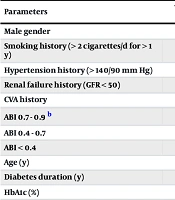
As shown in Table 1, the demographic and clinical characteristics of the participants were assessed. These variables were also described for patients with PAD according to impaired ABI during the acute and late phases after ACS.
Table 1.
Distribution of Demographic and Clinical Characteristics of Patients a
| Parameters | Total Patients (n = 100) | No Impaired ABI (n = 66) | Impaired ABI (Acute Phase, n = 37) | Impaired ABI (Late Phase, n = 34) |
|---|---|---|---|---|
| Male gender | 54 (54) | 42 (63.6) | 13 (35.1) | 12 (35.3) |
| Smoking history (> 2 cigarettes/d for > 1 y) | 48 (48) | 27 (40.9) | 23 (62.2) | 21 (61.8) |
| Hypertension history (> 140/90 mm Hg) | 47 (47) | 21 (31.8) | 33 (89.2) | 26 (76.5) |
| Renal failure history (GFR < 50) | 14 (14) | 5 (7) | 12 (32.4) | 9 (26.5) |
| CVA history | 3 (3) | 1 (1.5) | 2 (5.4) | 2 (5.9) |
| ABI 0.7 - 0.9 b | - | 58 (87.8) | 10 (27.02) | 8 (23.52) |
| ABI 0.4 - 0.7 | - | 49 (74.2) | 18 (48.64) | 17 (50) |
| ABI < 0.4 | - | 57 (86.3) | 9 (24.32) | 9 (26.47) |
| Age (y) | 68.7 ± 6.07 | 67.1 ± 2.4 | 71.43 ± 5.35 | 71.91 ± 5.3 |
| Diabetes duration (y) | 13.77 ± 4.3 | 11.22 ± 2.3 | 17.24 ± 4.58 | 18.05 ± 4.14 |
| HbA1c (%) | 7.82 ± 0.78 | 7.5 ± 0.72 | 8.33 ± 0.74 | 8.29 ± 0.69 |
| ABI (acute phase) | 0.83 ± 0.25 | 0.96 ± 0.32 | 0.54 ± 0.18 | - |
| ABI (late phase) | 0.85 ± 0.22 | 0.97 ± 0.08 | - | 0.62 ± 0.24 |
a Values are expressed as No. (%) or mean ± SD.
b Peripheral arterial disease severity was classified according to ABI values as mild (0.70 - 0.89), moderate (0.40 - 0.69), or severe (< 0.40).
Among the 100 patients with diabetes, 54% were male, and the mean age was 68.7 ± 6.07 years. The mean duration of diabetes was 13.77 ± 4.3 years, and the mean HbA1c was 7.82 ± 0.78%. During the acute phase, 37 patients had impaired ABI (< 0.9), including 10 (27.02%) with mild PAD (ABI, 0.7 - 0.9), 18 (48.64%) with moderate PAD (ABI, 0.4 - 0.7), and 9 (24.32%) with severe PAD (ABI < 0.4). During the late phase, 34 patients had impaired ABI, including 8 (23.52%) with mild PAD, 17 (50%) with moderate PAD, and 9 (26.47%) with severe PAD. In addition, 47% of patients had a history of hypertension, and 48% had a history of smoking. The mean ABI was 0.83 ± 0.25 during the acute phase and 0.85 ± 0.22 during the late phase.
4.2. Comparison of ABI Accuracy During the Acute and Late Phases of ACS
As shown in Table 2, ABI values measured during the acute and late phases of ACS were compared among patients with diabetes. A significant difference was observed between the two phases, with ABI values during the late phase being significantly higher than those during the acute phase (P = 0.02). ABI values were also compared between patients with impaired ABI (< 0.9) and those with normal ABI during both phases. Among patients with impaired ABI, no significant difference was observed between the acute and late phases (P = 0.08). However, among patients with normal ABI, a significant difference was observed, with higher values during the acute phase than during the late phase (P = 0.01).
Table 2.
Comparison of ABI Measurements Between the Acute and Late Phases of ACS in the Overall Study Population and According to Baseline ABI Status a
| Comparison | Acute phase (≤ 48 h) | Late phase (4 - 6 wk) | P-Value |
|---|---|---|---|
| All patients | 0.83 ± 0.25 | 0.85 ± 0.22 | 0.02 |
| Patients with abnormal ABI | 0.96 ± 0.32 | 0.97 ± 0.08 | 0.08 |
| Patients with normal ABI | 0.54 ± 0.18 | 0.62 ± 0.24 | 0.01 |
a Values are expressed as mean ± SD.
4.3. Diagnostic Performance of ABI for PAD
The diagnostic performance of ABI for identifying PAD was assessed using the parameters presented in Table 3. All patients diagnosed with severe PAD during the acute phase remained classified as having severe PAD during the late phase. One patient initially diagnosed with moderate PAD during the acute phase was reclassified as having mild PAD during the late phase. In addition, 3 patients identified as having mild PAD during the acute phase had normal ABI values during the late-phase evaluation. Table 3 showed 33 true-positive results (89.18%), 4 false-positive results (10.81%), 63 true-negative results (100%), and 0 false-negative results (0%). The corresponding diagnostic measures were sensitivity, 100%; specificity, 94.03%; positive predictive value, 69.55%; negative predictive value, 100%; and overall accuracy, 94.75%.
Table 3.
Diagnostic Performance of ABI for PAD in Patients with Diabetes and ACS a
| Parameters | Values |
|---|---|
| True positive | 33 (89.18) |
| False positive | 4 (10.81) |
| True negative | 63 (100) |
| False negative | 0 (0) |
| Metrics (%) | |
| Sensitivity | 100 |
| Specificity | 94.03 |
| Positive predictive value | 69.55 |
| Negative predictive value | 100 |
| Accuracy | 94.75 |
a Values are expressed as No. (%) or percentage.
These findings suggest that ACS negatively affects ABI in patients with diabetes and may produce falsely low values. ABI demonstrated robust performance in identifying severe PAD, with consistent classification across phases. However, in patients with mild or moderate PAD, ACS appeared to induce changes in ABI values, requiring cautious interpretation. This variability underscores the limitations of ABI as a diagnostic method during the acute phase of ACS, particularly for less severe PAD categories, and indicates the need for further investigation of its reliability in this context.
5. Discussion
The ABI is a simple, inexpensive, and noninvasive measurement that can estimate the extent of lower-extremity and systemic atherosclerotic involvement with high sensitivity and specificity (18, 19). However, its clinical implications in patients with ACS have not been adequately evaluated. Therefore, this study was designed to improve understanding of the effect of ACS on low ABI values in patients with diabetes.
Our analysis showed that 37% of patients with diabetes and ACS had an ABI < 0.9 during the early phase, decreasing slightly to 34% during the late phase. Nine patients diagnosed with severe PAD during the acute phase remained in the severe category during the late phase, indicating stability in advanced disease. In contrast, 1 patient with moderate PAD transitioned to mild PAD, and 3 patients with mild PAD had normal values during the late phase, suggesting potential reversibility or measurement variability in less severe disease. Acute-phase ABI had a sensitivity of 100% and a specificity of 94%. Mean ABI increased significantly from 0.83 ± 0.25 during the acute phase to 0.85 ± 0.22 during the late phase (P < 0.05). This increase suggests that ACS may cause a falsely low ABI during the acute period, possibly because of increased inflammation, catecholamine release, or vascular stiffness. The ABI performed reliably in severe PAD but varied in mild and moderate cases, requiring cautious interpretation in these subgroups. Although the difference was statistically significant using a paired t-test, the overall mean ABI values during the acute and late phases were very similar, and their standard deviations overlapped substantially. Therefore, the difference may have limited clinical relevance.
According to current European Society of Cardiology and American College of Cardiology/American Heart Association guidelines, PAD is diagnosed when the ABI is < 0.90 and is associated with calcification and consequent vascular stiffness (20, 21).
Sartore et al. (22) reported a PAD prevalence of 17% among patients with type 2 diabetes, consistent with international studies and approximately 3 times the prevalence in the general population with similar demographic characteristics (23-25). Diabetes is also more prevalent among patients with ACS, and patients with diabetes and PAD have substantially higher ACS-related mortality than those without PAD (26, 27). Current guidelines have therefore emphasized aggressive management strategies for this high-risk population with unstable ischemic heart disease (28, 29).
An ABI ≤ 0.9 is also common among patients with ACS, with a reported prevalence of 30% – 40%, and is significantly associated with a worse prognosis (27). In this study, the prevalence of ABI < 0.9 was higher than that reported in some previous studies (30-32) and was closer to this range because of the coexistence of clinical conditions associated with atherosclerosis in ACS.
Previous studies have described symptomatic or asymptomatic PAD as a strong and consistent independent predictor of cerebrovascular events and mortality in patients with coronary artery disease (28). Behar et al. reported that among patients experiencing myocardial infarction (MI), clinically diagnosed PAD was associated with an increased risk of in-hospital death but did not affect long-term mortality among discharged patients (33). Agnelli et al. reported that an abnormal ABI predicted adverse 1-year outcomes among patients with ACS (28).
Consistent with our study, Núñez et al. (34) reported a high prevalence of PAD, nearly 40%, among patients with ACS, although the disease was largely subclinical and mild. Other investigators reported much lower prevalence estimates. Froehlich et al. reported a prevalence of only 9.7% in a subanalysis of the Global Registry of Acute Coronary Events study, which included 41,108 hospitalized patients with ACS (31). The present findings similarly indicate that ABI measurement during the acute phase of ACS is not a completely reliable marker for evaluating PAD.
Although the populations of previous studies were not completely comparable with our study population, the findings of Chuter et al. regarding ABI diagnostic accuracy also suggest potential limitations in identifying PAD among patients with diabetes (35).
Other studies, including a meta-analysis, have suggested that ABI is a suitable diagnostic method, contrary to our findings (36). Using color Doppler ultrasonography as the reference standard, that study showed low sensitivity and high specificity for ABI in diagnosing PAD among patients with diabetes, indicating a high probability of false-negative results. A diagnostic method with a high probability of false-negative results has limited utility as a screening method. Thus, when ABI is used alone for screening, it may fail to identify a substantial proportion of patients with diabetes and PAD.
Chang et al. demonstrated the usefulness of ABI for predicting complex and diffuse coronary lesions, reporting a higher proportion of lesions at the bony level and in proximal segments among patients with ABI < 0.9 than among patients with ABI ≥ 0.9 (37). However, unlike the present study, they excluded patients with myocardial infarction or unstable angina, which may explain the different findings.
Arterial stiffness caused by calcification of the internal carotid artery may reduce ABI sensitivity (9). Therefore, the cutoff value for low ABI may be lower among patients with diabetes, and a normal ABI may be insufficient to identify subsequent adverse events in this population.
5.1. Study Limitations
Several limitations should be considered. A single ABI measurement may have introduced selection bias because dynamic changes over time were not captured. Medial arterial calcification, which is common in diabetes, likely reduces ABI sensitivity and may lower the effective cutoff value below 0.9 (38, 39). Normal ABI values of 0.9 - 1.3 may also mask PAD because of noncompressible vessels, a frequent concern among patients with diabetes (40). In addition, inconsistent adjustment for potential confounders, including antiplatelet therapy and other medications, may have affected the risk estimates. The inflammatory state and catecholamine surge during the acute phase may have further distorted ABI, a factor not fully addressed by the study design.
Although the difference in ABI between the acute and late phases reached statistical significance using a paired t-test, the absolute difference in mean ABI values was small, and the standard deviations overlapped considerably. Statistical significance may therefore have resulted, at least partly, from the paired analysis rather than from a clinically meaningful magnitude of change. The observed difference should be interpreted cautiously, and its clinical importance should not be overstated. Future studies should determine whether this degree of change is associated with meaningful clinical outcomes or affects patient management.
Future research should prioritize serial ABI assessments and evaluate alternative diagnostic methods, including toe systolic pressure and transcutaneous oxygen tension, which may detect PAD more accurately in the presence of calcification. The LABI method, which has been shown to improve sensitivity, and the toe-brachial index may complement ABI, particularly in high-risk populations such as patients with diabetes and ACS. Further investigation of the relationship between ACS-related inflammation and ABI accuracy may also refine its clinical use.
5.2. Conclusions
ABI values < 0.9 are widely used to identify PAD and increased cardiovascular risk. In this study, ABI measurements obtained during the acute phase of ACS differed from those obtained during the late phase, suggesting that assessment timing may influence ABI values among patients with type 2 diabetes. However, the absolute difference between phases was small and should be interpreted cautiously with respect to its clinical importance. Acute-phase ABI findings should therefore be considered within the overall clinical context. Further studies are needed to determine whether phase-related differences have meaningful implications for clinical practice.