
1. Background
Respiratory tract infection is the most common disease in children, and the identification of pathogens is very important for treatment and prognosis. The etiological detection methods for respiratory tract infection include isolation, antigen detection, antibody detection, and nucleic acid amplification technology (NAAT) (1, 2). To obtain test results quickly, outpatients often use NAATs and antigen detection, which have relatively low sensitivity, whereas NAATs have high sensitivity and high specificity (3). Multiple PCRs can simultaneously detect multiple pathogens (1). Capillary electrophoresis-based multiplex PCR (CEMP) can detect more than 10 common viruses and atypical bacteria involved in respiratory tract infection (4, 5). The upper respiratory tract samples used for virus detection include OPSs, nasopharyngeal swabs (NPSs), and nasopharyngeal aspirates. Upper respiratory tract samples are often collected from outpatients for testing (6). Owing to the convenience of specimen collection, there are also clinical reports of nasal swabs for respiratory virus testing (7, 8), but no comparisons of nasal swabs with other samples exist.
2. Objectives
In this study, CEMP was used to detect 13 respiratory pathogens in OPSs and nasal swabs of outpatients. On the one hand, understanding the pathogen distribution of outpatients with respiratory tract infections is helpful for clinical diagnosis, and on the other hand, evaluating the clinical application value of collecting nasal swabs for pathogen detection is important.
3. Methods
3.1. Subjects
Patients who presented to the Children’s Hospital of Zhejiang University School of Medicine from October 1, 2022, to June 30, 2023, met the inclusion criterion of fever (temperature ≥ 38°C) accompanied by cough or sore throat. The exclusion criteria were as follows: (1) Patients with a definite bacterial infection or a definite diagnosis of other diseases; (2) patients with active oral or nasal bleeding.
3.2. Specimen Collection
The OPSs and nasal swabs were collected from the same patient. (1) The OPS collection method: The collector held the sterile tongue plate in the left hand to press the tongue and inserted the sterile swab stick in the right hand through the mouth. The congested parts of the posterior pharyngeal wall and the tonsils were taken as the center, which was rubbed 3 times, mucosal epithelial cells were collected, and contact with saliva was avoided. (2) Nasal swab collection: The patient’s head was slightly tilted, and the sampling personnel gently supported the patient’s head with one hand and held the swab against the nostril for insertion, slowly and deeply along the lower nasal passage 1 - 1.5 cm, and then rotated it in the nasal cavity at least 4 times. After sampling with a special swab, it was quickly put into the sampling tube with 3 mL storage liquid, the tube cover was tightened, and it was closed for inspection. The storage solution contains protein stabilizers, antibiotics that prevent the growth of bacteria and fungi, and buffers.
Nasal and oropharyngeal swabs were collected within 5 minutes of each other, with no therapeutic interventions during sampling. All patients had no antiviral or antibiotic use within 24 hours before sampling.
3.3. Reagents and Instruments
An automatic nucleic acid extraction instrument (A96, Ningbo Haier shi Gene Technology, instrument serial No. 202102012) and a nucleic acid extraction or purification kit (Haier Shi, batch No. 211009002) were used. A PCR amplification kit (MA6000, Suzhou Molarray, instrument serial No. MA-010943) and an amplification kit [Haier Shi, 13 respiratory pathogens (PCR capillary electrophoresis fragment analysis), batch No. 211011019] were used. An electrophoresis instrument (ABI 3500Dx, USA, instrument series No. 31614-051) and an electrophoresis loading sample mixture (ABI HIDI, USA, lot No. 21050920) were used. A fluorescent internal standard dye (Haier Shi, SIZE-500 Plus lot No. 210812003) was used.
3.4. Nucleic Acid Extraction
The sampling tube was placed on a vortex mixer and vortexed fully for 10 s to wash the adhered virus and the cells containing the virus from the swab. After vortexing, samples were incubated for 5 minutes to ensure complete virus elution. According to the requirements of the nucleic acid extraction reagent, 300 μL samples were absorbed and added to the internal reference for RT‒PCR, and nucleic acid extraction and purification were performed with an automatic nucleic acid extraction instrument (A96).
3.5. Multiplex PCR Testing for 13 Respiratory Pathogens
3.5.1. Pathogen Detection
The 13 respiratory pathogens tested included influenza A (FluA, H7N9, H1N1, H3N2, H5N2), FluA H1N1 (2009), seasonal H3N2, influenza B (FluB, strains Victoria and Yamagata), adenovirus (ADV, Groups B, C and E), human bocavirus (HBoV), rhinovirus (HRV), parainfluenza (HPIV, types 1, 2, 3 and 4), coronavirus (HCoV, 229E, OC43, NL 63 and HKU1), respiratory syncytial viruses (RSV, Groups A and B), metapneumovirus (HMPV), Mycoplasma pneumoniae (MP), and Chlamydia (CH, Chlamydia trachomatis and Chlamydia pneumoniae). Among them, ADV, HPIV, HCoV, RSV, and CH were not typed.
3.5.2. Multiplex PCR amplification
Multiplex PCR amplification was performed in an amplification tube, including a prepared RT‒PCR system, sample addition, and RT‒PCR amplification. The reaction system was configured as follows: a total of 20 μL, including 14 μL of PCR mixture, 1 μL of PCR mixture containing the enzyme mixture, and 5 μL of nucleic acid. The 14 μL PCR mixture contains 10 μL 2× reaction buffer, 2 μL primer mix, and 2 μL enzyme mix. The following amplification procedure was used on the PCR instrument: 25°C for 5min, 50°C for 15 min, 95°C for 2 min, 94°C for 30 s, and 65→60°C for 30 s (65→60°C, 1°C for 6 cycles), 72°C for 1 min; 94°C for 30 s, 60°C for 30 s, and 72°C for 1 min; and 29 cycles at 72°C for 0 min, followed by storage at 4°C.
3.5.3. Separation of Capillary Electrophoresis Fragments
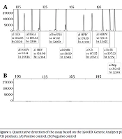
The following capillary electrophoresis system was prepared: sample mixture mixture (9 μL) (HiDi solution containing 2.5% 500 plus fluorescent internal label dye) + 1 μL of PCR product, and the PCR products of each sample were separated by capillary electrophoresis. Electrophoresis was performed at 15 kV for 25 minutes.
3.5.4. Analysis of the Results
The peaks (human RNA, human DNA, and reaction internal reference) appeared, indicating that the sample was not significantly degraded and that the experimental process was normal. The PCR product fragment length for all pathogens needs to be less than 1.5 bp from the reference size (9). A pathogen was considered positive if its characteristic peak had a fluorescence intensity ≥ 1000 RFU and fragment length deviation < 1.5 bp from the reference. Results were negative if fluorescence intensity < 1000 RFU or deviation ≥ 1.5bp. Indeterminate results (fluorescence intensity 500 - 1000 RFU) required retesting,If still indeterminate, results were classified as negative.
3.6. Statistical Analysis
Five samples were excluded (3 due to sampling failure, 2 due to detection errors) and not included in statistical analysis.
The classification data are expressed as frequencies (percentages), and the differences in the detection rates of different groups were analyzed via the chi-square test. Fisher’s exact probability was used when the number of cells with a theoretical frequency of less than 5 rows × lists exceeded 20%, and Bonferroni correction was used for pairwise comparisons. The above analyses were conducted in R version 4.2.3 software, and the Kappa coefficient consistency test was performed in SPSS 27.0. P < 0.05 was considered to indicate statistical significance.
4. Result
4.1. General Information
From October 1, 2022, to June 30, 2023, OPSs and nasal swabs were collected from 153 outpatients with respiratory tract infections. There were 75 males and 78 females, with a male-to-female ratio of 1:1.04. Age distribution: 70 patients were aged 0 - 3 years, 49 patients were aged > 3 - 6 years, and 34 patients were aged > 6 - 14 years. Mild nasal discomfort occurred in 2 patients (1.3%) during nasal swab collection, and 5 patients (3.3%) experienced nausea during oropharyngeal swab collection. No sample contamination or detection-related adverse events were observed.
4.2. Multiple PCR Results for Respiratory Pathogens
Among the 153 patients, 112 (73.2%, 95% CI: 65.7 - 79.6) were positive for at least one pathogen, 83 (54.2%, 95% CI: 46.3 - 61.9) were positive for one pathogen, and 29 (19%, 95% CI: 13.5 - 25.9) were coinfected. The pathogens detected included 30 cases (19.6%, 95% CI: 14.1 - 26.6) of various types of influenza, including FluA 15 (9.8%, 95% CI: 6.0 - 15.5), H1N1 (2009) 8 (5.2%, 95% CI: 2.7 - 10.0), seasonal H3N2 6 (3.9%, 95% CI: 1.8 - 8.3), FluB 1 (0.7%, 95% CI: 0.02 - 3.6), RSV 26 (17.0%, 95% CI: 11.9 - 23.7), MP 26 (17.0%, 95% CI: 11.9 - 23.7), HRV 25 (16.3%, 95% CI: 11.3 - 23.0), HMPV 11 (7.2%, 95% CI: 4.1 - 12.4), HADV 9 (5.9%, 95% CI: 3.1 - 10.8), HPIV 9 (5.9%, 95% CI: 3.1 - 10.8), HCoV 3 (2.0%, 95% CI: 0.4 - 5.6), HBoV 3 (2.0%, 95% CI: 0.4 - 5.6), and Ch 3 (2.0%, 95% CI: 0.4 - 5.6). Among the 29 coinfections, 28 (96.6%) involved 2 pathogens, and only 1 involved three pathogens. Coinfections are shown in Table 1.
Table 1.
Coinfections
| Coinfection Type | Number of Positive Cases | Positive Rate (%) | 95% CI |
|---|---|---|---|
| Flu A+seasonl H3N2 | 6 | 3.92 | 1.81 - 8.29 |
| Flu A+H1N1 (2009) | 8 | 5.23 | 2.67 - 9.98 |
| Flu A+HRV | 1 | 0.65 | 0.02 - 3.59 |
| ADV+MP | 1 | 0.65 | 0.02 - 3.59 |
| ADV+HMPV | 1 | 0.65 | 0.02 - 3.59 |
| ADV+ Ch | 1 | 0.65 | 0.02 - 3.59 |
| ADV+HBoV | 2 | 1.31 | 0.16 - 4.64 |
| ADV+HCoV | 1 | 0.65 | 0.02 - 3.59 |
| HRV+RSV | 2 | 1.31 | 0.16 - 4.64 |
| HRV+HMPV | 1 | 0.65 | 0.02 - 3.59 |
| HRV+MP | 2 | 1.31 | 0.16 - 4.64 |
| RSV+HCoV | 1 | 0.65 | 0.02 - 3.59 |
| HPIV+HRV | 1 | 0.65 | 0.02 - 3.59 |
| HPIV+HRV+MP | 1 | 0.65 | 0.02 - 3.59 |
| Total | 29 | 19.0 | 13.53 - 25.90 |
4.3. Detection of Pathogens by Sex and Age
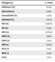
The pathogen detection rate was 73.3% (55/75) in males and 73.1% (57/78) in females, with no significant difference (χ² = 0.00, P = 1). The positive rates of pathogens in each age group are shown in Table 2. The overall detection rates of pathogens in different age groups were significantly different (χ² = 9.16, P = 0.01). There was no significant difference in the detection rate between 0 - 3 years and > 3 - 6 years (P > 0.05). The pathogen detection rates of 0 - 3 years and > 3 - 6 years were higher than those of > 6 - 14 years (P < 0.05). There was no significant difference in the coinfection rate among the three groups (χ² = 4.11, P = 0.13). The detection of various pathogens in the different age groups is shown in Table 3. The detection rates of RSV at 0 - 3 years and > 3 - 6 years were higher than those at > 6 - 14 years (P < 0.05). The infection rate of MP increased with age, and that of HRV decreased with age. For a comparison of the detection rates at different sampling sites, see Table 4. The positive detection rate of nasal swabs was 58.8% (90/153), and that of OPSs was 68.6% (105/153), with no significant difference (χ² = 3.63, P = 0.06). The total coincidence rate of the two samples was 80.4% (123/153), and the detection consistency was good (Kappa = 0.58, P < 0.05). The detection results of 13 pathogens in specimens from different sampling sites are shown in Table 5. The detection coincidence rates of each single pathogen in nasal swabs and oropharyngeal swabs were relatively high, and the detection consistency was good (P < 0.05). The positive rate of Mycoplasma pneumoniae in oropharyngeal swabs was 17.0%, which was significantly higher than that in nasal swabs 5.2% (χ² = 16.06, P < 0.05). No statistically significant differences in the detection rates of other pathogens between the two sites were observed (P > 0.05).
Table 2.
Pathogen Detection in Each Age Group a
| Groups (y) | The Number of Positive Cases | Coinfection |
|---|---|---|
| 0 - 3 (n = 70) | 55 (78.6) | 13 (18.6) |
| > 3 - 6 (n = 49) | 39 (79.6) | 13 (26.5) |
| > 6 - 14 (n = 34) | 18 (52.9) | 3 (8.8) |
| Total | 112 (73.2) | 29 (19.0) |
a Values are expressed as No. (%).
Table 3.
Pathogen detection of different age groups a
| Pathogen (n) | 0 - 3 Years | > 3 - 6 Years | > 6 - 14 Years | χ2 | P-Value |
|---|---|---|---|---|---|
| Influenza A (15) | 9 (12.9) | 4 (8.2) | 2 (5.9) | N/A | 0.57 |
| H1N1(2009) (8) | 6 (8.6) | 2 (4.1) | 0 (0.0) | N/A | 0.20 |
| Seasonal H3N2 (6) | 2 (2.9) | 2 (4.1) | 2 (5.9) | N/A | 0.86 |
| Influenza B (1) | 0 (0.0) | 1 (2.0) | 0 (0.0) | N/A | N/A |
| RSV (26) | 14 (20.0) | 12 (24.5) | 0 (0.0) | 9.36 | 0.01 |
| MP (26) | 2 (2.9) | 8 (16.3) | 16 (47.1) | 30.04 | < 0.01 |
| HRV (25) | 16 (22.9) | 8 (16.3) | 1 (2.9) | 6.37 | 0.01 |
| HMPV (11) | 6 (8.6) | 5 (10.2) | 0 (0.0) | N/A | 0.14 |
| HADV (9) | 3 (4.3) | 5 (10.2) | 1 (2.9) | N/A | 0.56 |
| HPIV (9) | 7 (10.0) | 2 (4.1) | 0 (0.0) | N/A | 0.12 |
| HCoV (3) | 1 (1.4) | 2 (4.1) | 0 (0.0) | N/A | N/A |
| HBoV (3) | 2 (2.9) | 1 (2.0) | 0 (0.0) | N/A | N/A |
| Ch (3) | 1 (1.4) | 2 (4.1) | 0 (0.0) | N/A | N/A |
Abbreviation: N/A, not available.
a Values are expressed as No. (%).
Table 4.
Comparison of Consistency Results of Samples from Different Sites a
| Nasal Swabs | OPSs | Total | |
|---|---|---|---|
| Positive | Negative | ||
| Positive | 82 | 7 | 89 |
| Negative | 23 | 41 | 64 |
| Total | 105 | 48 | 153 |
a Kappa = 0.58; P < 0.05.
Table 5.
Detection of 13 Types of Pathogens According to Specimen Type a
| Pathogen (n) | Nasal Swabs | OPSs | Coincidence Rate | Kappa | P-Value |
|---|---|---|---|---|---|
| Influenza A (15) | 15 (9.8) | 13 (8.5) | 98.7 | 0.92 | < 0.05 |
| H1N1(2009) (8) | 8 (5.2) | 6 (3.9) | 98.7 | 0.85 | < 0.05 |
| Seasonal H3N2 (6) | 5 (3.3) | 6 (3.9) | 99.3 | 0.91 | < 0.05 |
| Influenza B (1) | 1 (0.7) | 1 (0.7) | 100 | 1.0 | < 0.05 |
| RSV (26) | 25 (16.3) | 25 (16.3) | 98.7 | 0.95 | < 0.05 |
| MP (26) | 8 (5.2) | 26 (17.0) | 88.2 | 0.42 | < 0.05 |
| HRV (25) | 23 (15.0) | 17 (11.1) | 93.5 | 0.71 | < 0.05 |
| HMPV (11) | 9 (5.9) | 11 (7.2) | 98.7 | 0.89 | < 0.05 |
| HADV (9) | 4 (2.6) | 9 (5.9) | 96.7 | 0.60 | < 0.05 |
| HPIV (9) | 7 (4.6) | 9 (5.9) | 98.7 | 0.87 | < 0.05 |
| HCoV (3) | 3 (2.0) | 3 (2.0) | 100 | 1.0 | < 0.05 |
| HBoV (3) | 2 (1.3) | 3 (2.0) | 99.3 | 0.80 | < 0.05 |
| Ch (3) | 2 (1.3) | 2 (1.3) | 98.7 | 0.49 | < 0.05 |
a Values are expressed as No. (%).
5. Discussion
Multiple RT‒PCR combined with the capillary electrophoresis technique targets highly conserved sequences of 13 common respiratory pathogens. Thirteen sets of specific primers were designed, and one-step RT‒PCR was performed in an amplification tube. The results of pathogen detection were obtained via capillary electrophoresis of amplification products of different lengths. It is reported that 120 randomly selected samples detected by CEMP, reevaluated by a single RT‒PCR, showed a diagnostic agreement of 97.5% between the two methods (10). Li et al. compared the results to those obtained with the liquid chip-based LuminexxTAG Respiratory Viral Panel (RVP) Fast Kit (for viruses) and the agarose gel-based Seegene Pneumo Bacter ACE Detection Kit (for atypical bacteria); its sensitivity and specificity were 97.31% and 100%, respectively (11). In this study, among 153 outpatients, 112 (73.2%) had one positive pathogen, and 19% were positive for multiple pathogens, indicating that CEMP has unique advantages and high practical value in the diagnosis of pathogens. The co-infection rate of 19.0% in this study aligns with reports of 20% - 30% co-infection in pediatric pneumonia patients (12), highlighting the commonality of co-infections in children. Among the 13 pathogens, the top five were influenza virus, RSV, MP, HRV, and HMPV. The high detection rates of HRV and HMPV are consistent with findings on non-SARS-CoV-2 respiratory pathogens (13). Therefore, HRV and HMPV should be regarded as the main pathogens of respiratory tract infection in children. The clinical manifestations of HRV infection are fatigue, fever, and other symptoms, and severe cases may lead to damage to the spinal cord and other vital organs. Cough, runny nose, and fever are the main symptoms of HMPV infection. It can also lead to pharyngitis, bronchitis, and pneumonia. The incidence of pneumonia coinfection in children is reported to be 20% to 30%, and the younger the children are, the more likely they are to develop coinfection (2, 12, 14, 15). Bacterial infection was not evaluated in this group of patients, and the coinfection rate was 19%, indicating that high coinfection rates were also present in outpatients with influenza-like symptoms. The overall pathogen detection rate of the different age groups was 78.6% for those aged 0 - 3 years, and 79.6% of those aged > 3 - 6 years was greater than 52.9% of those aged > 6 - 14 years (P < 0.05). It has been reported that older children are more likely to be affected by MP or bacterial infection 2. The detection rate of RSV was 20.0% at 0 - 3 years and 24.5% at > 3 - 6 years. Fever caused by RSV infection is not uncommon in outpatients aged 3 - 6 years, which is even greater than the proportion of fever caused by influenza in this age group (18.4%). The infection rates of HRV were 22.9%, 16.3%, and 2.9% at 0 - 3 years, > 3 - 6 years, and > 6 - 14 years, respectively, and those of MP were 2.9%, 16.3%, and 47.1%, respectively, indicating that the infection rate of MP increased with age (trend χ² = 30.04, P < 0.01), whereas that of HRV decreased with age (trend χ² = 6.37, P = 0.01). Therefore, age has a suggestive effect on the pathogen diagnosis of respiratory tract infection in children. It is very important to choose respiratory samples for respiratory virus and atypical bacterial antigen or nucleic acid testing because they are related to the positive rate. Wang et al. tested 11 common respiratory tract pathogens in hospitalized children with lower respiratory tract infections via CEMP and compared the test results of OPSs and sputum. The positive rate of OPSs was 84%, whereas that of sputum was 88% (P = 0.007). Young patients are likely to have consistent results between the two samples (4). Shan et al. detected FluA, FluB, RSV, and ADV antigens in children with upper respiratory tract infections via gold immunoassays. The positive rate of NPSs was 52.3%, which was higher than that of OPSs (37.6%) (χ² = 16.49, P < 0.01) (6). Todsen et al. used RT‒PCR to detect SARS-CoV-2 in OPSs and NPSs (16). As a result, the virus detection rate in OPS was 78.7%, which was higher than the 72.7% in NPS (P = 0.049), while the sampling discomfort of the NPS was greater than that of the OPS. It is generally believed that the viral load of the nasopharynx is greater than that of the oropharynx (17). Owing to the low sensitivity of the colloid gold immunoassay for antigen detection, it is recommended to collect NPSs; however, the sensitivity of PCR is high, and the selection of NPSs is less important than that of the colloidal gold immunoassay for antigen detection. In this study, oropharyngeal swabs were compared with nasal swabs, and the total detection rates of 11 viruses, MP, and chlamydia were 58.8% for the nasal swabs and 69.3% for the oropharyngeal swabs, respectively, with no significant difference (χ² = 3.63, P = 0.06). The total coincidence rate of the two samples was 80.4%, and the test consistency was good (Kappa = 0.58, P < 0.05). The detection coincidence rates of each individual pathogen from nasal swabs and oropharyngeal swabs were high, and the test consistency was also good (P < 0.05). The positive rate of oropharyngeal swab for Mycoplasma pneumoniae was 17.0%, significantly higher than that of nasal swab at 5.2% (χ² = 16.06, P < 0.05). No statistically significant differences were observed in the detection rates of other pathogens between the two sites (P > 0.05). The comfort and convenience of nasal swabs are better than those of NPSs and OPSs. Therefore, for children who do not cooperate in collecting NPSs and OPSs, nasal swabs could be considered for testing for respiratory viruses and chlamydia. If the clinical diagnosis suggests Mycoplasma pneumoniae infection, then it is advisable to collect oropharyngeal swab for testing.
5.1. Study Limitations
This study only included outpatients with fever and respiratory symptoms, potentially excluding asymptomatic or mild cases (selection bias). The 13 pathogens tested do not cover all respiratory pathogens (e.g., bacteria), possibly missing co-infections. Sampling was conducted from October 2022 to June 2023, which may not reflect seasonal variations. Nasal and oropharyngeal swab collection depended on operator experience, which may have introduced variability.