Characterization of liposomes
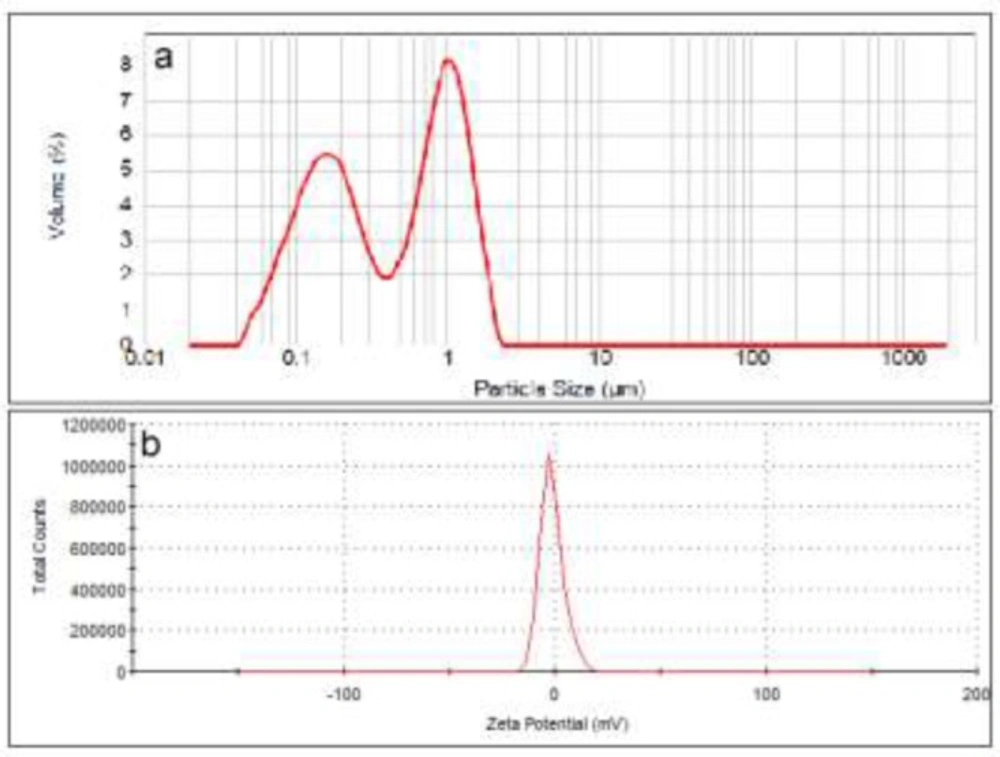
The particle size distribution curve of neutral liposomes showed two peaks at 200 and 1100 nm with particle range between 30-2000 nm. Zeta potential of this formulation was measured to be -1.38 mV (
Figure 1) and its D0.5 was measured to be about 450 nm with a span of 3.1 (n = 3).
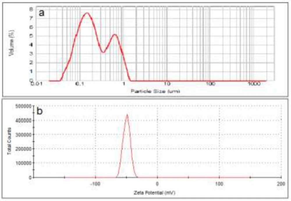
The negatively charged liposomes showed a D0.5 of about 200 nm with a span of 3.5 (n = 3). Range of size distribution in this formulation was from 30 nm to 1600 nm. The size distribution curve contained two peaks at 180 and 650 nm. Analyzing the zeta potential of these particles confirmed that they carry a negative charge of about -50 mV (
Figure 2).
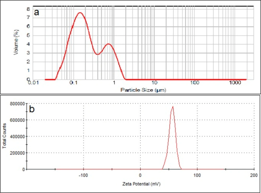
Particle size of the positively charged liposomes showed D0.5 of about 200 nm with a span of 4.2 (n = 3). Size distribution curve of these particles contains two peaks at 180 and 750 nm that covers range of between 30-1600 nm. Zeta potential of the particles measured to be about +56 mV (
Figure 3).
To investigate the effect of charges on liposomes separation, the particle size of the liposomal formulations was adjusted to be as similar as possible (
Figure 1-
3) while their zeta was different from each other. Also the lipid composition of the positively charged, negatively charged and neutral liposomes was almost the same, except for the charge determining lipids. Particle size distribution of all liposomal formulations were intentionally chosen to be wide, hence high spans, to have heterogeneous samples and higher possibility of size separation upon stagnation. The chosen size ranges cover the sizes usually employed or investigated in pharmaceutical studies and also covers the particle with high and low chances of Brownian motion.
Particle size distribution (a) and zeta potential (b) of neutral liposomes (DSPC:cholesterol).
Particle size distribution (a) and zeta potential (b) of anionic liposomes (DSPC: DSPG: cholesterol
Particle size distribution (a) and zeta potential (b) of cationic liposomes (DSPC: DOTAP: cholesterol
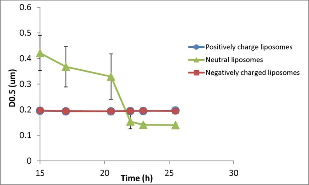
Changes in the particle size (D0.5) of liposomal formulation with different charges (n = 3) for samples taken at different times (resembling different depth in the burette). The zeta potential of initial (freshly prepared liposomes) are -50, -1.4 and +56 mV for negative, neutral and positive formulation respectively and their particle size range is 30-1600nm (negative liposomes), 30-2000 (neutral liposomes) and 30-1600 (positive liposomes
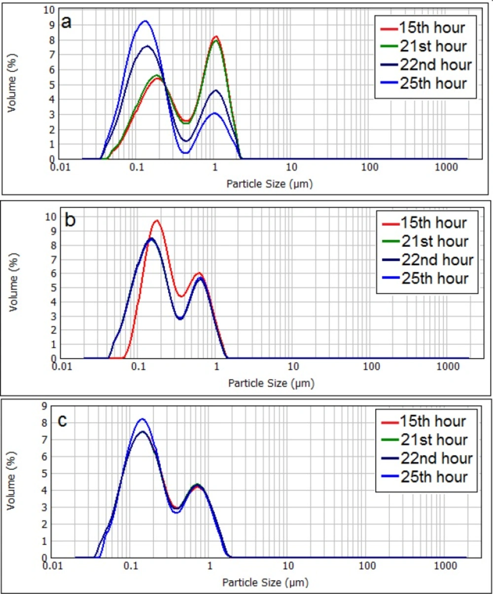
Changes in the particle size distribution of neutral liposomes (a), negatively charged (b), and positively charged (c) liposomes. The zeta potential of initial (freshly prepared liposomes) are -50, -1.4 and +56 mV for negative, neutral and positive formulation respectively and their particle size range is 30-1600nm (negative liposomes), 30-2000 (neutral liposomes) and 30-1600 (positive liposomes
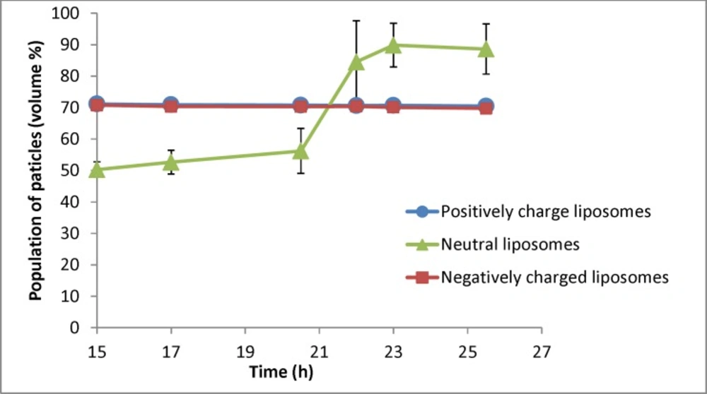
Changes in population of particles smaller than 400nm for liposomal formulation with different charges (n = 3) for samples taken at different times (resembling different depth in the burette). The zeta potential of initial (freshly prepared liposomes) are -50, -1.4 and +56 mV for negative, neutral and positive formulation respectively and their particle size range is 30-1600nm (negative liposomes), 30-2000 (neutral liposomes) and 30-1600 (positive liposomes
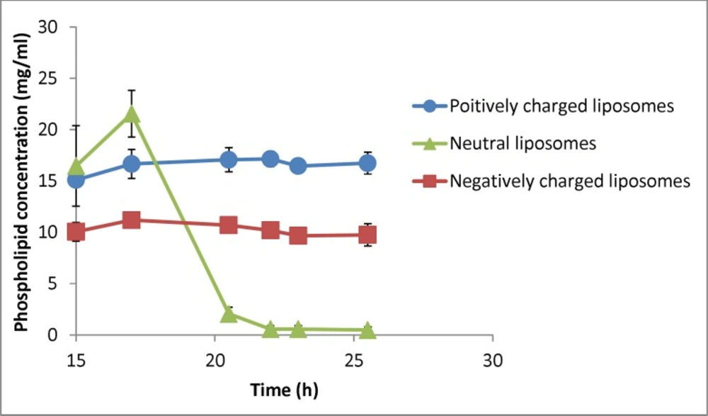
Changes in phospholipid concentration in liposomal formulation with different charges under gravity over time (n = 3). The zeta potential of initial (freshly prepared liposomes) are -50, -1.4 and +56 mV for negative, neutral and positive formulation respectively and their particle size range is 30-1600nm (negative liposomes), 30-2000 (neutral liposomes) and 30-1600 (positive liposomes
Separation of liposomes with different charges
Figure 4 shows the changes of D0.5 of three liposomal formulations encountering the gravity. The negatively and positively charged formulations showed no changes of particle size over time and among different samples, while the particle size of neutral liposomes got smaller over time. The size of neutral liposomes changed from 420 nm (15th h) to 140 nm (26
th hour). The separation factor (D0.5
last sample/D0.5
first sample) for this formulation was measured to be 0.33, while the same factor was measured to be 1.005 and 0.989 for positively and negatively liposomes respectively showing no size separation over time for charged liposomes. Please note that the sampling time resembles the sample depth as well.
The Stoke’s law discusses the free falling of particles at a constant rate and without any hindrance and uses the Anderson apparatus to measure the diameter of particles (
14) which is close to the sedimentation method of the current study. However, our model has employed narrower system and therefor can work with lower volume at the same depths. Our model provides much higher height as small volumes.
The main necessity for this law for the particles is absence of aggregation and inter-particular interactions such as electrostatic interactions. These interactions are more noticeable in particle with smaller size which is also the subject of present investigation. Sedimentation of nanoparticles smaller than 500 nm is almost negligible due to Brownian motion. Results of the present study show that sedimentation of particles occur even at sizes lower than what is supposed to be Brownian motion border (
Figure 4), probably due to aggregation or flocking. Our results also indicate that even large particles do not sediment in charged liposomes.
Particle size distribution curve of neutral liposomes shows that population of smaller sized liposomes grows significantly over time (
Figure 5), whilst the size distribution of charged liposomes showed no or little change during the experiment and the size distribution of charged liposomes remained almost unchanged (P > 0.05).
Figure 6 shows the percentage of particles smaller than 400 nm for different formulations. As shown in
Figure 6, there is no change for charged liposomes, while in neutral liposomes the population of liposomes less than 400 nm increases over time. The separation factor for this parameter in neutral, negative and positive liposomal formulation has been calculated as 1.76, 0.98 and 0.99 respectively. These results indicate that percentage of neutral liposomes with sizes smaller than 400 nm at the 26th hour is almost 2 times greater than the 15th h.
The above results showed that sedimentation of neutral liposomal formulations, even below the Brownian motion’s border, is considerable and there is a growth in nanoparticles population over time and in higher depths. Charged liposome acted completely different from the neutral particles and no sedimentation and size reduction occurred over time although they contain particles as large as 2 µM. This could be the effect of electrostatic repulsion between the particles which inhibited the sedimentation and separation of particles, and therefor size distribution of charged particles remained the same in all depths.
Gravitational, van der Waals and buoyancy are known to be three main forces in a suspension of particles. Van der Waals is the main force to influence the stability of these dispersions. If two particles approach each other up to a minimum distance, this force will bind them together and can cause sedimentation and instability of dispersion (
16). Increasing the charge density of these dispersions, which is known as electrostatic stabilization, prevents this phenomenon (
14). Regarding the present results, it seems that the electrostatic repulsion between the charged liposomal particles conquers gravity and prevents them from sedimentation, so the charged formulations remain uniformly distributed during the experiment. Lack or shortage of the electrostatic forces in the neutral formulation makes gravity the dominant forces in the system. In such conditions, any size separation (e. g. sedimentation) in the formulation led to increased percentage of smaller particles in the collected samples by increasing the time of sampling.
There is not such a data available on the effects of charge on separation of liposomes upon stagnation. However, Plessis
et al. (
17) investigated long-term (6 months) physical stability of different liposomes with different lipid compositions with positive zeta potentials of 75-100 mV. Their results showed that presence of cationic lipids decreases long-term physical stability of liposomes. They did not study phase separations upon stagnation or effect of charge on this phenomenon. The reported increased instability due to positive lipids might be related to higher chemical interaction of charged molecules. Other studies have suggested charge to be a promising factor on stability of liposomes (
12,
13) but there is lack of data about the effect of charge on their stagnation and separation behavior in such conditions.
Changes in the phospholipid concentration during the experiment were also investigated as an indicator of liposomes’ concentration (
Figure 7). Statistical analysis showed that phospholipid concentration of neutral liposomes in each time point was significantly different from the other samples (P < 0.05) and there was no difference in phospholipid concentration of charged liposomes during the experiment (P > 0.05). Separation factor for this parameter in neutral, negative and positive liposomal formulations encountering gravity was measured to be 0.03, 0.96 and 1.11. These results indicate that the charged particles with different sizes do not tend to separate under gravity force, whereas the neutral particles tend to behave the opposite way and the system cannot be considered homogenous considering the lipid composition.
These results show the importance of liposome formulation properties and indicate that the charge of liposomes is an effective factor on their sedimentation over time. The above result could be used in development new liposomal formulations, storage protocols, application considerations such as in infusion set or pumps, osmotic pumps, etc. Such separations are expected to affect other properties such as rheology of the system and also expected to be much more evident for heavy formulations such as magnetoliposomes (
7).
The findings of the present investigation can be used together with other of liposomal stabilization methods such as liposomal gel formulation (
18,
19). The results of the present investigation also indicate that instability can occur in short times and can be very important in application of liposomes such as in infusion systems.