


1. Introduction
2. Case Presentation
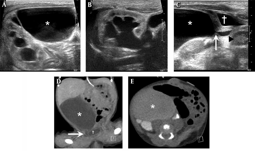
A one-day-old girl with a large cystic abdominal mass and right-sided hydroureteronephrosis discovered during an antenatal ultrasound examination. Abdominal ultrasound and abdominal contrast-enhanced CT. A, Axial ultrasonography (US) image shows a large cystic mass (asterisk) with multi-layered wall in the right upper abdomen; B, Ultrasound shows right pelvocalyceal dilatation and calyceal blunting with increased renal parenchymal echogenicity; C, Ultrasound identifies the continuity between the cystic mass (asterisk) and the cystic tubular structure (arrowhead). Abrupt luminal narrowing (white arrow) at the connection site is noted. The cystic tubular structure is connected with the urinary bladder (cross) with normal peristalsis. Peristalsis of the small cystic tubular structure is presented on video 1; D, Coronal view of contrast-enhanced CT scan demonstrates the cystic mass (asterisk) in the right lower abdomen and small cystic tubular structure (white arrow) which is between the cystic mass and urinary bladder (cross); E, Axial view of 2-hour delayed CT scan shows opacification of the urinary system and the cystic mass (asterisk) with excreted contrast.
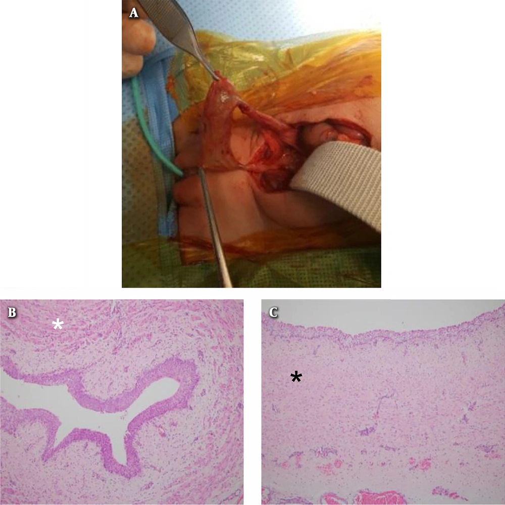
Intraoperative photograph and photomicrograph of the specimen. A, Intraoperative photograph shows the dilated segment of the right ureter of the congenital segmental giant megaureter; B and C, Photomicrograph of the cross-sectional view of the non-dilated segment; B, Exhibits normal distribution of the smooth muscle tissue (white asterisk) and photomicrograph of the cross-sectional view of the dilated segment; C, Shows abundant vascular elements and connective tissue under the urothelial epithelium, with marked dissociation of smooth muscle bundles (black asterisk) (hematoxylin and eosin, 100×).


