Cover image of the article: Unsuspected Pregnancy During Hysterosalpingography

Image Credit:I J Radiol

Unsuspected Pregnancy During Hysterosalpingography

Authors

Firoozeh Ahmadi1,*, Hadieh Haghighi1
1Department of Reproductive Imaging at Reproductive Biomedicine Research Center, Royan Institute for Reproductive Biomedicine, ACECR, Tehran, Iran
*Corresponding Author: Corresponding author: Firoozeh Ahmadi, Department of Reproductive Imaging at Reproductive Biomedicine Research Center, Royan Institute for Reproductive Biomedicine, ACECR, Tehran, Iran, E-mail: Email: f_ahmadi@royaninstitute.org

Iranian Journal of Radiology:Vol. 11, issue 3; e5033
Published online:Aug 01, 2014
Article type:Letter
Received:Apr 03, 2012
How to Cite:Ahmadi F, Haghighi H. Unsuspected Pregnancy During Hysterosalpingography. I J Radiol. 2014;11(3):e5033. doi: https://doi.org/10.5812/iranjradiol.5033

Dear Editor
Hysterosalpingography (HSG) is now increasingly used as the first-line investigation to assess the uterine cavity and tubal patency in infertility work-up. Most patients with a history of infertility do not use a contraceptive method and due to irregular menstrual cycles, menstrual history is not always reliable. In this letter, we emphasize on clinical implications for preventing inadvertently performed HSG during pregnancy. A 34-year-old woman with a four-year history of infertility was referred for routine base investigation by pelvic ultrasound before intrauterine insemination (IUI). HSG was carried out one month ago, 3 days after menstrual bleeding had stopped. Although she had regular menstruations, she had an abnormal bleeding one month ago and was unaware of her pregnancy and decidual bleeding was presumed as menstrual bleeding. Therefore, the patient was regarded as not pregnant and no β-HCG test was performed before the HSG procedure. HSG of this patient illustrated a large endometrial cavity with a filling defect, which was reported as myoma, and the patient was referred for ultrasound investigation of the uterine filling defect (Figure 1 A, B). Ultrasonography showed a 6 to 6.5-week gestational age embryo without a heart rate.Abortion occurred after a few days. Although there have been only a few case reports on undiagnosed pregnancy during (HSG) (1-7), it is important to emphasize on the unusual radiological features of this condition to alert radiologists and gynecologists reporting HSG. Early pregnancy during HSG may be recognized by a double outlined uterine cavity (DOUC) and the intrauterine filling defect denotes the fetal sac (1-7).However, a normal HSG cannot exclude early pregnancy during HSG (1). Contrast media infiltrating the decidual lining is responsible for the DOUC appearance (Figure 2). This sign can also suggest hyperplasia or late normal secretory endometrium. The filling defect can also be a sign of endometrial polyp, submucosal myomas or synechiae. Detection of DOUC sign should assist the operator to terminate the introduction of contrast medium and make a certain interpretation of early pregnancy (2). Although there have been several reports on the normal outcome in such cases (1-7), the potential concerns are teratogenic risks, miscarriage and displacement of the fertilized ovum (ectopic pregnancy,) due to the side-effect of the flushing effect of contrast media (3). The risk of mutation is low due to low exposure during HSG and termination is not recommended (4).
Inadvertently performed HSG during pregnancy can be prevented by:
1) Pelvic ultrasound evaluation before HSG (3)
2) Scheduling the procedure during the early proliferative phase
3) Maintaining a high index of suspicion, and not relying solely on the menstrual history. A pregnancy test on the day of the procedure,even in women with regular cycles and particularly for women with one irregular cycle or amenorrhea is mandatory.
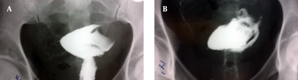
A and B, HSG illustrates a large endometrial cavity with a filling defect
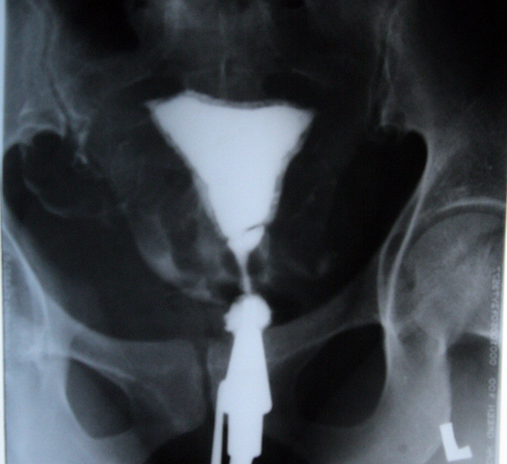
Figure 1.
A and B, HSG illustrates a large endometrial cavity with a filling defect
Outlined uterine cavity (DOUC) due to contrast material infiltrating into the endometrium during early pregnancy (arrows).
Figure 2.
Outlined uterine cavity (DOUC) due to contrast material infiltrating into the endometrium during early pregnancy (arrows).

Footnotes

  • Implication for health policy/practice/research/medical education:It is important to emphasize on the unusual radiological features of inadvertently performed HSG during pregnancy to alert radiologists and gynecologists reporting HSG.

References

  • 1.
    Cheung GW, Lok IH, Wong A, Yip SK. Unsuspected pregnancy at hysterosalpingography: a report of three cases with different outcomes. Hum Reprod. 2003;18(12):2608-9. [PubMed ID: 14645179].
  • 2.
    Isaacs I. Hysterographic double-outlined uterine cavity: a sign of unsuspected pregnancy. AJR Am J Roentgenol. 1978;131(2):305-6. [PubMed ID: 98013]. https://doi.org/10.2214/ajr.131.2.305.
  • 3.
    Akhigbe AO, Oyobere O, Ukah UR. Unsuspected pregnancy at hysterosalpingography: a case report and outcome. Niger J Clin Pract. 2009;12(3):327-9. [PubMed ID: 19803037].
  • 4.
    Jongen VHWM, Collins JMP, Lubbers JA, van Selm M. Unsuspected early pregnancy at hysterosalpingography. Fertility and Sterility. 2001;76(3):610-1. https://doi.org/10.1016/s0015-0282(01)01981-1.
  • 5.
    Justesen P, Rasmussen F, Andersen PEJr. Inadvertently performed hysterosalpingography during early pregnancy. Acta Radiol Diagn. 1986;27(6):711-3.
  • 6.
    Meamarzadeh MT, Shahrzad GH. Hysterosalpingography in unsuspected pregnancy: report of 6 cases and 2 relevant cases. Iran J Radiol. 2005;2(3):147-51.
  • 7.
    Damodaran S, Jones M, Mahmood TA. Rare finding in a common procedure: unsuspected pregnancy during hysterosalpingography. J Obstet Gynaecol. 2011;31(2):194-5. [PubMed ID: 21281049]. https://doi.org/10.3109/01443615.2010.533217.

Copyright

Copyright © 2014, Tehran University of Medical Sciences and Iranian Society of Radiology. This is an open-access article distributed under the terms of the Creative Commons Attribution-NonCommercial 4.0 International License (http://creativecommons.org/licenses/by-nc/4.0/) which permits copy and redistribute the material just in noncommercial usages, provided the original work is properly cited.

Similar Articles

30
Jun
2005
Hysterosalpingography in Unsuspected Pregnancy : Report of 6 Cases and 2 Relevant Cases

Hysterosalpingography in Unsuspected Pregnancy : Report of 6 Cases and 2 Relevant Cases

Mohammad Taghi Meamarzadeh,
G. H. Shahrzad

Meamarzadeh MT, Shahrzad GH. Hysterosalpingography in Unsuspected Pregnancy : Report of 6 Cases and 2 Relevant Cases. I J Radiol. 2005;2(3&4):e79128. doi:

7
Nov
2020
Effect of Hysterosalpingography (HSG) on Biochemical Pregnancy Rate in Women with Primary and Secondary Infertility

Effect of Hysterosalpingography (HSG) on Biochemical Pregnancy Rate in Women with Primary and Secondary Infertility

Sharareh Sanei Sistani,
Hamid Dahmardeh,
Roya Hasanzadeh,
Farahnaz Farzaneh

Sanei Sistani S, Dahmardeh H, Hasanzadeh R, Farzaneh F. Effect of Hysterosalpingography (HSG) on Biochemical Pregnancy Rate in Women with Primary and Secondary Infertility. Zahedan J Res Med Sci. 2020;22(4):e91725. doi: https://doi.org/10.5812/zjrms.91725

30
Dec
2006
Comparison of Sonohysterography and Hysterosalpingography with Hysteroscopy in the Diagnosis of Intrauterine Lesions

Comparison of Sonohysterography and Hysterosalpingography with Hysteroscopy in the Diagnosis of Intrauterine Lesions  

Shirin Qazizadeh

Qazizadeh S. Comparison of Sonohysterography and Hysterosalpingography with Hysteroscopy in the Diagnosis of Intrauterine Lesions  . I J Radiol. 2006;4(1):e79248. doi:

17
Aug
2012

Pregnancy rate after laparoscopy in the infertile women referring to

Jamali S,
Rasekh Jahromi A

S J, Jahromi A R. Pregnancy rate after laparoscopy in the infertile women referring to. Jundishapur J Chronic Dis Care. 2012;1(2):. doi:

12
Apr
2017

Womens Imaging, Us Imaging for Infertility Workup

Firoozeh Ahmadi

Ahmadi F. Womens Imaging, Us Imaging for Infertility Workup. I J Radiol. 2017;14(Special Issue):e48364. doi: https://doi.org/10.5812/iranjradiol.48364

More by these authors

Firoozeh AhmadiPubMedScholar
Hadieh HaghighiPubMedScholar
Share
Cited by
Metrics