Cover image of the article: A Primary Giant Hydatid Cyst of the Ovary

Image Credit:I J Radiol

A Primary Giant Hydatid Cyst of the Ovary

Authors

Abdurrahman Kaya1,*, Sibel Yildiz1, Reşat Özaras1, Ali Mert1
1Department of Infectious Diseases, Medical School of Cerrahpasa, Istanbul University, Istanbul, Turkey
*Corresponding Author: Corresponding author: Abdurrahman Kaya, Department of Infectious Diseases, Istanbul, Turkey. Tel.: +90-5066113328, Fax: +90-2126320050, E-mail: Email: dr.abdkaya@hotmail.com

Iranian Journal of Radiology:Vol. 9, issue 3; 165-166
Published online:Sep 17, 2012
Article type:Letter
Received:Jul 18, 2011
Accepted:Oct 16, 2011
How to Cite:Kaya A, Yildiz S, Özaras R, Mert A. A Primary Giant Hydatid Cyst of the Ovary. I J Radiol. 2012;9(3):165-166. doi: https://doi.org/10.5812/iranjradiol.7955
KeywordsCystsOvary

Dear Editor,
A 38-year-old female patient was admitted with right lower abdominal pain of 4 months duration. On her clinical examination, right lower abdominal tenderness was noted. Complete blood count including the numbers of eosinophils (80/mm3) was normal. Biochemistry was normal. An abdominal MRI showed an 11 × 9 × 5.5 cm sized cystic lesion in the right ovary surrounded by loculated fluid collection with septations (Figure 1). Echinococcal serology was positive (hydatid cyst antibodies by ELISA and indirect hemagglutination test dilutions up to 1/1280 were positive). Pathological examination of the material had a cuticle and germinative membrane of the hydatid cyst and around it chronic nonspecific inflammation leading to the formation of lymphoid follicles was seen. No other organ involvement was detected. Albendazole (10mg/kg/day) was prescribed. The cyst was removed surgically on the 30th day of albendazole treatment. Pathological examination of the cyst was compatible with hydatid cyst. The patient had regular follow-ups, with no recurrence of the cyst. Echinococcal disease is endemic in many countries (1). It involves multiple organs particularly the liver (50-70%) and the lung (20-30%), while primary involvement of the ovary is very rare (2). The liver and spleen cysts can rupture into the peritoneal cavity which is the way of secondary involvement (3). In our case, there were no cysts in other organs. Although ovarian cysts are usually single, this case showed multilocular cysts the treatment is surgical removal and albendazole is recommended before surgery.
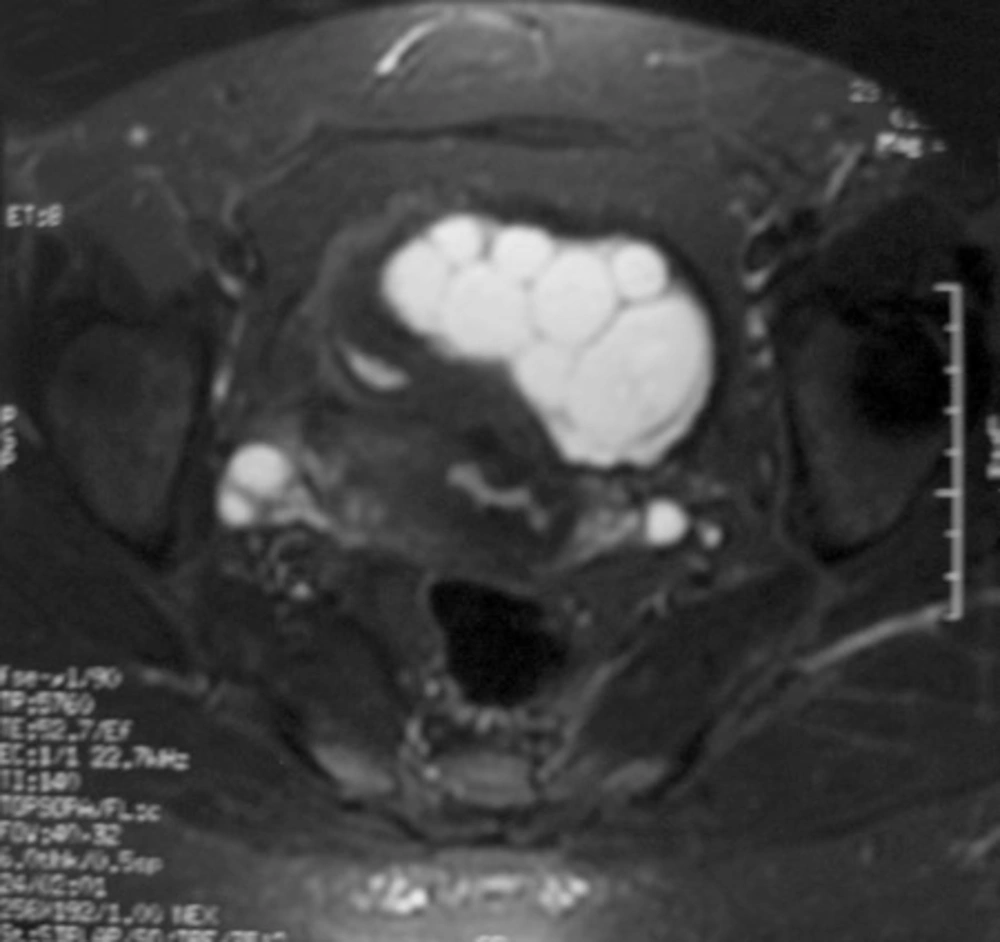
Pelvic MRI revealed a septated, lobulated and contrast enhancing cystic mass that is hyperintense on T2 and hypointense on T1 FLAIR sequences.
Figure 1
Pelvic MRI revealed a septated, lobulated and contrast enhancing cystic mass that is hyperintense on T2 and hypointense on T1 FLAIR sequences.

Footnotes

  • Please cite this paper as:Kaya A, Yildiz S, Özaras R, Mert A. A Primary Giant Hydatid Cyst of Ovary. Iran J Radiol. 2012;9(3):165-6.

  • Authors’ Contribution:Abdurrahman Kaya followed the patients and wrote the paper. Sibel Yıldız assisted in writing and took the photos. Reşat Özaras coordinated the follow-up and edited the paper. Ali Mert coordinated the follow-up and edited the paper.

  • Financial Disclosure:None declared.

References

  • 1.
    Nakayama DK. Survival in a fetus with sacrococcygeal teratoma and hydrops. Am J Obstet Gynecol. 1990;163(2):682.
  • 2.
    Fahy AM, Peters EE. Eccrine tumours of the hand: two cases. J Hand Surg Br. 1987;12(1):120-2. https://doi.org/10.1016/0266-7681(87)90075-1.
  • 3.
    Sing P, Mushtaq D, Verma N, Mahajan NC. Pelvic hydatidosis mimicking a malignant multicystic ovarian tumor. Korean J Parasitol. 2010;48(3):263-5. [PubMed ID: 20877508]. https://doi.org/10.3347/kjp.2010.48.3.263.

Copyright

Copyright © 2012, Tehran University of Medical Sciences and Iranian Society of Radiology. This is an open-access article distributed under the terms of the Creative Commons Attribution-NonCommercial 4.0 International License (http://creativecommons.org/licenses/by-nc/4.0/) which permits copy and redistribute the material just in noncommercial usages, provided the original work is properly cited.

Similar Articles

1
Mar
2019
Pericardial hydatid cyst in a Child: a case report

Pericardial hydatid cyst in a Child: a case report

Hafida Naoui,
Souad Azelmat,
Zohra Lemkente,
Siham Semlali,
Maryam Iken,
Jamal El Fenni
,et al.

Naoui H, Azelmat S, Lemkente Z, Semlali S, Iken M, et al. Pericardial hydatid cyst in a Child: a case report. Int Cardiovasc Res J. 2019;13(1):e70668. doi:

30
Jun
2015
Hydatid Cyst of Right Atrium: A Case Report

Hydatid Cyst of Right Atrium: A Case Report

Maral Mokhtari,
Hooriah Momen Zadeh

Mokhtari M, Momen Zadeh H. Hydatid Cyst of Right Atrium: A Case Report. Int Cardiovasc Res J. 2017;9(2):e11408. doi:

26
Mar
2016
Surgical Management of an Ovarian Bilateral Multiple Dermoid Cyst During Pregnancy: A Case Report Study

Surgical Management of an Ovarian Bilateral Multiple Dermoid Cyst During Pregnancy: A Case Report Study

Sanam Moradan

Moradan S. Surgical Management of an Ovarian Bilateral Multiple Dermoid Cyst During Pregnancy: A Case Report Study. Middle East J Rehabil Health Stud. 2016;3(2):e36097. doi: https://doi.org/10.17795/mejrh-36097

30
Jun
2013
Laparoscopic surgery of hepatic hydatid cyst: A case report

Laparoscopic surgery of hepatic hydatid cyst: A case report

A Aghaei,
SR. Mohamadi,
A Divanbeigi

Aghaei A, Mohamadi S, Divanbeigi A. Laparoscopic surgery of hepatic hydatid cyst: A case report. J Inflamm Dis. 2024;17(1):e155748. doi:

29
Mar
2013
The peritoneal benign multicystic mesothelioma simulating hydatid cyst manifestation

The peritoneal benign multicystic mesothelioma simulating hydatid cyst manifestation

Nasrin Jalilian,
Homa Badri,
Mahtab Rahbar

Jalilian N, Badri H, Rahbar M. The peritoneal benign multicystic mesothelioma simulating hydatid cyst manifestation. J Kermanshah Univ Med Sci. 2013;16(8):e77281. doi:

More by these authors

Abdurrahman KayaPubMedScholar
Sibel YildizPubMedScholar
Reşat ÖzarasPubMedScholar
Share
Cited by
Metrics