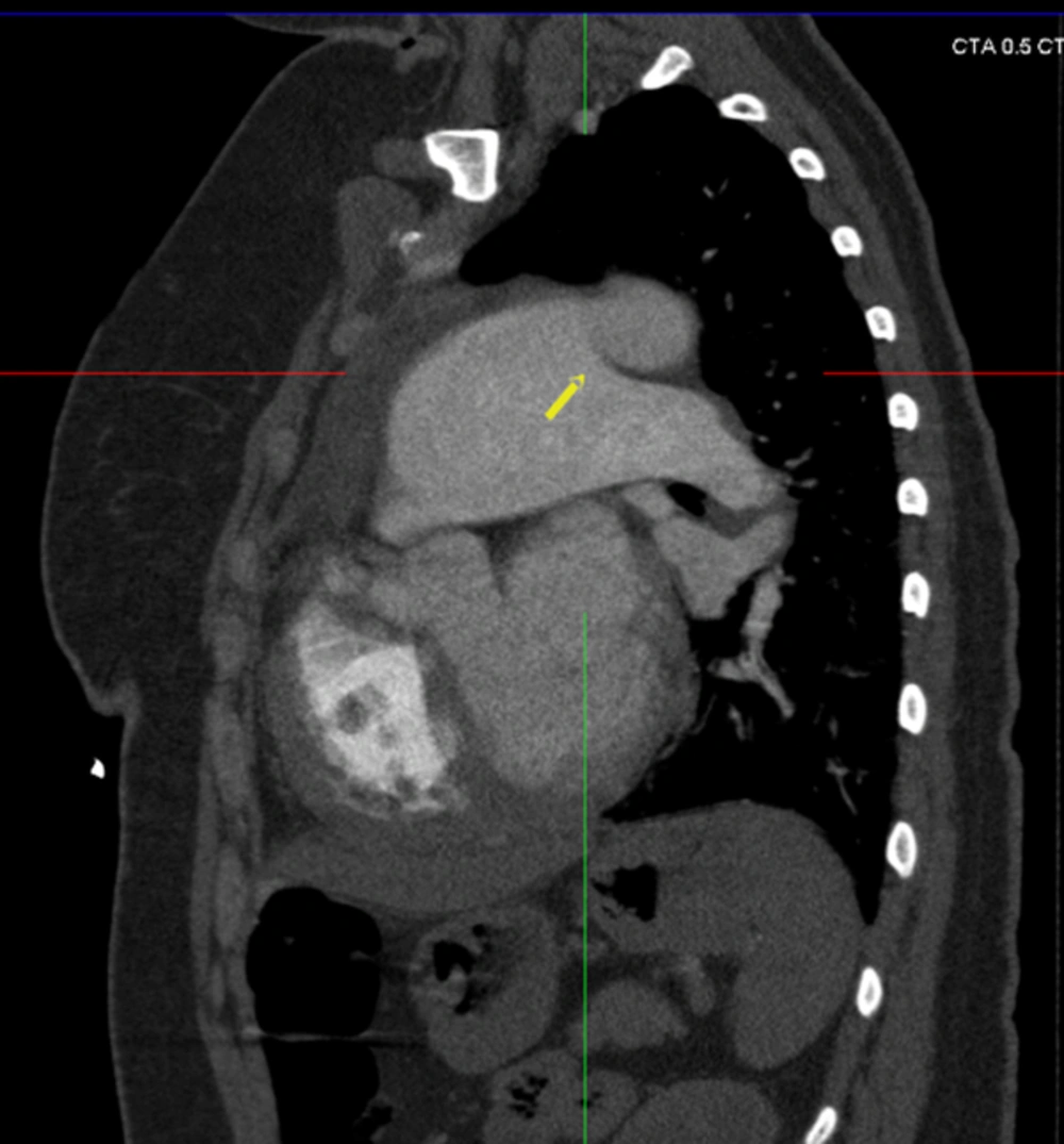
ALCAPA, also known as bland-white-garland syndrome, is an infrequent congenital cardiac anomaly. Normal coronary arteries are developed from: 1) endothelial sprouts localized in the walls of great arteries at the level of the sigmoidal values; 2) the right and left subepicardial vascular network; and 3) the intramyocardial sinusoids. Most of the ectopic coronary arteries are due to the alterations in the connection between these three embryonic elements. The deviation of one of the subepicardial vascular networks in the wrong way (in the direction of the pulmonary artery or the opposite valsalva sinus) will cause the development of endothelial sprouts, which will connect the abnormal connections and the anomalous origin of the coronary arteries (
3).
PDA is classified into two subtypes. These subtypes are the infantile subtype and the adult subtype. In the infantile subtype, the symptoms occur at ages younger than 12 months, and in the adult subtype the symptoms occur at ages older than 12 months (
4). Patients with this abnormality may present with subtle clinical complaints. Identifying these abnormalities is crucial in terms of medical and surgical management of patients. Poorly oxygenated blood from the coronary artery, with insufficient collateral circulation from the right coronary artery, and also the left coronary artery from the pulmonary artery, steals blood from the pulmonary circulation revealed by low coronary perfusion pressure due to the creation of myocardial ischemia symptoms.
Prenatally, there is no adverse effect that develops from ALCAPA because of the unique intrauterine fetal circulation; in the fetal circulation system, oxygen levels are equal between the main pulmonary artery and the aorta. Shortly after birth, a decrease in pressure, resistance and the oxygen content of the pulmonary artery leads to hypoperfusion of the myocardium by desaturated blood under low pressure. Without treatment, this syndrome is 90% fatal in the first year of life. Death is secondary to widespread myocardial ischemia, infarction, and at the end, developing of heart failure. Approximately 10% of cases are diagnosed in adulthood due to exertional chest pain and dyspnea caused by inadequate myocardial perfusion. In extremely rare cases, ALCAPA without symptoms is accidentally detected on cardiac screening in adults. But syncope, angina pectoris, dyspnea or cardiac arrest occurrence is more usual than an asymptomatic course with ALCAPA (
5-
7). After the development of a significant post-tricuspid shunt, PDA prevents the fall in the pulmonary artery pressure, and PDA also provides oxygenated blood to the left coronary artery system, thus preventing the dramatic changes seen in a child with associated ALCAPA.
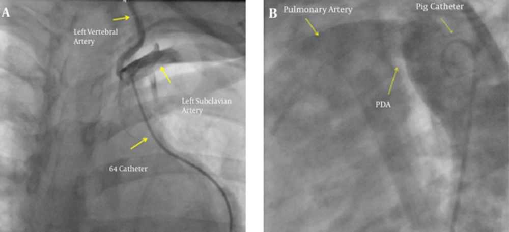
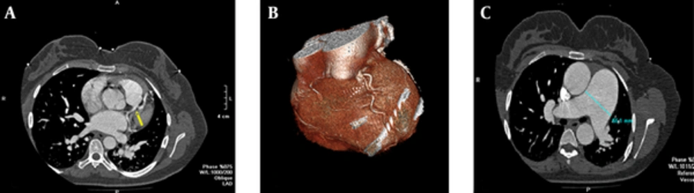
Conventional angiography is an invasive and limited diagnostic imaging modality for coronary artery anomalies. The sensitivity of conventional angiography is limited for the detection of a coronary artery anomaly because of its projection capability. CT coronary angiography is a beneficial non-invasive imaging method for demonstrating these types of coronary artery anomalies (
8). However, multi-slice computed tomography (MSCT) coronary angiography is an excellent three-dimensional (3D) imaging method. Using this imaging modality, we were able to demonstrate the coronary vessel anatomy with high quality. In addition, another beneficial imaging modality for detecting ALCAPA is two-dimensional Doppler echocardiography. This diagnostic method is considered the best diagnostic tool for detecting ALCAPA (
4).
Coronary artery anomalies originate from the left or right coronary artery, and both may be isolated or may be associated with different types of conotruncal and heart abnormalities (
9-
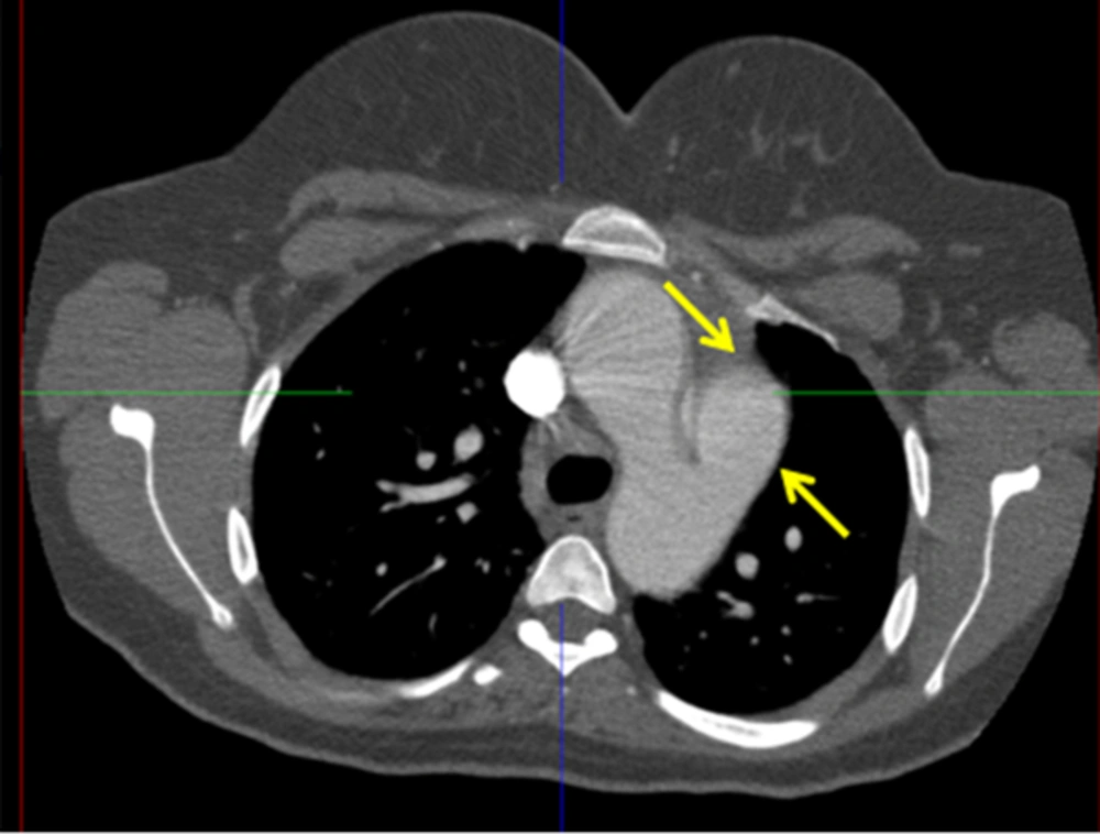
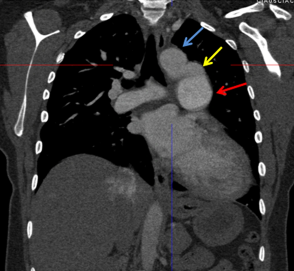
12). CT angiography is also a valuable tool for detecting other kinds of congenital cardiac abnormalities such as ventricular septal defect (VSD), atrial septal defect (ASD), PDA, tetralogy of Fallot, pulmonary atresia, Ebstein anomaly, transposition of the great arteries and tricuspid atresia. Ultrasonography is the best prenatal diagnostic imaging tool for intrauterine evaluation of the fetus. Also, congenital structural cardiac anomalies are easily detected by ultrasonography. However, coronary artery evaluation is not possible with ultrasonography.
The treatment of choice for ALCAPA syndrome is surgical repair. Since early repair is associated with improvement in ventricular function in neonates, early repair is vital in the first months of life. Conservative medical treatment in asymptomatic older patients is rarely a reasonable alternative. However, asymptomatic adults with only limited infarction and moderate myocardial ischemia have a favorable survival rate without surgical repair. Generally, however, even if no left to right shunt symptoms are presented surgical correction is suggested for asymptomatic patients with ALCAPA. The American College of Cardiology and the American Heart Association guidelines suggest that surgical repair in adult patients is necessary to provide sufficient myocardial blood supply (
13).
Treatment of our patient was not so straightforward. The patient had severe symptomatic pulmonary hypertension that warranted using an endothelin receptor antagonist, such as bosentan. We were not sure whether lowering the pulmonary artery pressure, at least on a small scale, would have a detrimental effect on left coronary pressure, resulting in left ventricular dysfunction. Since the patient had severe pulmonary hypertension, corrective surgery on coronary arteries carried a higher risk.
We think that this is the first case of adult type ALCAPA with PDA. Clinicians should be aware of this rare syndrome that may require a different approach in terms of designing and treatment modalities. Also, patients with possible diagnosis of primary pulmonary hypertension should be evaluated in terms of very rare congenital abnormalities and undergo routine left cardiac catheterization, as well as coronary artery angiography, which should be performed in order not to miss rare vascular abnormalities.