Full Text
Full text is available in PDF
An Innovative Journal in the Field of Radiology
Authors
 1,*, Deuk Jae Sung1, Min Ju Kim1, Sung Bum Cho1, Dong Sik Kim2, Jeong Hyeon Lee3
The patient is a previously healthy 52-year-old woman who presented with dyspepsia for two months. Multiple imaging modalities including ultrasound, computed tomography (CT), and magnetic resonance imaging (MRI) showed diffuse bile duct dilatation with an obstructive lesion of the distal extrahepatic biliary duct (EHD) as well as two masses in the peripancreatic area. The peripancreatic masses appeared cystic with posterior acoustic enhancement on ultrasound, low density on CT imaging, and high signal intensity on T2-weighted MRI. The lesion in the distal EHD exhibited similar characteristics on CT and MRI. A Whipple procedure was performed and histological specimens showed malignant cells with large mucin pools that was consistent with a diagnosis of colloid carcinoma of the EHD with metastatic lymphadenopathies.
Colloid carcinoma, also called mucinous carcinoma, is classified as a histologic variant of adenocarcinoma. Because the colloid carcinoma of the biliary tree is exceedingly rare, the imaging characteristics and the clinical features of colloid carcinoma remain relatively unknown. We report a case of colloid carcinoma of the common bile duct and its accompanied metastatic lymphadenopathies with characteristic imaging findings reflecting abundant intratumoral mucin pools.
© 2013, Author(s). This open-access article is available under the Creative Commons Attribution 4.0 (CC BY 4.0) International License (https://creativecommons.org/licenses/by/4.0/), which allows for unrestricted use, distribution, and reproduction in any medium, provided that the original work is properly cited.
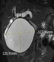
Zeinalpour A, Noori M, Gholizadeh B. An Unusual Type of Choledochal Cyst with Huge Dilation of Extrahepatic Bile Duct from Hilum to Ampulla of Vater; a Case Report and Literature Review. Int J Cancer Manag. 2019;12(10):e92136. doi: https://doi.org/10.5812/ijcm.92136
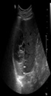
Song JS, Do Yang J, Sung Moon W. A Hepatic Cyst Complicated by Hemorrhage with Secondary Revascularization Mimicking Mucin-Producing Cystic Neoplasm: Radiologic-Pathologic Correlations. I J Radiol. 2018;15(2):e12992. doi: https://doi.org/10.5812/iranjradiol.12992
Mozafar M, Sobhiyeh M, Tadayon N, Bolouri N. Cervical Lymph Node Involvement by Medullary Carcinoma of Unknown Origin; a Rare Presentation. Int J Endocrinol Metab. 2010;8(1):. doi:
Barazandeh Ahmadabadi F, Siami Z, Seifi A, Ghiasvand F. A Case of Primary Biliary Tuberculosis with Subsequent Pulmonary Presentation. Arch Clin Infect Dis. 2018;13(6):e74374. doi: https://doi.org/10.5812/archcid.74374
Saif Andrabi SM, Bhat MH, Farhana B, Saba S, Saif Andrabi R, et al. Tuberculous Cervical Lymphadenitis Masquerding as Metastatis From Papillary Thyroid Carcinoma. Int J Endocrinol Metab. 2012;10(3):. doi: https://doi.org/10.5812/ijem.4500
Last Update: 2 weeks ago
Last Update: 2 weeks ago
Last Update: 2 weeks ago
Last Update: 6 days ago
Last Update: 1 hour ago
Last Update: 4 days ago