



Full Text
Full text is available in PDF
IJ Radiology
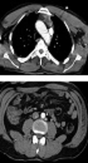
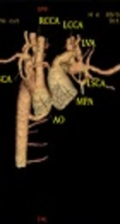
Anomalous retro aortic left brachiocephalic vein is a rare and less known vascular anomaly which may associate with complex heart disease and may be mistaken with normal or abnormal mediastinal structures.
We evaluated retrospectively all cardiovascular CTAs which were done in 3 referral centers between 2006 to 2010. CTA of 22 patients already diagnosed with anomalous retro aortic brachiocephalic vein were reevaluated by another expert radiologist in order to detect concomitant malformations. Chi square test was used for analysis of the data.
Patients' mean age was 12.9 years (from 2 month to 40 years). There were 12 males and 10 females. Among the 22 new cases, tetralogy of Fallot (TOF) component features (RVOT hypoplasia or atresia, overriding of aorta and RVH) were seen in 14 patients (63.6%). Abnormal aortopulmonary connections were detected in 14 cases (63.6%). Dilation of aortic arc was noted in 13 patients (59%). Right sided aortic arc was reported in 12 cases (54.5%) of which 5 had mirror image (22.7%). PDA was seen in 3 cases (13.6%). Hepatic hemangioma was incidentally detected in 2 patients (9.09%). In 1 patient, no other concomitant cardiovascular anomaly was seen (4.5%).
In our study, most common concomitant cardiovascular anomaly with anomalous retro aortic brachiocephalic vein was TOF. More than half of the patients had right sided aortic arc. In patients with pulmonary hypoplasia or aplasia, some parts of lung's blood supply were provided by aortic collaterals which had led to abnormal aortopulmonary connections. For a radiologist, it is important to differentiate this anomaly in cross-sectional imaging from a persistent left superior vena cava, partial anomalous pulmonary veins return, an elevated right pulmonary artery, and especially an enlarged lymph node. Investigations of this anomaly may lead to probable concomitant malformations and in isolated forms prevent further evaluations.
© 2014, Author(s). This open-access article is available under the Creative Commons Attribution 4.0 (CC BY 4.0) International License (https://creativecommons.org/licenses/by/4.0/), which allows for unrestricted use, distribution, and reproduction in any medium, provided that the original work is properly cited.
Sabouri S, Karimi MA, Mahdavirad H. Systemic Venous Anomalies in Patients with Pulmonary Atresia: A CT angiographic Study. I J Radiol. 2014;11(30th Iranian Congress of Radiology):e21433. doi: https://doi.org/10.5812/iranjradiol.21433
Mirdamadi A, Nejati M, Behjati M. A Case Report of Total Anomalous Pulmonary Vein Connection in Adult, through application of multi-modality imaging. Int Cardiovasc Res J. 2018;12(2):e11287. doi:
Sabouri S, Hosseini A, Shivaei SS. Retro-Aortic Inverted Left Renal Vein: A Rare Anomaly Found in a Renal Donor. I J Radiol. 2015;12(2):e11374. doi: https://doi.org/10.5812/iranjradiol.11374
Leblebisatan S, Gulek B, Soker G. Vena Cava and Left Renal Vein Variations and Association with Other Anomalies Including Horseshoe Kidney. I J Radiol. 2019;16(1):e79503. doi: https://doi.org/10.5812/iranjradiol.79503
Soleimantabar H, Sabouri S, Salajeghe S, Salajeghe S. Right Sided Aortic Arch with Type C Aortic Arch Interruption: CT Angiography Findings. I J Radiol. 2018;15(1):e57877. doi: https://doi.org/10.5812/iranjradiol.57877

Checking
Last Update: 2 weeks ago
Last Update: 2 weeks ago
Last Update: 2 weeks ago
Last Update: 4 days ago
Ordering Reprints
Articles are published under the Creative Commons license stated on each article. No permission or royalty fee is required for uses permitted by that license. CCC handles optional bulk and customized reprint orders. Any quotation covers production and delivery services only, not copyright permission. > Request Reprints from CCC
Author(s):