Full Text
Full text is available in PDF
An Innovative Journal in the Field of Radiology
Authors
Several lesions (Inflammatory, neoplastic and congenital) can involve in the skull base. The role of imaging is to detect the location and extension of lesion, determine mass and mass like lesions, differentiate benign from malignant tumors, detect invasion to adjacent structures and make decision in surgical planning. Skull base divides to anterior, middle and posterior cranial fossa and some pathologies are common in all spaces and a few are only seen in special sites. In this presentation we plan to explain the lesions in anterior skull base.
© 2017, Author(s). This open-access article is available under the Creative Commons Attribution 4.0 (CC BY 4.0) International License (https://creativecommons.org/licenses/by/4.0/), which allows for unrestricted use, distribution, and reproduction in any medium, provided that the original work is properly cited.
Jalal Shokouhi J. Skull Base (Middle Cranial Fossa Lesions). I J Radiol. 2017;14(Special Issue):e48350. doi: https://doi.org/10.5812/iranjradiol.48350
Sharifian H. Pre and Post-Operative Imaging Findings in Tumoral Lesions of Temporal Bone and Base of Skull. I J Radiol. 2014;11(30th Iranian Congress of Radiology):e21317. doi: https://doi.org/10.5812/iranjradiol.21317
Aghaghazvini L, Sedighi N, Karami P, Yeganeh O. Skull Base Aneurysmal Bone Cyst Presented with Foramen Jugular Syndrome and Multi-Osseous Involvement. I J Radiol. 2012;9(3):. doi: https://doi.org/10.5812/iranjradiol.7952

Rezaei-Kalantari K, Samimi K, Jafari M, Karimi MA, Ansari K, et al. Primary Diffuse Large B Cell Lymphoma of the Cranial Vault. I J Radiol. 2012;9(2):88-92. doi: https://doi.org/10.5812/iranjradiol.7734
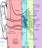
Mohebbi S, Daneshi A, Asghari A, Mohseni M, Arya Sadr M, et al. Surgical Landmarks of the Endoscopic Transcanal Transcochlear Approach: A Cadaveric Study. Arch Neurosci. 2020;7(1):e94763. doi: https://doi.org/10.5812/ans.94763
Last Update: 1 week ago
Last Update: 1 week ago
Last Update: 1 week ago
Last Update: 5 days ago
Last Update: 1 day ago
Last Update: 3 days ago