Cover image of the article: Pulmonary Alveolar Microlithiasis

Image Credit:I J Radiol

Outline

Pulmonary Alveolar Microlithiasis

Authors

H Ehteram1, H. R. Talari2, Ebrahim Razi3,*
1Department of Pathology, Kashan University of Medical Sciences, Kashan, Iran
2Department of Radiology, Kashan University of Medical Sciences, Kashan, Iran
3Department of Internal Medicine, Kashan University of Medical Sciences, Kashan, Iran
*Corresponding Author: Corresponding author: Ebrahim Razi, Department of Internal Medicine, Kashan University of Medical Sciences, Kashan, Iran, Tel: +98361 5550025-9 Email: ebrahimrazi@yahoo.com

Iranian Journal of Radiology:Vol. 3, issue 3; e79010
Published online:Jun 30, 2006
Article type:Case Report
Received:Oct 05, 2005
Accepted:Mar 16, 2006
How to Cite:Ehteram H, Talari HR, Razi E. Pulmonary Alveolar Microlithiasis. I J Radiol. 2006;3(3):e79010. doi:

Fulltext

The Authors' name and full text are available in PDF.

Copyright

© 2006, Author(s). This open-access article is available under the Creative Commons Attribution 4.0 (CC BY 4.0) International License (https://creativecommons.org/licenses/by/4.0/), which allows for unrestricted use, distribution, and reproduction in any medium, provided that the original work is properly cited.

Similar Articles

30
Jun
2010
Air Bronchogram Sign in Primary Pulmonary T-Cell Lymphoma with Multiple Nodules

Air Bronchogram Sign in Primary Pulmonary T-Cell Lymphoma with Multiple Nodules

W. J. Liang,
X. Y. Zhou,
Shun Liang Xu

Liang WJ, Zhou XY, Xu SL. Air Bronchogram Sign in Primary Pulmonary T-Cell Lymphoma with Multiple Nodules. I J Radiol. 2010;7(2):e79453. doi:

20
Nov
2012
Multiplanar Reconstructed Thoracic CT Bronchoscopy in Endobronchial Tuberculosis

Multiplanar Reconstructed Thoracic CT Bronchoscopy in Endobronchial Tuberculosis

Fariba Rezaeetalab,
Donya Farrokh,
Behrouz Zandiee

Rezaeetalab F, Farrokh D, Zandiee B. Multiplanar Reconstructed Thoracic CT Bronchoscopy in Endobronchial Tuberculosis. I J Radiol. 2012;9(4):234-6. doi: https://doi.org/10.5812/iranjradiol.8726

4
Jul
2016
Assessment of Independent Risk Factors of Developing Pneumothorax During Percutaneous Core Needle Lung Biopsy: Focus on Lesion Depth

Assessment of Independent Risk Factors of Developing Pneumothorax During Percutaneous Core Needle Lung Biopsy: Focus on Lesion Depth

Ha-Yeon Lee,
In Jae Lee

Lee H, Lee IJ. Assessment of Independent Risk Factors of Developing Pneumothorax During Percutaneous Core Needle Lung Biopsy: Focus on Lesion Depth. I J Radiol. 2016;13(4):e30929. doi: https://doi.org/10.5812/iranjradiol.30929

10
Jun
2013
The Prevalence of Active Tuberculosis Among Patients With Fibrotic Lesion in Chest CT-Scan

The Prevalence of Active Tuberculosis Among Patients With Fibrotic Lesion in Chest CT-Scan

Seyed Ali Javad Moosavi,
Hanieh Raji,
Mahshid Talebi-Taher,
Shadi Ghourchian

Moosavi SAJ, Raji H, Talebi-Taher M, Ghourchian S. The Prevalence of Active Tuberculosis Among Patients With Fibrotic Lesion in Chest CT-Scan. Jundishapur J Microbiol. 2013;6(4):e5179. doi: https://doi.org/10.5812/jjm.5179
12
Apr
2020
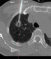
CT-Guided Percutaneous Core Needle Biopsy for Lung Lesions: A 14-Year Review of Needle Angle and Lesion Depth

CT-Guided Percutaneous Core Needle Biopsy for Lung Lesions: A 14-Year Review of Needle Angle and Lesion Depth

Jee Hyeon Lee,
In Jae Lee

Lee JH, Lee IJ. CT-Guided Percutaneous Core Needle Biopsy for Lung Lesions: A 14-Year Review of Needle Angle and Lesion Depth. I J Radiol. 2020;17(2):e96221. doi: https://doi.org/10.5812/iranjradiol.96221

More by these authors

H EhteramPubMedScholar
H. R. TalariPubMedScholar
Ebrahim RaziPubMedScholar
Share
Cited by
Metrics