


1. Background
2. Objectives
3. Patients and Methods
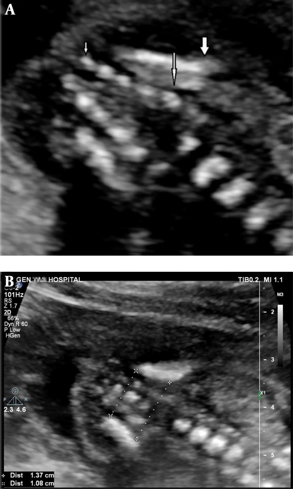
A, Ultrasound scan at 19 weeks of gestation in the coronal section in spine up position shows 5 sacral ossification nuclei. Thick arrow: upper contour of iliac wing; large thin arrow: S1; small thin arrow: S5; B, Method of bi-iliac distance measurement in the same section in another 19 weeks gestation fetus
4. Results
| Variables | Values |
|---|---|
| Age | |
| Mean ± SD | 29.9 ± 5.1 |
| Range | 18 - 44 |
| Inter-quartile range | 8 |
| Gravidity | |
| 1 | 172 (54.4) |
| 2 | 91 (28.8) |
| 3 | 45 (14.2) |
| 4 | 8 (2.5) |
| Parity | |
| 0 | 183 (57.9) |
| 1 | 103 (32.6) |
| 2 | 27 (8.5) |
| 3 | 3 (0.9) |
| Fetal gender | |
| Male | 155 (49.1) |
| Female | 161 (50.9) |
Abbreviation: SD, standard deviation.
a Values are expressed as No. (%) unless otherwise indicated.
| Gestational age (week-week+day) | Number of cases | S1 | S2 | S3 | S4 | S5 | |||||
|---|---|---|---|---|---|---|---|---|---|---|---|
| Body | Arch | Body | Arch | Body | Arch | Body | Arch | Body | Arch | ||
| 14 - 14 + 6 | 6 | 5 (83) | 5 (83) | 5 (83) | 3 (50) | 3 (50) | 0 | 0 | 0 | 0 | 0 |
| 15 - 15 + 6 | 9 | 9 (100) | 9 (100) | 9 (100) | 6 (66) | 6 (66) | 0 | 0 | 0 | 0 | 0 |
| 16 - 16 + 6 | 9 | 9 (100) | 9 (100) | 9 (100) | 6 (66) | 7 (77) | 2 (22) | 1 (11) | 1 (11) | 0 | 0 |
| 17 - 17 + 6 | 28 | 28 (100) | 28 (100) | 28 (100) | 28 (100) | 28 (100) | 16 (57) | 19 (67) | 5 (18) | 0 | 0 |
| 18 - 18 + 6 | 96 | 96 (100) | 96 (100) | 96 (100) | 96 (100) | 96 (100) | 79 (82) | 92 (96) | 56 (57) | 4 (4.2) | 2 (2.1) |
| 19 - 19 + 6 | 72 | 72 (100) | 72 (100) | 72 (100) | 72 (100) | 72 (100) | 68 (94.5) | 71 (98.5) | 59 (80) | 5 (7) | 4 (4) |
| 20 - 20 + 6 | 35 | 35 (100) | 35 (100) | 35 (100) | 35 (100) | 35 (100) | 35 (100) | 35 (100) | 35 (100) | 2 (5.7) | 2 (5.7) |
| 21 - 21 + 6 | 23 | 23 (100) | 23 (100) | 23 (100) | 23 (100) | 23 (100) | 23 (100) | 23 (100) | 23 (100) | 6 (26) | 5 (21) |
| 22 - 22 + 6 | 13 | 13 (100) | 13 (100) | 13 (100) | 13 (100) | 13 (100) | 13 (100) | 13 (100) | 13 (100) | 5 (38) | 4 (30) |
| 23 - 23 + 6 | 9 | 9 (100) | 9 (100) | 9 (100) | 9 (100) | 9 (100) | 9 (100) | 9 (100) | 9 (100) | 2 (22) | 1 (11) |
| 24 - 24 + 6 | 8 | 8 (100) | 8 (100) | 8 (100) | 8 (100) | 8 (100) | 8 (100) | 8 (100) | 8 (100) | 5 (62) | 3 (37) |
| 25 - 25 + 6 | 7 | 7 (100) | 7 (100) | 7 (100) | 7 (100) | 7 (100) | 7 (100) | 7 (100) | 7 (100) | 7 (100) | 3 (42) |
| Gestational weeks | Mean ± SD | IQR | Percentiles | Range | ||||
|---|---|---|---|---|---|---|---|---|
| 5th | 25th | 50th | 75th | 95th | ||||
| 14 w - 14 w + 6 d (n = 6) | ||||||||
| Upper contour | 6.9 ± 1.39 | 2.13 | 6 | 6 | 6.15 | 8.13 | 9.4 | 6 - 9.4 |
| Lower contour | 5.8 ± 1.27 | 2.33 | 4.8 | 4.89 | 5.15 | 7.2 | 7.8 | 4.8 - 7.8 |
| 15 w - 15 w + 6 d (n = 9) | ||||||||
| Upper contour | 8.1 ± 1.06 | 1.45 | 6.4 | 7.45 | 7.7 | 8.9 | 9.9 | 6.4 - 9.9 |
| Lower contour | 6.73 ± 0.58 | 1.15 | 6.2 | 6.25 | 6.4 | 7.4 | 7.6 | 6.2 - 7.6 |
| 16 w - 16 + 6 d (n = 9) | ||||||||
| Upper contour | 9.11 ± 0.98 | 1.95 | 8 | 8.15 | 9 | 10.1 | 10.4 | 8 - 10.4 |
| Lower contour | 7.31 ± 0.87 | 1.15 | 5.5 | 6.95 | 7.3 | 8.1 | 8.4 | 5.5 - 8.4 |
| 17 w - 17 w + 6 d (n = 28) | ||||||||
| Upper contour | 10.62 ± 1.05 | 1.42 | 8.84 | 9.85 | 10.5 | 11.28 | 12.69 | 8.7 - 13 |
| Lower contour | 8.96 ± 0.65 | 0.8 | 7.44 | 8.7 | 9 | 9.5 | 10 | 7.3 - 10 |
| 18 w - 18 w + 6 d (n = 97) | ||||||||
| Upper contour | 11.84 ± 1.45 | 1.45 | 10 | 11.3 | 11.7 | 12.75 | 14 | 2.6 - 14.3 |
| Lower contour | 9.92 ± 1.11 | 1.15 | 8.59 | 9.35 | 10 | 10.5 | 11.35 | 2.5 - 12 |
| 19 w - 19 w + 6 d (n = 72) | ||||||||
| Upper contour | 12.73 ± 1.15 | 1.38 | 10.90 | 12 | 12.9 | 13.38 | 15 | 10 - 15.7 |
| Lower contour | 10.71 ± 0.88 | 1.30 | 9.33 | 10 | 10.65 | 11.3 | 12.24 | 8.9 - 12.5 |
| 20 - 20 + 6 d (n = 35) | ||||||||
| Upper contour | 13.96 ± 1.11 | 2 | 11.84 | 13 | 14.2 | 15 | 15.42 | 11.2 - 15.5 |
| Lower contour | 11.63 ± 0.9 | 1.20 | 9.98 | 11 | 11.6 | 12.2 | 13.02 | 9.9 - 13.5 |
| 21 w - 21 w + 6 d (n = 23) | ||||||||
| Upper contour | 15.3 ± 1.56 | 2.5 | 11.62 | 14.1 | 15.3 | 16.6 | 17.66 | 11.2 - 17.7 |
| Lower contour | 12.49 ± 1.3 | 1.80 | 9.84 | 11.7 | 12.5 | 13.5 | 14.84 | 9.69 - 15 |
| 22 w - 22 w + 6 d (n = 13) | ||||||||
| Upper contour | 15.72 ± 1.29 | 2.1 | 13.1 | 14.65 | 16 | 16.75 | 17.6 | 13.1 - 17.6 |
| Lower contour | 13.19 ± 0.86 | 1.20 | 12 | 12.35 | 13.4 | 13.55 | 14.9 | 12 - 14.9 |
| 23 w - 23 w + 6 d (n = 9) | ||||||||
| Upper contour | 17.47 ± 1.04 | 1.85 | 15.9 | 16.6 | 17.2 | 18.45 | 18.7 | 15.9 - 18.7 |
| Lower contour | 14.33 ± 1.03 | 1.80 | 12.7 | 13.4 | 14.5 | 15.2 | 15.8 | 12.7 - 15.8 |
| 24 w - 24 w + 6 d (n = 8) | ||||||||
| Upper contour | 18.45 ± 1.44 | 2.38 | 17.2 | 17.4 | 17.65 | 19.78 | 21 | 17.2 - 21 |
| Lower contour | 16.18 ± 1.13 | 2.10 | 14.9 | 15.18 | 15.8 | 17.28 | 17.9 | 14.9 - 17.9 |
| 25 w - 25 w + 6 d (n = 7) | ||||||||
| Upper contour | 19.46 ± 1.44 | 1.2 | 18.2 | 18.7 | 18.9 | 19.9 | 22.5 | 18.2 - 22.5 |
| Lower contour | 16.1 ± 0.5 | 0.7 | 15.5 | 15.8 | 15.9 | 16.5 | 17 | 15.5 - 17 |
Abbreviation: d, day; IQR, interquartile range; SD, standard deviation; w, week.
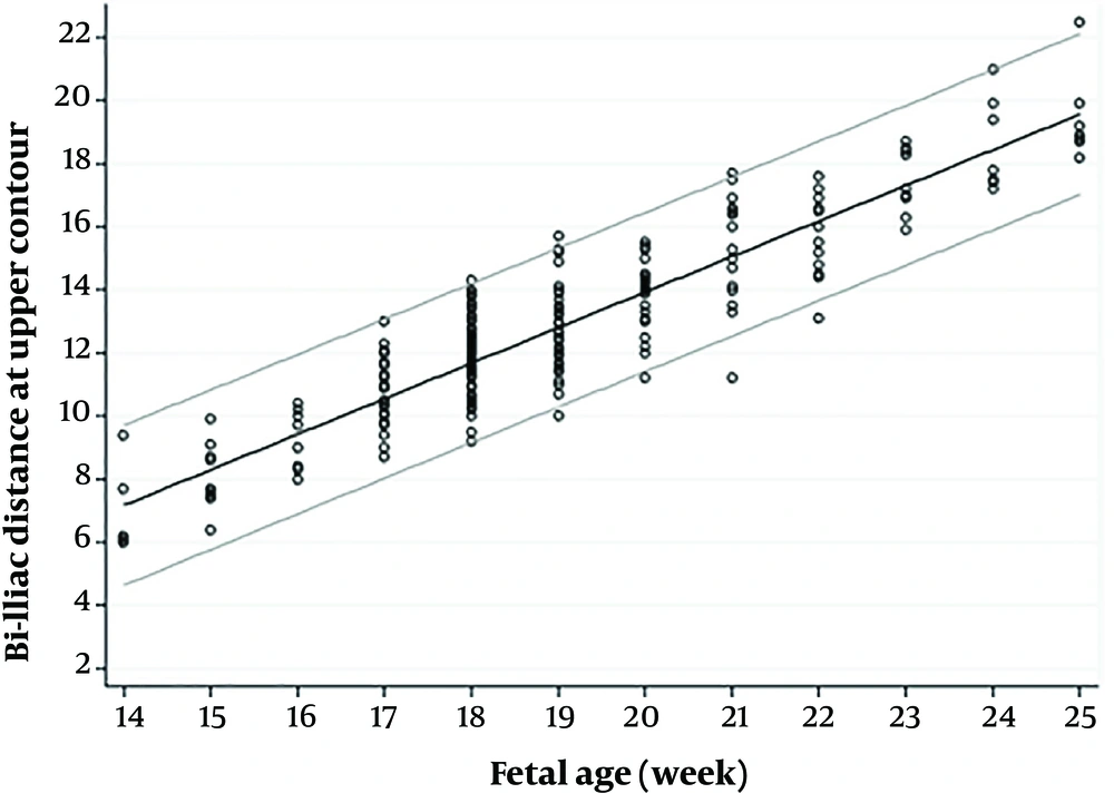
Scatter diagram of bi-iliac distance plotted on 5th and 95th percentiles in the upper iliac contour
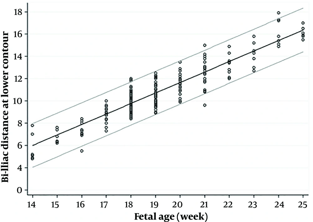
Scatter diagram of bi-iliac distance plotted on 5th and 95th percentiles in the lower iliac contour
| Case | MA | Clinical history | GA | Fetal gender | Associated US finding | Outcome | Confirmation |
|---|---|---|---|---|---|---|---|
| 1-Sirenomelia | 25 | - | 17 w + 4 d | ? | SUA, bilateral renal agenesis, dilated cardiomyopathy, pericardial effusion, severe oligohydramnios, blocked vertebrae | TOP | External examination |
| 2-Sirenomelia | 28 | - | 17 w | ? | SUA, bilateral renal cystic disease, hemivertebra and mild scoliosis, mild ventriculomegaly, severe oligohydramnios | TOP | External examination |
| 3- Sacral agenesis | 35 | DM, IVF | 18 w + 3 d | F | Decreased fetal feet movement, Unilateral club foot, Soft tissue wasting in lower limbs | TOP | External examination & graphy |
| 4- Sacral agenesis | 28 | - | 18 w + 2 d | M | Unilateral club foot | TOP | Not performed |
| 5- Sacral agenesis | 30 | DM | 17 w | F | Corpus callosum agenesis, omphalocele, sacral diastematomyelia, blocked vertebrae in sacrum, soft tissue wasting in lower limbs | TOP | External examination and graphy |
Abbreviations: d, day; DM, diabetes mellitus; F, female; GA, gestational age; IVF, in vitro fertilization; M, male; MA, maternal age; SUA, single umbilical artery; TOP, termination of pregnancy; US, ultrasonography; w, week.

A, A 17 weeks + 4 days case of sirenomelia (case 1) with sacral agenesis and close contact of both iliac wings (small arrow) and only a single bone was present (large arrow), with no legs or feet; B, In case 3 with sacral agenesis, the last ossification center was L3 in the arch and L5 in the body. Bodies of L4 and L5 are fused together (large arrow). Both iliac wings articulated with the last vertebra and bi-iliac distance were very small especially in the lower contour (small arrow); C, Case 4 with mild sacral agenesis had small bi-iliac distance (9.8 mm and 5.7 mm for upper and lower contours, respectively) under the 5th percentile based on our result; D, Case 5 with diastematomyelia (horizontal arrow) and partial unilateral sacral agenesis with two ossification centers on the left side (small vertical arrow: S2) and four ossification nuclei on the other side with blocked vertebra of S3 and S4 (wide vertical arrow). The fetus also had a small bi-iliac distance in the lower contour (7 mm) and normal in upper contours (10 mm).


