Primary tumor volume is an important prognostic factor that affects survival compared to the stages of the tumor (
19). Some studies reported that calculation of the tumor volume with pre-catchment imaging methods may be useful to assess the surgical treatment options, and response of the tumor to the treatments such as radiation therapy, affects the anatomical structure of the brain (
20,
21). Tumor volume is also used to determine tumor location, relationship with adjacent structures and its directions (
22).
Volumetric measurement of the tumor area in magnetic resonance imaging (MRI) is clinically important for diagnosis and treatment assessment in patients with brain tumors.
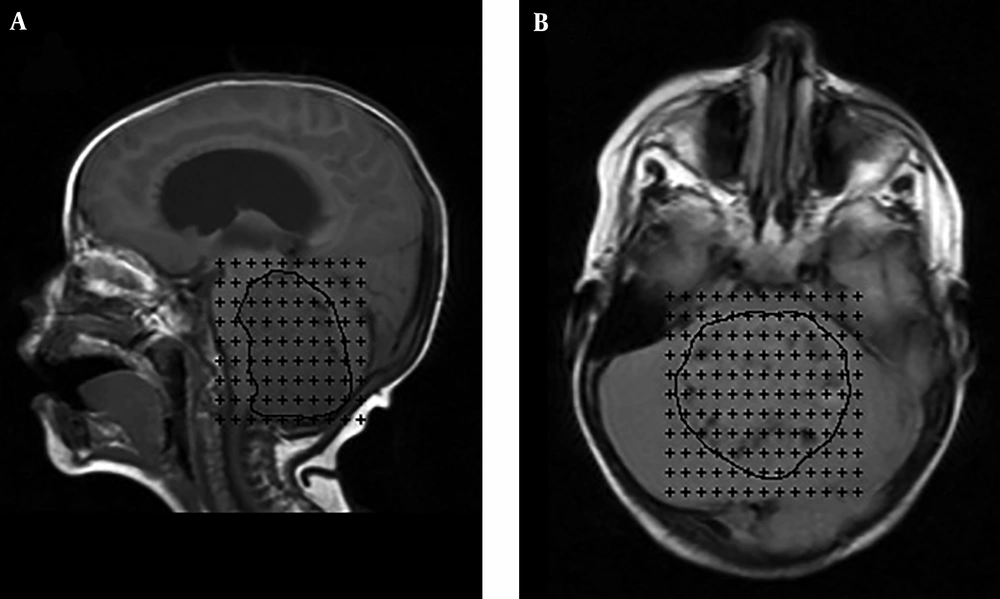
Moreover, stereology is used to calculate unbiased properties of structures existing in three dimensions (3D) from two-dimensional (2D) medical images. An unbiased estimate of organ volume can be obtained using the Cavalieri principle, considered a stereological method (
23).
In fact, this method has the following advantages for the researcher: (1) The structure under study needs no preconditioning since the Cavalieri method is a design-based method but not a model based approach; (2) The actual properties of the structure, such as section thickness, are taken into consideration; and (3) The sampling or estimating system can be easily modified to obtain an appropriate coefficient of variation. Furthermore, the reliability and efficiency of the Cavalieri method for volume determination have been confirmed frequently (
24).
Applying the Cavalieri principle in studies aimed at obtaining quantitative data on irregularly shaped three-dimensional objects suggests benefits such as consequential quantitative data, application of strict sampling procedures, easily reproducible data, and a well-established theoretical background, making the reliability of the data easy for examination (
25-
27). Therefore, this method accompanied with MR imaging data can be potentially applied as a simple, reliable and quantitative method for diagnosis and follow-up of the treatment options in patients with brain tumors.
There has been growing recent interest in quantitative techniques for measuring volume using CT scans or MRI. Some proposed geometric methods for assessing the volumetric properties of radiological images based on direct geometric dimensions such as length, diameter, or the largest diameter measurements (
28,
29). In such methods, it has been presumed that the structure under investigation has an ellipsoidal, spherical, or known geometric shape. However, the assumption of such smooth geometric properties in biological tissues is not always correct (
23). In addition, the other techniques such as computer-based 3D volumetric reconstructions, and planimetry methods, are also used for volume estimations (
30-
33). The planimetric methodologies give more precise and accurate results compared to the above-mentioned geometric techniques (
28,
30,
31).
However, several studies showed that there are also assumptions in the planimetric methods resulting in volume measurement over-estimation and some degree of systematic bias (
28,
29,
34,
35). In addition, the computerized reconstructions generally require quite expensive equipment and trained personnel for their routine application, and these features make these applications unsuitable for most clinics or research centers (
36,
37).
Point counting techniques represent a more reliable and efficient approach than a planimetric methods (
38-
42); therefore, results obtained through point counting will be a direct and assumption-free estimate of the total volume of the interest structure, and hence very valuable in monitoring brain tumors.
In fact, the volume of any structure may be considered using histological sections, CT or MR images through Cavalieri method (
22). Other advantages of this method are cost and time efficiency (
43).
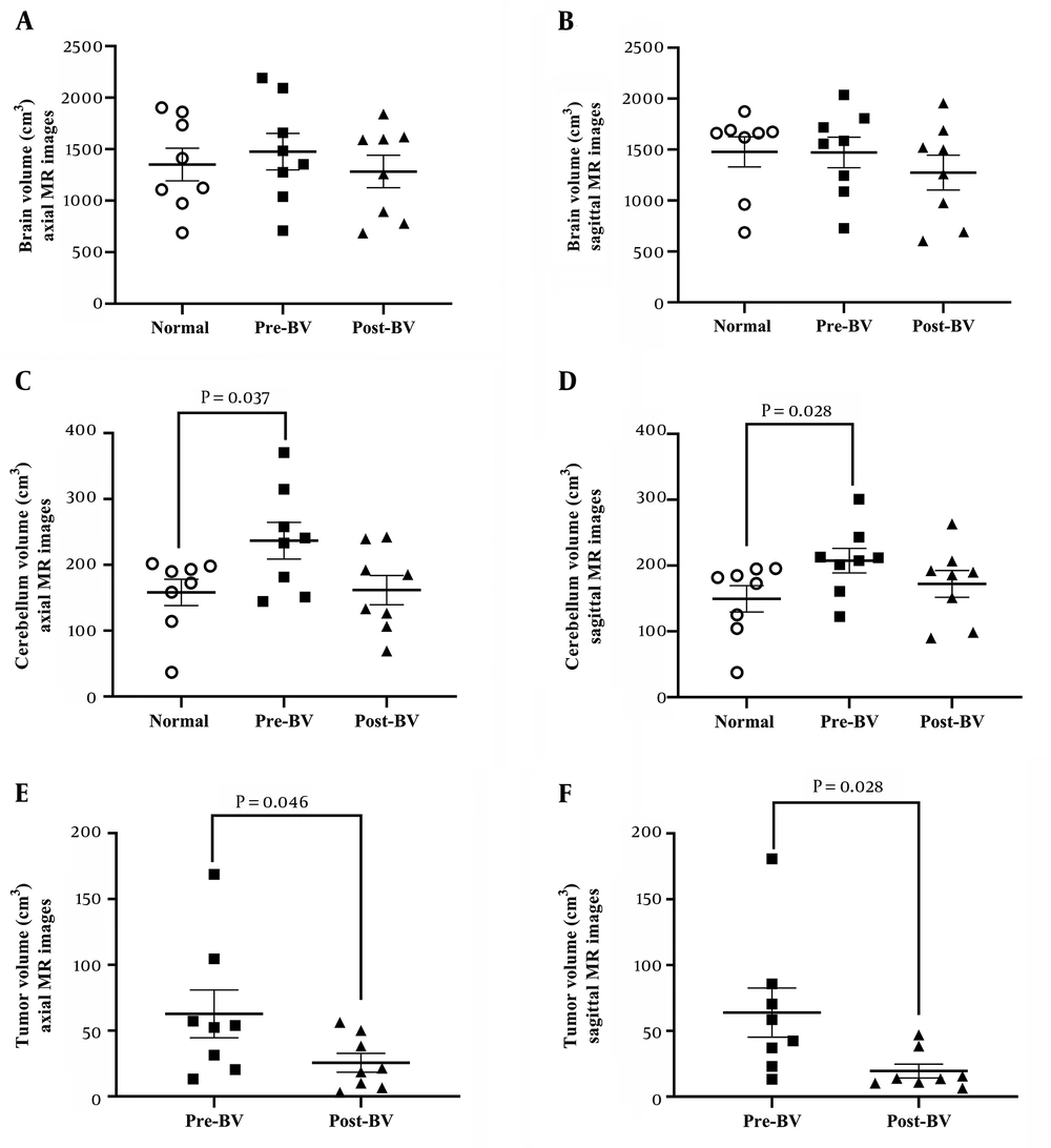
Results showed that the tumor volumes in axial/sagittal sections after surgery were significantly lower than those before surgery. In pediatric population, the spread of residual surgical resection has been shown to influence the survival outcomes. Excision of more than 90% of the tumor is correlated with improved survival rate in children older than three years (
44). In our study, in four patients, more than 90% of the tumor was removed, suggesting an increase in survival rate. The results of the present study also showed differences in the volume of the cerebellum between the control and patient groups before surgery. According to this result, the cerebellum volume in axial and sagittal MRI images in patients with medulloblastoma tumor were significantly higher compared to that of the control group. This agreement with structural MR scanning studies revealed that medulloblastoma tumor can cause bilateral olivary hypertrophy in the human cerebellum which is known as hypertrophic olivary degeneration (HOD) (
45).
A common problem in the stereological method is an over-projection or under-projection effect, an artifact that results from section thickness. This problem also exists when MRI or CT scans are used for volume estimations (
23,
24,
46). There are several solutions for management of the under-projection or over-projection effects of radiological imaging (
47,
48). A previous study showed that the approach formulized as Equation 1 eliminates possible over-estimation due to over-projection (
24). Therefore, we used this method in the present study.
Some brain tumors such as MB do not show a smooth demarcation with brain on MRI or CT scans. Therefore, it is almost impossible to obtain reliable quantitative data on tumor size using the diameter or length of the tumor on MRI. However, the surgeon needs the exact volume of the tumor for comparison of the pre-operative and post-operative scans (
28).
During the operation, the whole tumor mass cannot be resected totally to avoid excision of the pathological structures with the surrounding healthy tissues. Thus, assessment of the exact tumor volume not only provides important data for the assessment of the size of the structures to be excised during the operation but also allows treatment planning and post-operative follow up (
28,
34,
49).
In addition, Cavalieri method using automated software is currently available and could be performed easily and rapidly. Stereological measurement can therefore provide additional useful data to supplement MR measurements, especially in borderline and controversial cases.
It could be concluded that the combination of MRI and the Cavalieri principle could estimate tumor volume that may be useful in evaluating the efficiency of surgical treatment and prognostication of tumor regression rate. The presented method does not require any change in routine procedures and can be performed on any complete set of MRI scans. The method can be efficiently used without any need for additional equipment and expert personnel that is required for routine MRI.