
1. Introduction
In the Indian interventional pain-medicine community, the term “minimally invasive spine endoscopic pain procedure” (MisEpp) is used as an umbrella term for pain-physician-led spinal procedures performed via an endoscopic route. This terminology has not yet been universally standardized internationally; therefore, in this report, we retain MisEpp to describe the endoscopic pain-procedure framework used for the present intervention.
Lumbar disc herniation at L5–S1 is a common cause of lumbosacral radicular pain. Compression of the traversing S1 nerve root classically produces posterior leg symptoms and may be accompanied by plantar-flexion weakness, sensory changes, and a reduced Achilles reflex. Endoscopic or surgical decompression is effective in appropriately selected patients when conservative management fails (1-4).
Epidural interventions, including caudal, transforaminal, and targeted S1 approaches, have also been investigated in patients with lumbar spinal stenosis and radicular pain (5-7).
Anatomical factors beyond the disc fragment may contribute to the degree and persistence of neural compression. Lumbar facet orientation varies by spinal level, and asymmetric facet orientation, or facet tropism, has been associated in observational studies with disc degeneration and disc herniation (8-12). However, these associations do not establish a direct causal relationship in an individual patient.
In addition to their potential biomechanical associations, hypertrophied or medially positioned facet/articular processes can directly contribute to lateral recess narrowing and compression of a traversing nerve root (13). In a patient with a paracentral disc extrusion, the coexistence of anterior disc compression and posterior or posterolateral facet-related bony encroachment can therefore create two anatomically distinct sites of constraint around the same nerve root.
In this report, we use the term “double compression” as a descriptive concept for simultaneous anterior compression by disc material and posterolateral compression by a medially encroaching facet-related bony structure. We do not propose it as a new diagnostic entity. We present a case in which this configuration was suspected on magnetic resonance imaging (MRI) and confirmed during endoscopic decompression, with both components addressed through an interlaminar minimally invasive approach.
2. Case Presentation
A woman in her fifth decade presented with a 1-year history of progressively worsening left-sided low back and leg pain. The pain radiated from the posterior thigh and lateral calf to the dorsum of the left foot and was described as lancinating and burning, with a numeric pain rating scale (NRS) score of 7 - 8/10. Symptoms were aggravated by lumbar flexion, prolonged standing, and walking and were partially relieved by positional change. The patient reported a walking tolerance of approximately 80 - 100 m. The predominant clinical presentation was unilateral lumbosacral radicular pain; the distal distribution included the dorsum of the foot, which can overlap with L5 radicular symptoms.
She did not report subjective paresthesias. Neurological examination, however, demonstrated reduced light-touch sensation in the left S1 distribution, mild weakness of left plantar flexion (grade 4/5 on the Medical Research Council muscle-strength grading scale), and a diminished left Achilles deep-tendon reflex. The straight-leg-raise test was positive at 30° on the left, with reproduction of concordant leg symptoms. The contralateral test was negative. Lumbar range of motion was globally restricted, with pain provocation during flexion and left lateral flexion. The preoperative Oswestry Disability Index (ODI) was 54%.
Eight months of structured conservative management with physiotherapy, nonsteroidal anti-inflammatory drugs, and pregabalin provided only transient relief lasting approximately 4 - 6 weeks, followed by recurrence of symptoms. An epidural steroid injection had been performed previously at another healthcare facility without significant symptomatic improvement.
2.1. Imaging Findings
Lumbar MRI, including axial T2-weighted sequences, demonstrated a large left paracentral L5–S1 disc protrusion with superimposed extrusion occupying the left lateral recess, contacting and displacing the traversing left S1 nerve root. The thecal sac was displaced toward the right.
The left L5 – S1 facet complex was markedly more sagittally oriented than the contralateral side, consistent with asymmetric facet orientation/tropism. The medial margin of the left articular process complex projected into the lateral recess, producing marked focal narrowing at the level of the traversing S1 nerve root. The narrowing is described qualitatively because a prospectively standardized measurement protocol was not documented in the case record.
The combination of the paracentral disc extrusion and posterior/posterolateral bony encroachment created two anatomically distinct potential sites of S1 nerve-root constraint. This configuration was designated descriptively as a “double compression” pattern in the present report.
2.2. Procedural Planning and Approach Selection
Because MRI demonstrated both a medial disc extrusion and marked facet-related lateral recess encroachment, the MisEpp interlaminar endoscopic approach was selected to permit direct visualization of the traversing S1 nerve root, lateral recess, disc material, and adjacent bony structures through a single posterior corridor.
The L5 – S1 interlaminar window provided access to the posterior epidural space. The approach was also considered suitable for the paracentral location of the disc fragment and for targeted removal of the medially encroaching bony margin without wide facet resection.
Written informed consent was obtained from the patient for the procedure.
2.3. Procedural Technique
Fluoroscopic anteroposterior and lateral views were used to confirm the L5 – S1 level. A paramedian skin incision approximately 8 mm long was made approximately 1 cm lateral to the midline on the left side, over the L5 – S1 interlaminar space. Dilation was performed through the musculature under fluoroscopic guidance, followed by introduction of the working cannula and endoscope.
Under continuous saline irrigation, the epidural space was accessed after controlled resection of the ligamentum flavum. The traversing left S1 nerve root was identified as congested, edematous, and flattened. It was compressed anteriorly by the extruded disc fragment and posterolaterally by the medially projecting bony margin of the left L5 inferior articular process, confirming the two-site compression pattern suspected on MRI.
Targeted undercutting of the medially encroaching bony margin was performed, with preservation of the outer cortical portion of the facet complex. The adjacent hypertrophied superior articular process was also partially decompressed where it contributed to the lateral recess narrowing. The aim was to enlarge the lateral recess while limiting the extent of facet resection.
Following bony decompression, the extruded disc fragment was removed with gentle medial mobilization of the S1 nerve root. The final endoscopic survey demonstrated a freely mobile and pulsatile nerve root, no visible residual disc material, and no residual bony compression of the lateral recess. No dural violation, epidural hematoma, or nerve-root injury was observed (Figures 1 and 2).
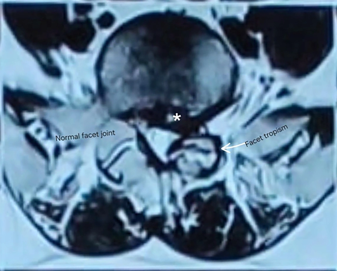
Figure 1.
Axial T2-weighted MRI at L5 – S1 demonstrating a large left paracentral disc extrusion occupying the left lateral recess, asymmetric/sagittalized left facet orientation, and medial facet-related encroachment. The figure illustrates the anatomical configuration described as “double compression.”
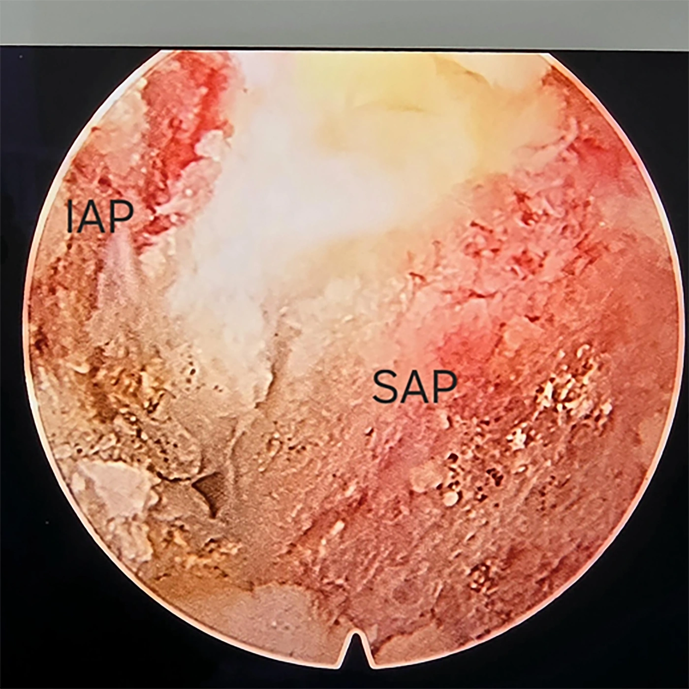
Figure 2.
Intraoperative endoscopic view showing the inferior and superior articular processes (IAP and SAP) and the facet-related bony anatomy after targeted decompression.
2.4. Postprocedure Course and Outcome
The patient was mobilized 6 hours after the procedure and discharged the following day with oral analgesics. At the 2-week follow-up, she reported complete resolution of radicular leg pain (NRS 0/10), with mild residual axial low back discomfort (NRS 2/10). Plantar-flexion power had returned to 5/5, and sensory examination in the S1 distribution had normalized.
At 3 months, the ODI had improved from 54% preoperatively to 14%, representing a 40-percentage-point improvement. The patient had resumed activities of daily living and occupational duties without restriction.
3. Discussion
Facet morphology has been studied in relation to lumbar disc degeneration and disc herniation. Earlier anatomical and radiographic work suggested that asymmetric facet orientation may influence local spinal mechanics (8-12). These studies provide a rationale for considering facet orientation as one component of the anatomical environment in which disc pathology develops; however, they do not establish that sagittalization directly causes annular failure in an individual patient.
In the present case, the clinically relevant finding was not facet orientation alone, but rather the combination of asymmetric/sagittalized facet morphology and clear medial bony encroachment into the lateral recess. The latter provided an anatomically plausible second site of compression of the traversing S1 root, distinct from the paracentral disc extrusion. This distinction is important because facet orientation and facet-related lateral recess stenosis are related but not synonymous findings.
The term “double compression” is therefore used here only as a descriptive anatomical label for the coexistence of anterior disc-related and posterolateral facet-related neural constraint. The intraoperative findings supported this anatomical interpretation because the S1 root was visibly compressed by both structures before decompression and became freely mobile after both components were addressed.
However, the clinical outcome should not be interpreted as evidence that facet decompression is universally necessary or superior to discectomy alone. Both components were treated in the same patient; therefore, their individual contributions to symptom resolution cannot be distinguished. Likewise, improvement after combined decompression cannot establish that discectomy alone would have failed.
From a procedural perspective, the MisEpp interlaminar endoscopic corridor at L5 – S1 can provide direct visualization of the traversing S1 nerve root, the lateral recess, and adjacent articular processes. In selected patients with a paracentral disc extrusion and concomitant lateral recess narrowing, this visualization may allow the operator to determine whether a bony component remains after removal of the disc material and to perform targeted decompression when clinically appropriate (13-20).
Targeted undercutting should be distinguished from complete facetectomy. In this case, the resection was limited to the medially encroaching bony margin while preserving the outer cortical portion of the facet complex. Nevertheless, the present case cannot establish long-term preservation of segmental stability, particularly because follow-up was limited to 3 months.
The case also highlights the importance of correlating MRI findings with the clinical and neurological examination. The patient’s pain extended to the dorsum of the foot, which can overlap with an L5 distribution. Nevertheless, the combination of plantar-flexion weakness, reduced Achilles reflex, S1 sensory findings, and direct intraoperative identification of the traversing S1 root as the compressed neural structure supported predominant S1 involvement.
3.1. Limitations
This report has several limitations. First, it describes a single patient and therefore cannot establish the prevalence, causality, or treatment superiority associated with the proposed dual-compression pattern. Second, follow-up was limited to 3 months, precluding conclusions regarding long-term recurrence, durability of functional improvement, or segmental stability following facet undercutting. Third, because disc and bony decompression were performed together, the independent contribution of each component to the clinical outcome cannot be determined. Finally, the lateral recess measurement was not obtained using a prospectively standardized measurement protocol; therefore, the narrowing is described qualitatively without categorical numerical thresholds.
3.2. Conclusions
This case illustrates a potential dual-compression pattern at L5 – S1 in which a paracentral disc extrusion coexists with facet-related medial encroachment of the lateral recess and compression of the traversing S1 nerve root. The term “double compression” is used descriptively rather than as an established diagnostic entity. In this patient, endoscopic identification and targeted decompression of both the disc and the bony lateral recess component were followed by substantial clinical improvement. A single case cannot demonstrate that combined decompression is necessary or superior to discectomy alone; however, the findings support careful preoperative assessment of the lateral recess and facet morphology in selected patients with lumbar disc herniation.