
1. Introduction
Mitral regurgitation (MR) is a significant challenge in cardiovascular health, affecting approximately 10% of the population (1). The MR is a prevalent form of mitral valve (MV) dysfunction with various etiologies, including degenerative causes such as valve prolapse and ischemic causes resulting from coronary artery disease. In both developed and developing countries, rheumatic problems are also major contributors to this condition (2). In recent years, mitral valve diseases, particularly MR, have impacted a substantial number of adults globally, making MR the second most common reason for valve surgery (3). Studies in the United States indicate that MR is the most common cause of moderate to severe valvular heart disease among adults. The severity of MR varies among patients and depends on several factors, including left ventricular loading conditions, heart rhythm, conduction system disease, and myocardial ischemia (4).
Over the past decades, the demand for mitral valve re-surgery has steadily increased, a trend expected to continue in the future (5, 6). Mitral valve replacement (MVR) surgery is a complex and delicate procedure requiring significant technical expertise from the medical team. Re-surgery of the heart is generally more challenging than primary surgery due to various factors (5). This complexity is further exacerbated when the patient has severe heart failure, making the anesthesia management of such individuals critically important during cardiac surgery.
Right ventricular failure (RVF) following cardiac surgery is a significant contributor to morbidity and mortality (7). Effective management of RVF during and after complex cardiac operations is therefore essential. A recent cohort study by Levy et al. reported RVF in 2.9% of 3,826 patients undergoing cardiac surgery with cardiopulmonary bypass (CPB) (8). In this case report, we describe the successful anesthesia management of MVR in a 46-year-old woman with severe right ventricular failure, a history of anemia, drug allergy, and chronic hypertension.
2. Case Presentation
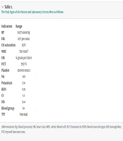
A 46-year-old female presented to the emergency department with complaints of shortness of breath, excessive fatigue, and severe dizziness persisting for the last three days. She was scheduled for redo MVR and insertion of atrioventricular polar leads. Her surgical history included an atrioventricular septal defect (AVSD) repair and MVR performed 28 years prior, at age 17, as well as mitral valve repair and large ventricular septal defect (LVSD) closure 27 years prior. Additionally, the patient had a known history of chronic hypertension, atrial fibrillation (AF) rhythm, complete heart block, anemia, and an allergy to valsartan. Pulmonary function tests (PFTs) indicated a forced expiratory volume (FEV1) of 40%. Vital signs are summarized in Table 1.
Table 1.
The Vital Signs of the Patient and Laboratory Criteria Were as Follows
| Indication | Range |
|---|---|
| Blood pressure (mmHg) | 10/71 |
| Heart rate (/min) | 107 |
| O2 saturation (%) | 92 |
| White blood cell (mm3) | 730 |
| HB (g/L) | 9 |
| HCT (%) | 29.7 |
| Platelet (mm3) | 131000 |
| Na | 141 |
| Potassium | 3.6 |
| BUN | 0.9 |
| Cr | 1.1 |
| Hb | 9.4 |
| Blood group | A+ |
| TFT | Normal |
The patient was taken to the operating room, and full monitoring was implemented, including pulse oximetry, electrocardiogram (ECG), invasive blood pressure (IBP), and bispectral index (BIS). General anesthesia was induced with 7 mg midazolam, 500 mcg sufentanil, and 70 mg rocuronium. Intubation was performed using a size 7.5 endotracheal tube. Capnography and cerebral oximetry monitoring were applied. A right internal jugular central venous (CV) line and two peripheral 16-gauge intravenous (IV) lines were placed and secured.
Due to the patient's small femoral arteries and veins, both sides of the sternum were carefully opened. The patient was placed on CPB. Following removal of the previous mitral valve, a Carbomedix 27 valve was implanted. Two leads were placed in the right atrium (RA) and right ventricle (RV), but they were nonfunctional. The electrophysiology service (EPS) scheduled the installation of an endocardial pacemaker. Prolonged pump time was required due to the reoperation and significant adhesions.
Upon weaning from CPB, the patient experienced persistent hypotension. Epinephrine infusion was initiated and titrated up to 0.2 µg/kg/min. However, the patient did not respond to the increasing epinephrine dosage, and low blood pressure persisted. Esophageal echocardiography was performed, revealing acute right ventricular failure. Consequently, epinephrine was discontinued, and norepinephrine and vasopressin infusions were started.
During the surgery, the patient exhibited continuous bleeding, low central venous pressure (CVP: 5 - 6 mmHg), and hypotension, necessitating massive transfusion. The patient received six units of packed red blood cells, six units of fresh frozen plasma (FFP), and six units of platelets.
An increase in blood pressure was observed following intervention. At the end of the operation, the patient was transferred to the intensive care unit (ICU) with high-dose inotropes due to severe RVF, including norepinephrine at 0.2 µg/kg/min and vasopressin at 2 units per hour, with acceptable arterial blood gas (ABG) values. Table 2 presents the latest patient information before transfer to the ICU.
Table 2.
Vital Information of the Patient Before Transfer to Intensive Care Unit
| Vital Signs | Data Range |
|---|---|
| BP | 117/67 |
| HR | 70 |
| SpO2(%) | 100 |
| Hb | 8.1 |
| HCT | 26 |
| Na | 141 |
| K | 4.1 |
| pH | 7.43 |
| PO2 | 186 |
| PCO2 | 34 |
| HCO3 | 23 |
| BE | -1 |
Abbreviations: BP, blood pressure; HR, heart rate.
Upon arrival in the ICU, the patient was fully awake and responsive. Chest radiography (CXR) revealed a white lung on the right side, attributed to reoperation, extensive adhesions, and atelectasis. The patient had a mediastinal drain and a left chest tube in place. In the ICU, the patient experienced frequent ventricular tachycardia (V-tach) and went into shock. Lidocaine and mexiletine were administered twice to manage the arrhythmias. Metabolic alkalosis was identified and corrected.
The patient presented with high CVP at 20 mmHg, low blood pressure, and reduced urine output. Echocardiography confirmed severe RV dysfunction and RV failure. To address hypotension and volume deficits caused by bleeding, the patient received packed red blood cells, colloids, gelatin, and albumin. Vasopressin and norepinephrine inotropes were continued, and dobutamine was added; however, dobutamine exacerbated tachycardia and ventricular tachycardia (VT), prompting its discontinuation. Pulmonary edema and increased pulmonary hypertension were managed through ventilator adjustments and pharmacological treatment.
After 24 hours of inotropic support (norepinephrine, vasopressin, and milrinone) and volume compensation with diuretics and blood products, the patient's hemodynamic status stabilized. A permanent pacemaker (PPM-DR) with DDD mode (dual-chamber pacing) and a heart rate of 70 bpm was successfully installed by the electrophysiology (EP) service.
Despite being awake and alert, the patient could not tolerate ventilator weaning due to tachypnea and respiratory distress. Wheezing and shortness of breath were observed, likely due to pre-existing respiratory issues. Ventilator settings were adjusted to assist-control ventilation (ACV) mode, with increased positive end-expiratory pressure (PEEP) and respiratory rate to maintain hemodynamic stability. Combivent and Pulmicort nebulizers were administered based on the patient's respiratory status.
After six days, the patient's respiratory condition improved, and the chest X-ray findings were acceptable. The patient was extubated successfully with stable respiratory parameters. She was discharged from the institute 12 days after her initial emergency room visit in good general condition.
3. Discussion
Mitral regurgitation is a widespread heart valve disease globally, drawing significant attention from cardiologists and anesthesiologists. Patients with MR require special consideration due to the elevated risk of cardiovascular complications during and after both cardiac and non-cardiac surgeries (9). Cardiac reoperations present greater complexity compared to initial surgeries, as the presence of mediastinal and pericardial adhesions increases with shorter intervals between surgeries and a higher number of prior operations, thereby amplifying the potential for damage to adjacent critical anatomical structures (5).
Mitral valve replacement poses numerous challenges, particularly when patients experience severe right ventricular failure. In such complex and high-risk cases, early diagnosis, rapid intervention, and coordinated teamwork in the operating room are crucial for ensuring a successful outcome. The heart team plays an essential role in managing patients who require valve reinterventions. During these intricate surgeries, anesthetic considerations are especially critical. This includes selecting an appropriate anesthesia protocol, implementing meticulous monitoring methods in the operating room, devising hemodynamic management strategies to stabilize the patient during surgery, and ensuring comprehensive post-operative care.
This article highlights the successful management of a 46-year-old female patient with a complex medical history, including chronic hypertension, anemia, and drug sensitivity, who underwent MVR surgery during her teenage years. A study evaluating MVR outcomes in adult patients emphasizes the importance of making well-informed surgical decisions and administering the most appropriate interventions during MVR procedures (10). The findings of this study align with previous research and underscore the critical importance of proper management, timely diagnosis, and interdisciplinary collaboration among cardiologists, heart surgeons, and cardiac anesthesiologists. A separate study comparing MVR surgery to the Transcatheter Mitral Valve-in-Valve procedure highlighted that adopting a patient-centered approach and conducting comprehensive preoperative assessments are essential for tailoring treatment decisions to individual patients (11). In our case, due to the need for reoperation, surgical methods were prioritized. This report emphasizes the significance of meticulous consideration of the patient’s condition and seamless cooperation among the medical team.
During cardiac surgery, acute right ventricular dysfunction (RVD) can arise from various factors, including fluid overload, intrinsic myocardial contractile dysfunction caused by hypoxia or myocardial ischemia, microemboli, air emboli, arrhythmias, extended CPB times, reperfusion lung injury with secondary pulmonary hypertension (PH), preexisting pulmonary vascular disease, or sepsis-associated myocardial depression. Acute RV failure can ultimately lead to systemic congestion and circulatory collapse (12).
Given the patient’s history of repeat surgery, there was a heightened risk of right ventricular (RV) failure. A recent cohort study by Levy et al. reported RV failure in 2.9% of 3,826 patients undergoing cardiac surgery with CPB (8). In this case, evaluating hemodynamics and fluid volume management was a critical priority. During the initial stages of surgery, hypovolemia is often observed in patients with RV involvement, particularly when significant bleeding is present (13). This scenario was evident in the current case, and the medical team implemented a comprehensive treatment strategy to restore the lost volume. This approach included the administration of blood, fresh frozen plasma (FFP), platelets, gelatin, and serum. These interventions were effectively managed, resulting in a positive response from the patient.
However, the necessity of additional blood transfusion to address significant bleeding posed a risk of exacerbating RV dysfunction. In the postoperative period, the possibility of hypervolemia becomes a concern, further complicating the management of RV dysfunction. In this patient, invasive monitoring was conducted using an arterial line and CVP, ensuring precise assessment and management of fluid status.
However, the use of advanced monitoring techniques, such as Hemosphere, FloTrac, and central venous oxygen saturation, is highly recommended. It is suggested that volume management for such patients be guided by goal-directed therapy methods (14), which were not utilized in this case. In this patient, hypervolemia resulted in pulmonary edema, which was effectively managed with diuretics. The medical team successfully addressed the patient's fluid overload and stabilized the condition.
During the treatment of such patients, the use of inotropic agents often becomes imperative. It is essential to select inotropes that provide targeted support to the RV free wall. Among commonly used inotropes, Dobutamine is particularly noted for its ability to reduce pulmonary vascular resistance (PVR) and enhance contractility (15, 16). Consequently, Dobutamine therapy was initiated for this patient in the ICU. However, the patient developed tachycardia as a side effect of Dobutamine, leading to its discontinuation.
Research indicates that mitral surgery in patients with a preoperative left ventricular ejection fraction (LVEF) of less than 50% is associated with a higher risk of postoperative RV failure (8). Additionally, these patients are at risk for reduced cardiac output following surgery, primarily due to a marked increase in afterload from the cessation of left atrial reverse flow. In this case, after valve replacement, a sudden influx of blood from the right atrium into the right ventricle caused acute RV failure due to the abrupt rise in ventricular volume. Unlike the left ventricle, the RV adheres more closely to Frank-Starling's law, making it highly sensitive to deviations in preload and afterload, which can precipitate dysfunction.
In the setting of RV failure, decreased cardiac output initiates a maladaptive cycle, further compromising RV function. Following MVR, this patient developed such a cycle, leading to RV failure, reduced coronary perfusion, impaired pressure regulation, and further RV dysfunction.
One significant challenge for anesthesiologists and cardiac surgeons after mitral valve re-replacement surgery is the use of inotropes. Norepinephrine and vasopressin are particularly suitable for RV failure as they do not negatively affect PVR. Both were initiated for this patient. Subsequently, milrinone, an inodilator that reduces RV afterload, was added to the treatment regimen. Milrinone improves cardiac contractility (inotropy), enhances cardiac relaxation (lusitropy), and induces vasodilation. However, caution is required when prescribing inotropes due to the potential for tachycardia, which can lead to complications such as rupture of the atrioventricular groove (AV groove) post-surgery.
Epinephrine and norepinephrine are frequently required in patients with low ejection fraction (EF), but their use demands strict precautionary measures. Past studies have demonstrated that advanced hemodynamic monitoring significantly aids anesthesiologists in the judicious use of inotropes (17). In this case, advanced hemodynamic monitoring was not employed during surgery. Instead, the patient was managed using CVP measurements and invasive blood pressure waveforms. These tools, though less advanced, provided sufficient guidance to stabilize the patient.
3.1. Conclusions
This article highlights a successful case of mitral valve reimplantation in a 46-year-old patient with severe right ventricular failure, achieved through the coordinated efforts of a multidisciplinary valve replacement team, including a cardiologist, heart surgeon, cardiac anesthesiologist, cardiovascular perfusionist, and cardiac rehabilitation specialist. Heart valve replacement surgeries are inherently challenging, particularly when compounded by pre-existing conditions like chronic hypertension and RV failure.
In this case, invasive monitoring, such as arterial line and CVP, was utilized. However, incorporating advanced monitoring techniques, including goal-directed therapy tools like Hemosphere, FloTrac, and central venous oxygen saturation, is strongly recommended. These advanced tools would have been invaluable in managing the significant risks associated with repeat surgery, such as bleeding and the need for substantial fluid and product replacement. The case underscores the importance of continuous, detailed teamwork and advanced monitoring in achieving successful outcomes in complex cardiac surgeries.