Cover image of the article: Morphology, Viability, and Expression of Cardiac Markers in Human Cardiomyocytes After Exposure to Methamphetamine

Image Credit:Int Cardiovasc Res J

Morphology, Viability, and Expression of Cardiac Markers in Human Cardiomyocytes After Exposure to Methamphetamine

Authors

Hossein Kargar JahromiHossein Kargar Jahromi ORCID1, Khatere RoozbehiKhatere Roozbehi ORCID2, Faezeh Ebrahimi-ZadehFaezeh Ebrahimi-Zadeh ORCID3, Vida MirzaeiVida Mirzaei ORCID4, Fatemeh ParsaeiFatemeh Parsaei ORCID5, Vida KhalafiVida Khalafi ORCID3, Mohammad Javad ZibaeenezhadMohammad Javad Zibaeenezhad ORCID5, Iman Razeghian-JahromiIman Razeghian-Jahromi ORCID5,*
1Research Center for Noncommunicable Diseases, Jahrom University of Medical Sciences, Jahrom, Iran
2School of Medicine, Yasuj University of Medical Sciences, Yasuj, Iran
3Student Research Committee, Jahrom University of Medical Sciences, Jahrom, Iran
4Department of Cardiology, Shiraz University of Medical Sciences, Shiraz, Iran
5Cardiovascular Research Center, Shiraz University of Medical Sciences, Shiraz, Iran
*Corresponding Author: Cardiovascular Research Center, Shiraz University of Medical Sciences, Shiraz, Iran. Email: razejahromi@yahoo.com

International Cardiovascular Research Journal:Vol. 20, issue 1; e166605
Published online:Nov 01, 2026
Article type:Research Article
Received:Sep 24, 2025
Accepted:Oct 17, 2026
How to Cite:Kargar Jahromi H, Roozbehi K, Ebrahimi-Zadeh F, Mirzaei V, Parsaei F, et al. Morphology, Viability, and Expression of Cardiac Markers in Human Cardiomyocytes After Exposure to Methamphetamine. Int Cardiovasc Res J. 2026;20(1):e166605. doi: https://doi.org/10.69107/icrj-166605

Abstract

Background:

Methamphetamine (METH), a highly addictive amphetamine derivative, is associated with severe cardiovascular complications, including cardiomyopathy. However, its precise effects on human cardiomyocytes (HCM) remain poorly understood.

Objectives:

This study aimed to evaluate the biological activity of HCM, including viability and cardiac-related marker expression, after exposure to METH.

Methods:

Cultured HCM were treated with 0.6 mM METH or phosphate-buffered saline vehicle. Cell morphology was assessed by inverted microscopy on days 1, 3, and 6. Viability was measured using the trypan blue exclusion method over 8 days. Expression of GATA-binding protein 4 (GATA4) and creatine kinase transcripts was measured by real-time polymerase chain reaction (PCR) on days 1, 3, and 6, with β-actin as the endogenous control. Experiments were performed in 3 biological replicates, each assessed in technical triplicate. Independent-samples t tests, analysis of variance (ANOVA), and post hoc tests were used for statistical comparisons between the METH-treated and vehicle-treated groups. A P < 0.05 was considered statistically significant.

Results:

Unlike vehicle-treated cells, METH-treated HCM appeared unhealthy, with long, thin cytoplasmic processes on days 3 and 6. The number of viable cells increased transiently by day 4 but declined sharply by day 6 compared with vehicle-treated cells. GATA4 expression peaked significantly on day 1 (4.53 ± 0.74; 95% CI, 2.69 - 6.37; P = 0.011), decreased by day 3 (1.17 ± 1.43; 95% CI, 0.00 - 4.72), and was downregulated by day 6 (0.10 ± 0.23; 95% CI, 0.00 - 0.67) compared with the vehicle-treated group. Creatine kinase transcript expression showed a similar, statistically nonsignificant trend. Expression increased to 1.81 ± 0.71 (95% CI, 0.05 - 3.57) on day 1 and then was 1.47 ± 1.46 (95% CI, 0.00 - 5.10) on day 3. By day 6, expression had dropped sharply to 0.11 ± 0.38 (95% CI, 0.00 - 1.05).

Conclusions:

Methamphetamine induces morphological changes, increases cell number, and dynamically alters the expression of cardiac-related markers in HCM, possibly reflecting early stress followed by dysfunction. These findings highlight changes in cardiomyocyte biological activity after METH exposure that may adversely affect cellular function. This study also demonstrates the potential of HCM models for investigating cardiomyopathy associated with exogenous substances.

1. Background

Methamphetamine (METH), also known as “speed” or “crystal,” is a highly addictive amphetamine derivative that is widely abused for its ability to induce intense euphoria and increase wakefulness, as well as for other effects, such as decreased appetite (1-3). Its additional methyl group facilitates passage across the blood-brain barrier, contributing to its effects (4-6). Methamphetamine also impairs normal cardiovascular function. Associated complications include malignant hypertension, arrhythmias, heart failure, and myocardial infarction secondary to acute vasospasm unresponsive to vasodilator therapy. These cardiovascular complications constitute the second leading cause of mortality in METH overdose cases (7-9). Chronic METH exposure has also been reported to exacerbate myocardial damage (4, 7). Histological examinations and autopsy reports of long-term METH users have revealed hypertrophy, fibrosis, necrosis, and, ultimately, cardiomyopathy (10).
Nevertheless, the precise effects of METH on cardiac cells remain incompletely understood. Most studies of its cardiovascular effects have used animal models or human autopsy findings (11, 12). Understanding the behavior of METH-treated human cardiomyocytes (HCM) may help guide appropriate interventions to counteract adverse effects. Cell models provide an opportunity to investigate the biological activity of HCM.

2. Objectives

This study aimed to evaluate morphology, viability, and the expression of cardiac-related markers, including GATA-binding protein 4 (GATA4) and the creatine kinase transcript, in HCM after exposure to METH.

3. Methods

This controlled in vitro experimental study used cultured HCM. Cells were obtained under the designation “HCM” (human cardiac myocytes) from a local branch of the Pasteur Institute (https://www.pasteur.ac.ir/; invoice number 10967; catalogue number 1578; NCBI code C599). These fibroblast-like cells were originally derived from heart tissue. Donor information was not provided by the Pasteur Institute. According to the supplier’s quality-control documentation, the cells had been tested and confirmed to be free of mycoplasma, bacterial, and fungal contamination. Therefore, no in-house contamination testing was performed. The supplier did not specify whether the cells had been assessed for fibroblast contamination.
Human cardiomyocytes were cultured in a specialized medium containing Ham’s F12 and DMEM (1:1, v/v), supplemented with insulin (5 μg/mL) and basic fibroblast growth factor (bFGF; 50 ng/mL), in a humidified atmosphere at 37 °C with 5% CO2. Cells were passaged at 80% confluence using 0.05% trypsin/EDTA; a seeding density of 7500 cells/cm2 was recommended for the subsequent passage. Passage-3 cells were used for the experiments. All experiments were performed in 3 biological replicates, each assessed in technical triplicate.
Human cardiomyocytes were cultured in T25 flasks until they reached 50% confluence. The METH-treated group was exposed to METH (Sigma-Aldrich, USA) at a final concentration of 0.6 mM in a total volume of 5 mL. Vehicle-control cells received culture medium with a matched phosphate-buffered saline (PBS) vehicle in a total volume of 5 mL. Cell morphology was evaluated using an inverted microscope equipped with a digital camera on days 1, 3, and 6. Viability was assessed using the trypan blue exclusion method. Briefly, METH-treated and vehicle-treated HCM were seeded in 24-well plates at 50 000 cells per well. After 24 h, cells from 3 wells in each plate were treated with 0.05% trypsin/EDTA. Following centrifugation at 1200 rpm for 10 min, a defined volume of the cell pellet was mixed with an equal volume of 0.04% trypan blue dye. Live cells were counted using a Neubauer counting chamber. This procedure was repeated for 8 consecutive days.
Cell groups were prepared in T25 flasks for gene expression analysis on days 1, 3, and 6. On each specified day, cells were harvested, and RNA was extracted from the cell pellets using RNX Plus solution according to the manufacturer’s instructions (CINAGEN, Iran). RNA quality was assessed using the 260/280 and 260/230 absorbance ratios measured with a NanoDrop instrument. Genomic DNA contamination was removed by DNase I treatment following RNA extraction. RNA was then used as the template for cDNA synthesis (AddScript cDNA Synthesis Kit, Adbio, South Korea). Real-time polymerase chain reaction (PCR) was performed using RealQ Plus 2x Master Mix Green (Ampliqon, Denmark) and specific primers for GATA4 and the creatine kinase transcript (Table 1). β-actin was used as the endogenous control. Data were analyzed using the 2-ΔΔCt method. Quantitative PCR efficiency was approximately 99%. No-template controls and no-reverse-transcription controls were included in every run.
Table 1.
Primer Sequences Used in Real-Time PCR a
TargetForward (5’→3’)Reverse (5’→3’)Size (bp)
GATA4CCCAATCTCGTAGATATGGCTGAGGCTTGATGAG172
Creatine kinaseATGACCACTTCCTGTTCATCTCCTCAATCTTCTGC223
β-actinCTGGAACGGTGAAGGTGACAGTCCTCGGCCACATTGTGAA70
a Abbreviations: GATA4, GATA-binding protein 4; PCR, polymerase chain reaction; bp, base pairs.
Continuous variables were expressed as mean ± standard deviation (SD). The experimental unit for all quantitative analyses was an independent biological replicate. Technical triplicates were averaged to obtain a single representative value for each biological replicate before inferential statistical analysis. Because formal tests of normality and homoscedasticity have limited power at small sample sizes, data normality was visually assessed using distribution plots, and equal variance was assumed in accordance with standard cell culture analytical conventions. Statistical comparisons were performed using the independent-samples t-test or one-way analysis of variance (ANOVA), with Tukey’s post hoc test for pairwise comparisons. All statistical analyses were conducted using SPSS Statistics, version 16 (IBM Corp., USA). A two-tailed P < 0.05 was considered statistically significant.

4. Results

4.1. Cell Morphology

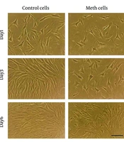
Although the morphology of METH-treated and vehicle-treated cells did not appear to differ significantly over 6 days, long, thin processes extending from the cytoplasm were prominent in the METH-treated group. This appearance was particularly evident on day 3 and was absent in the vehicle-treated group. On day 6, METH-treated cells also appeared unhealthy compared with the distinct fibroblast-like morphology characteristic of vehicle-treated HCM (Figure 1).
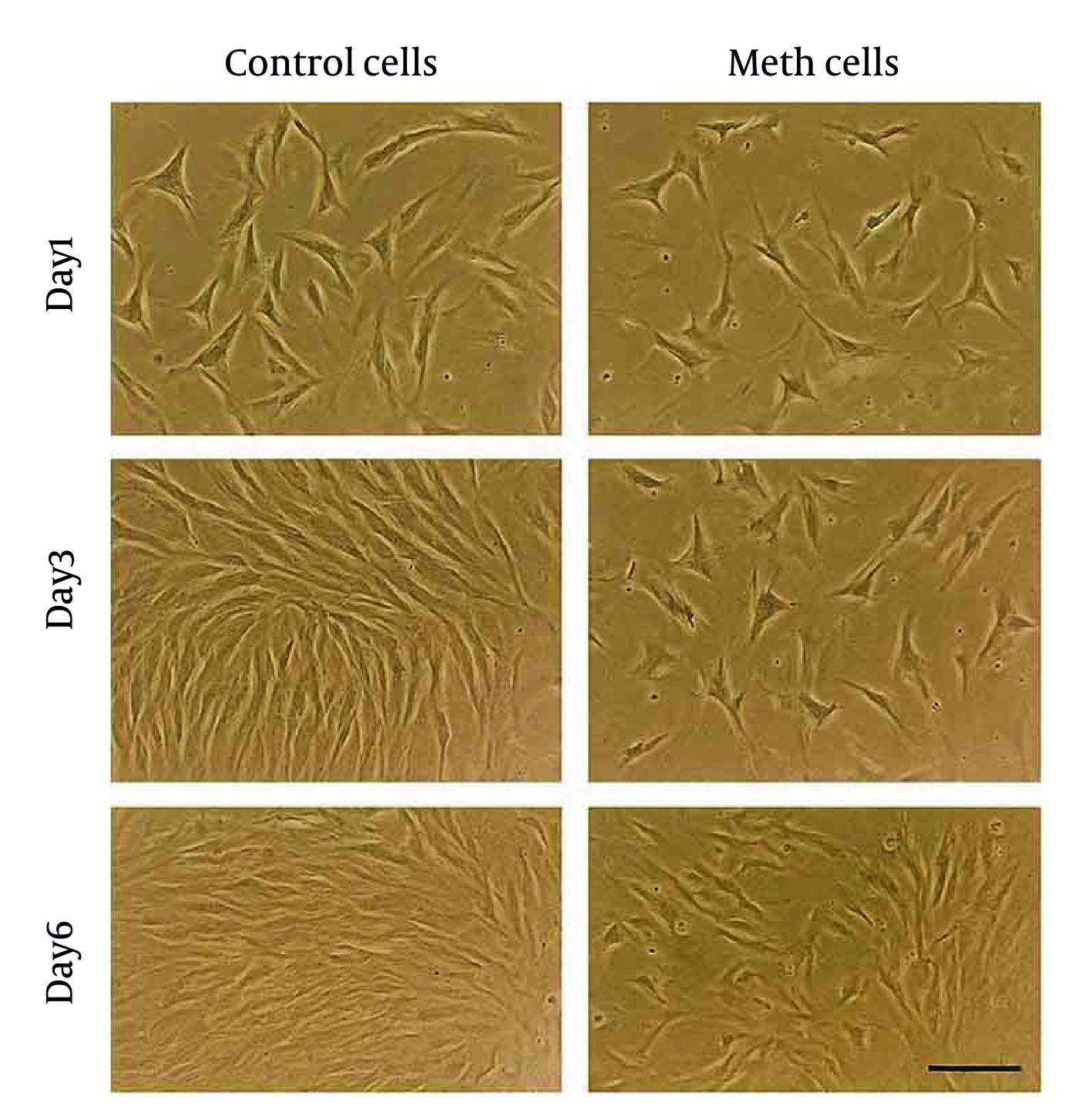
Morphology of methamphetamine-treated and vehicle-treated human cardiomyocytes over 6 days. Magnification: 360×; scale bar = 100 μm.
Figure 1.
Morphology of methamphetamine-treated and vehicle-treated human cardiomyocytes over 6 days. Magnification: 360×; scale bar = 100 μm.

4.2. Cell Growth Curve

Growth curve analysis revealed that METH-treated HCM had a significantly increased viable cell number compared with vehicle-treated cells. This increase was more pronounced on day 4. However, the difference in cell number was statistically nonsignificant on all days. Vehicle-treated cells maintained an increasing growth pattern until day 6, whereas METH-treated cells experienced a sharp decline thereafter (Figure 2).
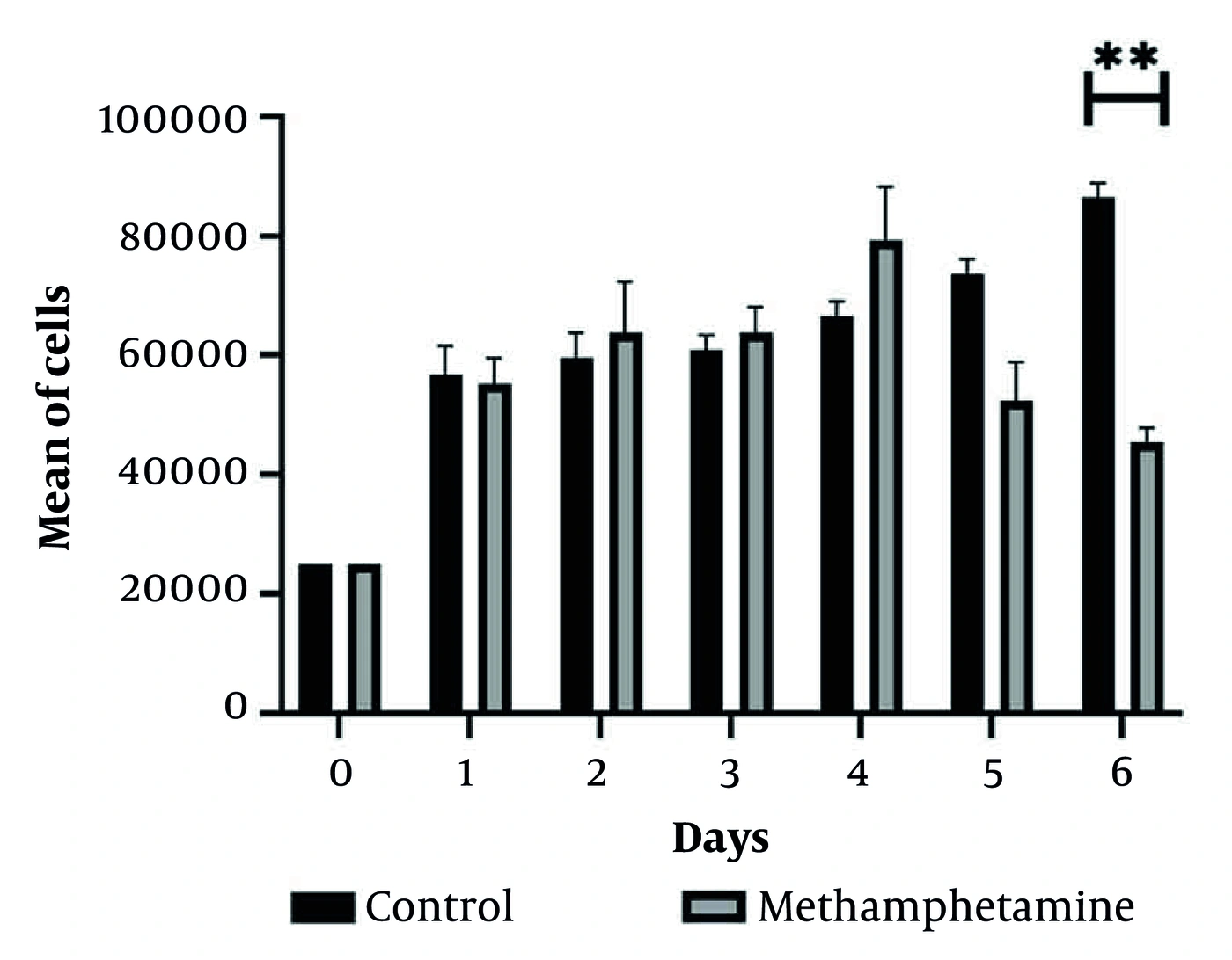
Growth pattern of cardiomyocytes treated with methamphetamine or phosphate-buffered saline vehicle. The experiment was performed in 3 biological replicates, each assessed in technical triplicate. A statistically significant difference between treated and vehicle-treated groups was evident only on day 6. Error bars represent the standard deviation. Analysis of variance (ANOVA) was used, with P &lt; 0.05 indicating statistical significance.
Figure 2.
Growth pattern of cardiomyocytes treated with methamphetamine or phosphate-buffered saline vehicle. The experiment was performed in 3 biological replicates, each assessed in technical triplicate. A statistically significant difference between treated and vehicle-treated groups was evident only on day 6. Error bars represent the standard deviation. Analysis of variance (ANOVA) was used, with P < 0.05 indicating statistical significance.

4.3. Gene Expression

GATA4 expression peaked significantly on day 1 (4.53 ± 0.74; P = 0.011) in METH-treated HCM, decreased to near vehicle-treated levels on day 3 (1.17 ± 1.43), and was significantly downregulated by day 6 (0.10 ± 0.23; P = 0.002) compared with vehicle-treated cells. A significant difference in GATA4 expression was also observed between days 1 and 3 (P = 0.011). A similar descending pattern was observed for creatine kinase transcript expression, which increased to 1.81 ± 0.71-fold on day 1, followed by 1.47 ± 1.46-fold on day 3. By day 6, expression had dropped sharply to 0.11 ± 0.38. Differences in creatine kinase transcript expression between METH-treated and vehicle-treated cells were nonsignificant throughout the 6 days (P = 0.152) (Table 2). Treatment-group values are expressed as fold changes relative to controls and were calculated using the 2-ΔΔCt method. Control values were normalized to 1. Comparisons were performed across days 1, 3, and 6 and between the specified pairs of time points; the corresponding P values are presented.
Table 2.
Cardiac-Related Marker Expression Ratios in the METH-Treated Group Relative to Vehicle-Treated Controls a
MarkerDay 1Day 3Day 6P value bP value (days 1, 3) cP value (days 1, 6) cP value (days 3, 6) c
GATA44.53 ± 0.741.17 ± 1.430.10 ± 0.230.0020.0110.0020.400
Creatine kinase1.81 ± 0.711.47 ± 1.460.11 ± 0.380.152---
a Values are expressed as mean ± SD. Control values were set to 1. Treatment-group values represent fold changes relative to controls, calculated using 2-ΔΔCt analysis. Abbreviations: METH, methamphetamine; GATA4, GATA-binding protein 4; SD, standard deviation.
b P value indicates the comparison across days 1, 3, and 6.
c P value indicates comparisons between the specified pairs of time points. A P < 0.05 was considered statistically significant. This experiment was performed in 3 biological replicates, each assessed in technical triplicate.

5. Discussion

This study examined the effects of METH on the biological activity of human cardiac cells. Morphology, growth kinetics, and expression of cardiac-related markers, including GATA4 and the creatine kinase transcript, showed notable changes in HCM following METH exposure. By day 3, METH-treated HCM exhibited long, thin cytoplasmic processes and, by day 6, an unhealthy appearance, unlike the typical fibroblast-like morphology of vehicle-treated cells. These changes are consistent with previous studies linking METH to oxidative stress, mitochondrial dysfunction, and disruption of autophagy-lysosome pathways in cardiac tissue (7, 13).
The growth curve showed a statistically nonsignificant increase in proliferative capacity by day 4, followed by a sharp decline by day 6, in contrast to sustained growth in vehicle-treated cells. This biphasic pattern is consistent with previous reports of early compensatory responses to METH-induced stress (2, 14), followed by cell death driven by senescence and apoptosis (7, 15). These findings support the hypothesis that cellular responses to METH toxicity follow a complex, multistage trajectory. Because cell number was determined using trypan blue exclusion and serial cell counting, the observed changes represent alterations in viable cell number and growth kinetics rather than direct evidence of increased cellular proliferation. The early increase in cell number may reflect changes in proliferation, survival, attachment, or other cellular processes.
The influence of METH on HCM was also investigated by examining cardiac-related gene expression. The myocardial band isoenzyme of creatine kinase is found primarily in cardiac muscle and is released into the bloodstream following myocardial injury, making it a biomarker of cardiac damage (16, 17). GATA4 is another protein expressed by cardiomyocytes (18). A previous study showed that METH exposure causes cardiomyocyte senescence by increasing GATA4 levels (19). In the present study, GATA4 expression initially increased and subsequently decreased. Creatine kinase transcript expression also showed a trend toward change. Together, these findings suggest dynamic responses to METH, reflecting early stress and subsequent cellular responses in HCM. However, a retrospective study by Richards et al. suggested that the diagnostic utility of biomarkers in methamphetamine-associated cardiomyopathy (MACM) is likely comparable to that in the general heart failure population. Among METH-positive patients with elevated B-type natriuretic peptide levels, creatinine levels were significantly increased, whereas troponin I levels were not (20).
The METH concentration used in this study (0.6 mM) should be interpreted as an experimental exposure concentration rather than as a direct representation of circulating METH levels in humans. Previous in vitro studies investigating the direct effects of METH on cardiomyocytes have used concentrations ranging from micromolar to millimolar levels, including 0.05 - 1.0 mM in cultured adult rat ventricular cardiomyocytes (21) and 0.1 - 5.0 mM in isolated murine cardiomyocytes (2). These studies demonstrate that relatively high experimental concentrations can be used to investigate direct cellular responses to METH. However, concentrations measured in the blood of METH users are generally in the low-micromolar range (22). Therefore, the 0.6 mM concentration used here is substantially higher than typical circulating concentrations and should not be considered physiologically representative. Future dose-response studies incorporating a broader range of clinically and toxicologically relevant concentrations are required to determine whether these responses also occur at lower METH concentrations.
Previous studies have shown that the mechanisms underlying MACM are complex (23). Although they were not directly examined in this study, possible mechanisms include myofibrillar disorganization caused by sarcomeric protein degradation, mitochondrial dysfunction, impaired calcium homeostasis, disruption of cellular degradation systems, including the ubiquitin-proteasome system and autophagy-lysosome pathway, and altered myosin gene expression. These changes may promote cellular senescence and apoptosis (2, 13-15, 22). Such mechanisms impair contractile function and promote inflammation. Over time, persistent inflammation, myocyte loss, and replacement fibrosis lead to chamber dilation and reduced systolic function, manifesting clinically as dilated cardiomyopathy and heart failure with reduced ejection fraction (24-26).
Methamphetamine-associated cardiomyopathy is increasingly recognized as a distinct clinical entity amid the escalating METH epidemic (27, 28). Affected individuals are typically younger than those with cardiomyopathy of other etiologies (28). These patients often present with advanced heart failure, characterized by reduced ejection fraction and New York Heart Association class III - IV symptoms (25, 29). Several case reports have documented acute myocardial infarction in patients with MACM, frequently attributed to vasospasm or coronary thrombosis in the absence of preexisting coronary artery disease (30, 31). Their severe presentation is often associated with a poor prognosis. Nevertheless, a key characteristic of MACM is the potential reversibility of cardiac dysfunction following sustained abstinence from METH use (32). Cessation of METH use for 1 year is associated with improved global longitudinal strain and a lower risk of heart failure-related hospitalization (33). Optimal management requires a multidisciplinary approach involving primary care providers, psychiatry, social services, and substance use counselors.

5.1. Limitations

This study has several limitations. First, although the in vitro model provides a controlled environment, it may not fully replicate the complex physiological conditions of chronic METH exposure in vivo. Second, only 2 cardiac-related markers were evaluated, which may not capture the full spectrum of molecular changes associated with METH-induced cardiomyopathy. Additional markers, including complementary cardiac and cell-death markers, should be measured. Third, the single METH concentration used may not reflect the range of doses encountered in human use, limiting insight into dose-dependent effects. Potential sources of bias should also be considered. Measurement and observer bias cannot be completely excluded because morphological assessment was performed without explicit blinding to treatment allocation. Experimental bias may arise from using a single source of HCM. Authentication of the cardiomyocyte preparation represents an additional limitation. The limited number of independent biological replicates may restrict statistical power and the assessment of run-to-run experimental variability.

5.2. Conclusions

This study demonstrates that METH exposure induces morphological changes, alters growth kinetics, and dynamically modulates cardiac-related gene expression in HCM under in vitro conditions. Further studies are needed to clarify the underlying mechanisms and determine whether these findings translate into clinically relevant cardiovascular effects.
Footnotes

  • AI Use Disclosure:The authors declare that no generative AI tools were used in the creation of this article.

  • Authors' Contribution:Study Concept and Design: H. K. G. and I. R. J.; Data Acquisition: K. R., F. E., F. P., V. K., and V. M.; Data Analysis and Interpretation: I. R. J.; Manuscript Drafting: K. R. and I. R. J.; Critical Revision for Important Intellectual Content: All authors; Statistical Analysis: I. R. J. and M. J. Z.; Administrative, Technical, and Material Support: H. K. J. and V. M.; Study Supervision: I. R. J.

  • Conflict of Interests Statement:The authors declare no conflicts of interest for this study.

  • Data Availability:The dataset presented in the study is available on request from the corresponding author during submission or after publication.

  • Ethical Approval:This study was approved under the ethical approval code of IR.JUMS.REC.1402.033 by the Research Ethics Committees of Jahrom University of Medical Sciences.

  • Funding/Support:This study was supported in part by grant 5678 from Jahrom University of Medical Sciences.

References

  • 1.
    Panenka WJ, Procyshyn RM, Lecomte T, MacEwan GW, Flynn SW, Honer WG, et al. Methamphetamine use: a comprehensive review of molecular, preclinical and clinical findings. Drug Alcohol Depend. 2013;129(3):167-79. [PubMed ID: 23273775]. https://doi.org/10.1016/j.drugalcdep.2012.11.016.
  • 2.
    Turdi S, Schamber RM, Roe ND, Chew HG, Jr, Culver B, et al. Acute methamphetamine exposure inhibits cardiac contractile function. Toxicol Lett. 2009;189(2):152-8. [PubMed ID: 19481142]. [PubMed Central ID: PMC2730117]. https://doi.org/10.1016/j.toxlet.2009.05.015.
  • 3.
    Mohammad Aghaei A, Gholami J, Amin-Esmaeili M, Baheshmat S, Rostam-Abadi Y, Rahimi-Movaghar A. Six-Year Follow-up of People Who Use Methamphetamine in Iran: A Case Series Study. Int J High Risk Behav Addict. 2023;12(1). e131659. https://doi.org/10.5812/ijhrba-131659.
  • 4.
    Fleckenstein AE, Volz TJ, Riddle EL, Gibb JW, Hanson GR. New insights into the mechanism of action of amphetamines. Annu Rev Pharmacol Toxicol. 2007;47(1):681-98. [PubMed ID: 17209801]. https://doi.org/10.1146/annurev.pharmtox.47.120505.105140.
  • 5.
    Homer BD, Solomon TM, Moeller RW, Mascia A, DeRaleau L, Halkitis PN. Methamphetamine abuse and impairment of social functioning: a review of the underlying neurophysiological causes and behavioral implications. Psychol Bull. 2008;134(2):301-10. [PubMed ID: 18298273]. https://doi.org/10.1037/0033-2909.134.2.301.
  • 6.
    Robertson SD, Matthies HJG, Galli A. A closer look at amphetamine-induced reverse transport and trafficking of the dopamine and norepinephrine transporters. Mol Neurobiol. 2009;39(2):73-80. [PubMed ID: 19199083]. [PubMed Central ID: PMC2729543]. https://doi.org/10.1007/s12035-009-8053-4.
  • 7.
    Kevil CG, Goeders NE, Woolard MD, Bhuiyan MS, Dominic P, Kolluru GK, et al. Methamphetamine Use and Cardiovascular Disease. Arterioscler Thromb Vasc Biol. 2019;39(9):1739-46. [PubMed ID: 31433698]. [PubMed Central ID: PMC6709697]. https://doi.org/10.1161/ATVBAHA.119.312461.
  • 8.
    Nabaei G, Oveisgharan S, Ghorbani A, Fatehi F. Impaired arterial smooth muscle cell vasodilatory function in methamphetamine users. J Neurol Sci. 2016;370:107-11. [PubMed ID: 27772737]. https://doi.org/10.1016/j.jns.2016.09.027.
  • 9.
    Paratz ED, Cunningham NJ, MacIsaac AI. The Cardiac Complications of Methamphetamines. Heart Lung Circ. 2016;25(4):325-32. [PubMed ID: 26706652]. https://doi.org/10.1016/j.hlc.2015.10.019.
  • 10.
    Zhao SX, Kwong C, Swaminathan A, Gohil A, Crawford MH. Clinical Characteristics and Outcome of Methamphetamine-Associated Pulmonary Arterial Hypertension and Dilated Cardiomyopathy. JACC Heart Fail. 2018;6(3):209-18. [PubMed ID: 29496022]. https://doi.org/10.1016/j.jchf.2017.10.006.
  • 11.
    Ago M, Ago K, Hara K, Kashimura S, Ogata M. Toxicological and histopathological analysis of a patient who died nine days after a single intravenous dose of methamphetamine: a case report. Legal medicine. 2006;8(4):235-9. [PubMed ID: 16793314]. https://doi.org/10.1016/j.legalmed.2006.03.002.
  • 12.
    Ibrahim Kurawa M, Torkaman-Boutorabi A, Shafaghi L, Hassanzadeh G, Zahmatkesh M, Vousooghi N, et al. The Effects of Subchronic Methamphetamine Administration on the NLRP3 Inflammasome, Memory Function, and Hippocampal Morphology. Arch Neurosci. 2024;11(4). e145644. https://doi.org/10.5812/ans-145644.
  • 13.
    Funakoshi-Hirose I, Aki T, Unuma K, Funakoshi T, Noritake K, Uemura K. Distinct effects of methamphetamine on autophagy-lysosome and ubiquitin-proteasome systems in HL-1 cultured mouse atrial cardiomyocytes. Toxicology. 2013;312:74-82. [PubMed ID: 23933405]. https://doi.org/10.1016/j.tox.2013.07.016.
  • 14.
    Sugimoto K, Okamura K, Tanaka H, Takashima S, Ochi H, Yamamoto T, et al. Methamphetamine directly accelerates beating rate in cardiomyocytes by increasing Ca(2+) entry via L-type Ca(2+) channel. Biochem Biophys Res Commun. 2009;390(4):1214-20. [PubMed ID: 19878660]. https://doi.org/10.1016/j.bbrc.2009.10.124.
  • 15.
    Won S, Hong RA, Shohet RV, Seto TB, Parikh NI. Methamphetamine-associated cardiomyopathy. Clin Cardiol. 2013;36(12):737-42. [PubMed ID: 24037954]. [PubMed Central ID: PMC4319790]. https://doi.org/10.1002/clc.22195.
  • 16.
    Katrukha IA. Human cardiac troponin complex. Structure and functions. Biochemistry (Moscow). 2013;78(13):1447-65. [PubMed ID: 24490734]. https://doi.org/10.1134/S0006297913130063.
  • 17.
    Thygesen K, Alpert JS, Jaffe AS, Chaitman BR, Bax JJ, Morrow DA, et al. Fourth universal definition of myocardial infarction (2018). Eur Heart J. 2019;40(3):237-69. [PubMed ID: 30165617]. https://doi.org/10.1093/eurheartj/ehy462.
  • 18.
    Oka T, Maillet M, Watt AJ, Schwartz RJ, Aronow BJ, Duncan SA, et al. Cardiac-specific deletion of Gata4 reveals its requirement for hypertrophy, compensation, and myocyte viability. Circulation research. 2006;98(6):837-45. [PubMed ID: 16514068]. https://doi.org/10.1161/01.RES.0000215985.18538.c4.
  • 19.
    Zhao X, Lu J, Zhang C, Chen C, Zhang M, Zhang J, et al. Methamphetamine induces cardiomyopathy through GATA4/NF-κB/SASP axis-mediated cellular senescence. Toxicology and Applied Pharmacology. 2023;466. 116457. [PubMed ID: 36914120]. https://doi.org/10.1016/j.taap.2023.116457.
  • 20.
    Richards JR, Harms BN, Kelly A, Turnipseed SD. Methamphetamine use and heart failure: Prevalence, risk factors, and predictors. The American Journal of Emergency Medicine. 2018;36(8):1423-8. [PubMed ID: 29307766]. https://doi.org/10.1016/j.ajem.2018.01.001.
  • 21.
    Maeno Y, Iwasa M, Inoue H, Koyama H, Matoba R. Methamphetamine induces an increase in cell size and reorganization of myofibrils in cultured adult rat cardiomyocytes. International journal of legal medicine. 2000;113(4):201-7. [PubMed ID: 10929235]. https://doi.org/10.1007/s004149900088.
  • 22.
    Abdullah CS, Aishwarya R, Alam S, Morshed M, Remex NS, Nitu S, et al. Methamphetamine induces cardiomyopathy by Sigmar1 inhibition-dependent impairment of mitochondrial dynamics and function. Communications biology. 2020;3(1). 682. [PubMed ID: 33203971]. [PubMed Central ID: PMC7673131]. https://doi.org/10.1038/s42003-020-01408-z.
  • 23.
    Ziaolhagh SJ, Rajabi S, Nosrati Z. Examining the cardioprotective effects of aerobic exercise combined with crocin or berberine on methamphetamine-induced heart damage in female rats. Int J High Risk Behav Addict. 2024;13(3). e147435. https://doi.org/10.5812/ijhrba-147435.
  • 24.
    Darke S, Duflou J, Kaye S. Prevalence and nature of cardiovascular disease in methamphetamine-related death: a national study. Drug and alcohol dependence. 2017;179:174-9. [PubMed ID: 28787694]. https://doi.org/10.1016/j.drugalcdep.2017.07.001.
  • 25.
    Schürer S, Klingel K, Sandri M, Majunke N, Besler C, Kandolf R, et al. Clinical characteristics, histopathological features, and clinical outcome of methamphetamine-associated cardiomyopathy. JACC: Heart Failure. 2017;5(6):435-45. [PubMed ID: 28571597]. https://doi.org/10.1016/j.jchf.2017.02.017.
  • 26.
    Karch SB. The unique histology of methamphetamine cardiomyopathy: a case report. Forensic science international. 2011;212(1 - 3):e1-e4. [PubMed ID: 21664081]. https://doi.org/10.1016/j.forsciint.2011.04.028.
  • 27.
    Neeki MM, Kulczycki M, Toy J, Dong F, Lee C, Borger R, et al. Frequency of methamphetamine use as a major contributor toward the severity of cardiomyopathy in adults ≤ 50 years. The American journal of cardiology. 2016;118(4):585-9. [PubMed ID: 27374605]. https://doi.org/10.1016/j.amjcard.2016.05.057.
  • 28.
    Sliman S, Waalen J, Shaw D. Methamphetamine-associated congestive heart failure: increasing prevalence and relationship of clinical outcomes to continued use or abstinence. Cardiovasc Toxicol. 2016;16(4):381-9. [PubMed ID: 26661075]. https://doi.org/10.1007/s12012-015-9350-y.
  • 29.
    Wijetunga M, Seto T, Lindsay J, Schatz I. Crystal methamphetamine-associated cardiomyopathy: tip of the iceberg? J Toxicol Clin Toxicol. 2003;41(7):981-6. [PubMed ID: 14705845]. https://doi.org/10.1081/CLT-120026521.
  • 30.
    Khaheshi I, Mahjoob MP, Esmaeeli S, Eslami V, Haybar H. Simultaneous thrombosis of the left anterior descending artery and the right coronary artery in a 34-year-old crystal methamphetamine abuser. Korean Circ J. 2015;45(2):158-60. [PubMed ID: 25810738]. [PubMed Central ID: PMC4372982]. https://doi.org/10.4070/kcj.2015.45.2.158.
  • 31.
    Al Shehri MA, Youssef AA. Acute myocardial infarction with multiple coronary thromboses in a young addict of amphetamines and benzodiazepines. J Saudi Heart Assoc. 2016;28(3):180-4. [PubMed ID: 27358538]. [PubMed Central ID: PMC4917712]. https://doi.org/10.1016/j.jsha.2015.11.004.
  • 32.
    Reddy PKV, Ng TMH, Oh EE, Moady G, Elkayam U. Clinical characteristics and management of methamphetamine-associated cardiomyopathy: state-of-the-art review. J Am Heart Assoc. 2020;9(11). e016704. [PubMed ID: 32468897]. [PubMed Central ID: PMC7428977]. https://doi.org/10.1161/JAHA.120.016704.
  • 33.
    Bhatia HS, Nishimura M, Martinez A, Vanam S, Kahn AM, DeMaria A, et al. Systolic dysfunction in patients with methamphetamine use and heart failure with preserved ejection fraction. International journal of cardiology. 2022;348:90-4. [PubMed ID: 34921901]. [PubMed Central ID: PMC10174667]. https://doi.org/10.1016/j.ijcard.2021.12.024.

Copyright

Copyright © 2026, Kargar Jahromi et al. This open-access article is available under the Creative Commons Attribution 4.0 (CC BY 4.0) International License (https://creativecommons.org/licenses/by/4.0/), which allows for unrestricted use, distribution, and reproduction in any medium, provided that the original work is properly cited.

Similar Articles

31
Oct
2019
Postmortem Study of Molecular and Histological Changes in the CA1 Hippocampal Region of Chronic Methamphetamine User

Postmortem Study of Molecular and Histological Changes in the CA1 Hippocampal Region of Chronic Methamphetamine User

Gholam-Reza Mahmoudiasl,
Hojjat Allah Abbaszadeh,
Mostafa Rezaei-Tavirani,
Mohammad-Amin Abdollahifar,
Yousef Sadeghi,
Maryam Sadat Khoramgah
,et al.

Mahmoudiasl G, Abbaszadeh HA, Rezaei-Tavirani M, Abdollahifar M, Sadeghi Y, et al. Postmortem Study of Molecular and Histological Changes in the CA1 Hippocampal Region of Chronic Methamphetamine User. Iran J Pharm Res. 2019;18(4):e126261. doi: https://doi.org/10.22037/ijpr.2019.15483.13123

15
Oct
2013
Effects of Methamphetamine on Pituitary Gonadal Axis and Spermatogenesis in Mature Male Rats

Effects of Methamphetamine on Pituitary Gonadal Axis and Spermatogenesis in Mature Male Rats

Malihe Heidari-Rarani,
Ali Noori,
Arash Ghodousi

Heidari-Rarani M, Noori A, Ghodousi A. Effects of Methamphetamine on Pituitary Gonadal Axis and Spermatogenesis in Mature Male Rats. Zahedan J Res Med Sci. 2014;16(12):37-42. doi:

17
May
2026
The Impact of Marijuana (Cannabis sativa) on Viability and Expression of Cardiac Markers in Human Cardiomyocytes

The Impact of Marijuana (Cannabis sativa) on Viability and Expression of Cardiac Markers in Human Cardiomyocytes

Hossein Kargar Jahromi,
Vida Khalafi,
Vida Mirzaei,
Faezeh Ebrahimi-Zadeh,
Pouria Azami,
Tara Abdolahy
,et al.

Kargar Jahromi H, Khalafi V, Mirzaei V, Ebrahimi-Zadeh F, Azami P, et al. The Impact of Marijuana (Cannabis sativa) on Viability and Expression of Cardiac Markers in Human Cardiomyocytes. Iran J Pharm Res. 2026;25(1):e168324. doi: https://doi.org/10.5812/ijpr-168324

16
Sep
2024
The Effects of Subchronic Methamphetamine Administration on the NLRP3 Inflammasome, Memory Function, and Hippocampal Morphology

The Effects of Subchronic Methamphetamine Administration on the NLRP3 Inflammasome, Memory Function, and Hippocampal Morphology

Musa Ibrahim Kurawa,
Anahita Torkaman-Boutorabi,
Lida Shafaghi,
Gholamreza Hassanzadeh,
Maryam Zahmatkesh,
Nasim Vousooghi
,et al.

Ibrahim Kurawa M, Torkaman-Boutorabi A, Shafaghi L, Hassanzadeh G, Zahmatkesh M, et al. The Effects of Subchronic Methamphetamine Administration on the NLRP3 Inflammasome, Memory Function, and Hippocampal Morphology. Arch Neurosci. 2024;11(4):e145644. doi: https://doi.org/10.5812/ans-145644

30
Apr
2021
Protective Effect of Cinnamaldehyde on METH-induced Neurotoxicity in PC12 Cells via Inhibition of Apoptotic Response and Oxidative Stress

Protective Effect of Cinnamaldehyde on METH-induced Neurotoxicity in PC12 Cells via Inhibition of Apoptotic Response and Oxidative Stress

Roghayeh Rashidi,
Seyed Adel Moallem,
Mohammad Moshiri,
Farzin Hadizadeh,
Leila Etemad

Rashidi R, Moallem SA, Moshiri M, Hadizadeh F, Etemad L. Protective Effect of Cinnamaldehyde on METH-induced Neurotoxicity in PC12 Cells via Inhibition of Apoptotic Response and Oxidative Stress. Iran J Pharm Res. 2021;20(2):e124508. doi: https://doi.org/10.22037/ijpr.2020.111891.13411

More by these authors

Hossein Kargar JahromiPubMedScholar
Khatere RoozbehiPubMedScholar
Faezeh Ebrahimi-ZadehPubMedScholar
Vida MirzaeiPubMedScholar
Fatemeh ParsaeiPubMedScholar
Vida KhalafiPubMedScholar
Mohammad Javad ZibaeenezhadPubMedScholar
Iman Razeghian-JahromiPubMedScholar
Share
Cited by
Metrics