
Fulltext
The full text is available in PDF.
International Cardiovascular Research Journal
An annual journal in scientific affiliation with the Cardiovascular Research Center, Shiraz University of Medical Sciences
Image Credit:Int Cardiovasc Res J
Authors
Background:
Coronary Artery Disease (CAD) is a major cause of morbidity and mortality world-wild. Thus, its early diagnosis and treatment could be life-saving. Mitral Valve (MV) and mitral annular velocities as well as left atrial and left ventricular wall strain have been claimed to be helpful for prediction of the presence of CAD.
Objectives:
This case-control study aimed to assess the predictive value of these echocardiographic parameters for detection of CAD.
Methods:
Eighty consecutive individuals referring with acute angina-like chest pain and requiring diagnostic coronary angiography and had a concomitant echocardiographic Left Ventricular Ejection Fraction (LVEF) ≥ 50% were divided into two groups: those with significant CAD (n = 45) and those with non-significant CAD (n = 35). Tissue Doppler Imaging (TDI) was employed for all participants. Peak early and late MV velocities (E and A, respectively), peak longitudinal systolic myocardial velocity (mitral S′), peak early and late diastolic velocities (e′ and a′, respectively) from medial mitral annulus, E/e′ ratio, peak Left Atrial (LA) strain, and Left Ventricular Global Longitudinal Strain (LV GLS) were measured and compared in the two groups. Chi-square and t-test were used to compare the two groups as appropriated. Multiple logistic regression was also performed to detect the impact of categorical variables on the predictors of the presence of CAD. Receiver operating characteristic curves were constructed as needed. A P value ≤ 0.05 was considered to be statistically significant.
Results:
Mitral S′ and E/e′ were significantly different in the two groups. Mitral S′ was lower and E/e′ was higher in the patients with CAD compared to the control group (P = 0.01 and P = 0.03, respectively). Considering 5.3 cm/sec as the cut-off point for mitral S′, sensitivity and specificity for significant CAD were 67.40% and 68.60%, respectively with an odds ratio of 0.275. The cut-off point for mitral E/e′ was defined as 12.65, with sensitivity and specificity for significant CAD to be 52.17% and 77.14%, respectively with an odds ratio of 1.249.
Conclusion:
This study showed that mitral S′ and E/e′ could predict the presence of significant CAD in patients with chest pain and preserved LVEF. Thus, TDI could be used as a simple, reliable, and non-invasive tool to achieve this goal.
© 2019, Author(s). This open-access article is available under the Creative Commons Attribution 4.0 (CC BY 4.0) International License (https://creativecommons.org/licenses/by/4.0/), which allows for unrestricted use, distribution, and reproduction in any medium, provided that the original work is properly cited.
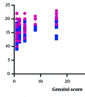
Safi M, Bayat F, Gachkar L, Shahri MR. Correlation of LVEDP with Severity of Coronary Artery Involvement in Patients Referred to Modarres Hospital with Coronary Artery Disease. Int J Cardiovasc Pract. 2025;10(1):e159763. doi: https://doi.org/10.5812/intjcardiovascpract-159763
Zheng XZ, Yang B, Wu J. Comparison of the Efficacy of Conventional Echocardiographic Parameters in the Diagnosis of Significant Coronary Artery Stenosis. I J Radiol. 2015;12(1):e11405. doi: https://doi.org/10.5812/iranjradiol.11405
Esmaeilzadeh M, Hamidzad M, Kiavar M, Bakhshandeh Abkenar H, Esmaeilzadeh F. Non-Invasive Assessment of Left Ventricular End-Diastolic Pressure in Patients with Chronic Aortic Regurgitation, Comparison of the Sensitivity and Specificity of CW Doppler Echocardiography with Angiography. Int Cardiovasc Res J. 2009;3(2):e68465. doi:
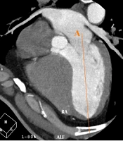
Faghihi T, Ehsani A, Shojaeian F, Safavi Naini SAA. Relationship Between the Coronary Artery Calcium (CAC) Score and the Angle Between the Aortic Valve and the Left Ventricular Inflow Long Axis (AV-LV) as Cardiovascular Risk Factors. I J Radiol. 2021;18(4):e110421. doi: https://doi.org/10.5812/iranjradiol.110421
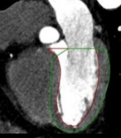
Kadiyoran C, Yilmaz PD. Monocyte-to-High-Density Lipoprotein Ratio may be Related to the Coronary Artery Disease Reporting and Data System Score and Increased Left Ventricular Mass. I J Radiol. 2022;19(1):e118837. doi: https://doi.org/10.5812/iranjradiol.118837
Last Update: 1 month ago
Last Update: 1 month ago
Last Update: 1 month ago
Last Update: 8 hours ago
Last Update: 11 hours ago
Last Update: 4 days ago
