Cover image of the article: A Neonate with Scimitar Syndrome, Absent Right Pulmonary Artery, Imperforated Anus, and Coarctation of the Aorta

Image Credit:Int Cardiovasc Res J

A Neonate with Scimitar Syndrome, Absent Right Pulmonary Artery, Imperforated Anus, and Coarctation of the Aorta

Authors

Mehrdad Rezaei1, Amirmasoud Borghei1, Nazanin Farahbakhsh1, Hamid AmoozgarHamid Amoozgar ORCID2, 3,*
1Department of Pediatrics, Shiraz University of Medical Sciences, Shiraz, IR Iran
2Neonatology Research Center, Shiraz University of Medical Sciences, Shiraz, IR Iran
3Cardiovascular Research Center, Shiraz University of Medical Sciences, Shiraz, IR Iran
*Corresponding Author: Corresponding author: Hamid Amoozgar, Department of Pediatrics, Nemazee Hospital, Shiraz, Iran, PO Box: 71937-11351. Tel: +98-9173111877, Fax: +98-7116474298, E-mail: Email: amozgah@sums.ac.ir

International Cardiovascular Research Journal:Vol. 10, issue 4; e9817
Published online:Oct 01, 2016
Article type:Case Report
Received:Nov 24, 2014
Accepted:Apr 08, 2015
How to Cite:Rezaei M, Borghei A, Farahbakhsh N, Amoozgar H. A Neonate with Scimitar Syndrome, Absent Right Pulmonary Artery, Imperforated Anus, and Coarctation of the Aorta. Int Cardiovasc Res J. 2016;10(4):e9817. doi:

Abstract

Background: Scimitar syndrome is a rare and complex congenital defect that often involves hypoplasia of the right lung, partial anomalous pulmonary venous connection of the right lung draining into the inferior vena cava, and a systemic collateral artery from the descending aorta supplying the right lower lobe (lobar sequestration). Our case was a male newborn with scimitar syndrome associated with an absent right pulmonary artery, imperforated anus, and coarctation of the aorta. Hemodinamically, the patient was asymptomatic and diagnosed accidentally. This combination has not been reported before. The pathological process of the scimitar syndrome is unclear. However, it appears to originate from a basic developmental disorder of the complete pulmonary bud early in embryogenesis. This rare syndrome with different presentations can occur alone or in combination with cardiac and /or non-cardiac anomalies.

Highlights

1. Introduction

Scimitar syndrome is a rare and complex congenital defect that often involves hypoplasia of the right lung and partial anomalous pulmonary venous connection of the right lung draining into the inferior vena cava. The right pulmonary artery is usually hypoplastic; however, it is fully absent (1-3). Systemic collateral artery from the descending aorta supplies the right lower lobe (lobar sequestration).
The term “scimitar” was derived from the shadow created by the abnormal right lower pulmonary vein on the chest X-ray that connected to inferior vena cava resembles an arched Turkish sword (4). Up to now, several associations have been described with scimitar syndrome, including hypoplasia of the right lung, anomalies of the bronchial system, horseshoe lung, hypoplasia of the right pulmonary artery, anomalous arterial connection to the right lung from the aorta, pulmonary sequestration, and secondary dextrocardia due to lung disease (5).

2. Case Presentation

A male neonate was noted to have imperforated anus soon after birth. He was the product of normal vaginal delivery, gravid 4, live 3, and abortion 1 from a 27-year-old mother. The patient had no cardiopulmonary symptoms, but right-sided white lung and abnormal heart shadow were detected in pre-operation chest radiography (Figure 1). After the operation, he was referred to our neonatal intensive care unit for cardiopulmonary evaluation due to his imperforated anus. On physical examination, he was 3.5 kg with no cyanosis and his right hand arterial saturation was 90% at room temperature. His respiratory rate was 50/min and his respiratory pattern was normal. Nonetheless, he had a decreased breathing sound and dullness to percussion on the right hemithorax. His heart rate was 165/min and the blood pressure of his right hand was 70/45 mmHg. In addition, the pulse of extremities was present, but his lower extremities were slightly cold with weak pulse. Moreover, the heart sound was heard in the right mid-clavicular line and an ejection systolic murmur of grade 3/6 could also be heard.
Chest Radiography Showed White Right Hemi Thorax with Right Mediastinal Shift
Figure 1.
Chest Radiography Showed White Right Hemi Thorax with Right Mediastinal Shift
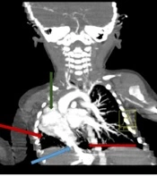
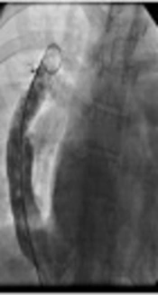
Chest radiography showed white right hemithorax with right mediastinal shift. On echocardiography, the patient had coarctation of the aorta, right pulmonary artery stenosis, and right to left ductus arteriosus flow. Chest computerized tomography scan also revealed right lung hypoplasia, absence of right pulmonary artery, and drainage of the small remnant of the right lung directly into the inferior venacava (Figure 2). Besides, cardiac catheterization revealed normal filling of the main and left pulmonary arteries. Right pulmonary artery filling was absent. Furthermore, aortogram showed coarctation of the aorta in the juxtaductal area with 30 mmHg pressure gradient between the ascending and descending aorta and small patent ductus arteriosus (PDA). Sequestration of the lower lobe of the right lung with partial anomalous drainage to inferior vena cava was seen, as well (Figure 3). After all, scimitar syndrome was diagnosed considering the results of tomography and angiography.
Chest Computerized Tomography Scan Revealed Right Lung Hypoplasia, Absence of Right Pulmonary Artery, and Drainage of the Small Remnant of the Right Lung Directly into the Inferior Venacava
Figure 2.
Chest Computerized Tomography Scan Revealed Right Lung Hypoplasia, Absence of Right Pulmonary Artery, and Drainage of the Small Remnant of the Right Lung Directly into the Inferior Venacava
Angiography Showed Coarctation of the Aorta in the Juxtaductal Area, Sequestration of the Lower Lobe of the Right Lung with Partial Anomalous Drainage to the Inferior Vena Cava, Severe Right Pulmonary Artery Stenosis, and Normal Size and Perfusion of the Left Pulmonary Artery
Figure 3.
Angiography Showed Coarctation of the Aorta in the Juxtaductal Area, Sequestration of the Lower Lobe of the Right Lung with Partial Anomalous Drainage to the Inferior Vena Cava, Severe Right Pulmonary Artery Stenosis, and Normal Size and Perfusion of the Left Pulmonary Artery
During the hospital course, balloon angioplasty was successfully done to correct the coarctation of the aorta. Post-angioplasty, pressure gradient between the ascending and descending aorta became 10 mmHg. The patient was discharged in a good condition on the 22nd day of his birth.

3. Discussion

Partial anomalous pulmonary venous connection (PAPVC) refers to a situation where some, but not all, venous drainage enters the left atrium and the remaining veins connect to the right-sided circulation. Scimitar syndrome is a kind of PAPVC in which, an anomalous pulmonary vein descends from the right lung and drains into the inferior vena cava (6). The abnormal pulmonary vein draining into the inferior vena cava can often be seen as a classic curvilinear density on the right chest on plain radiography and forms a sort of a Turkish sword or scimitar, but this may not always be seen. Thus, this syndrome is also named congenital pulmonary veno-lobar syndrome, hypogenic lung syndrome, vena cava bronchovascular syndrome, or epibronchial right pulmonary artery syndrome (1, 3). Schramel recognized two different types of scimitar vein: 1- the simple classic vein running from the middle of the right lung to the cardiophrenic angle and 2- double arched vein in the upper and lower lung zones, with drainage into the left atrium and inferior caval vein (7). The infantile form of scimitar syndrome can be symptomatic soon after birth. The clinical course is usually presented by severe pulmonary hypertension and sign of right heart failure. Thus, its management can be accompanied with high mortality (8). Since there was no similar report of the association between imperforated anus and asymptomatic scimitar syndrome, this case report was presented.

3.1. Conclusion

This case report indicated the importance of careful and repeated examination of even asymptomatic neonates for early diagnosis of neonatal problems. Moreover, one congenital anomaly should make us really suspicious about the existence of other anomalies.
Acknowledgments

Footnotes

  • Authors’ Contribution:All the authors contributed to writing the manuscript, data gathering, and reading the final version.

  • Financial Disclosure:There is no financial disclosure.

  • Funding/Support:There is no support.

  • Implication for Health Policy/ Practice/ Research/ Medical Education:This rare case report indicated that combination of multiple anomalies was associated with scimitar syndrome.

References

  • 1.
    Grech V, Xuereb R, Xuereb M, Manche A, Schembri K, Degiovanni J. Late presentation and successful treatment of classical scimitar syndrome. Images Paediatr Cardiol. 2003;5(3):49-62. [PubMed ID: 22368630].
  • 2.
    Patel AM, Joshi R, Vaghela D, Shah HR, Shah U, Talsania B. A face of scimitar syndrome. Indian J Radiol Imaging. 2004;14(4):401.
  • 3.
    Rokade ML, Rananavare RV, Shetty DS, Saifi S. Scimitar syndrome. Indian J Pediatr. 2005;72(3):245-7. [PubMed ID: 15812122].
  • 4.
    Chun P, Lee HD, Kim YD. A rare case of scimitar syndrome accompanied with an imperforate anus in a newborn. J Korean Soc Neonatol. 2011;18(2):404-8.
  • 5.
    Takawira FF, Omar F. Unusual variant of scimitar syndrome associated with an absent right pulmonary artery, stenosis of the inferior vena cava, hemi-azygous continuation and the VACTERL association. Cardiovasc J Afr. 2013;24(3):3-6.
  • 6.
    van de Woestijne PC, Verberkmoes N, Bogers AJ. Partial anomalous pulmonary venous connection (including scimitar syndrome). Multimed Man Cardiothorac Surg. 2013;2013:mmt001. [PubMed ID: 24413000]. https://doi.org/10.1093/mmcts/mmt001.
  • 7.
    Schramel FM, Westermann CJ, Knaepen PJ, van den Bosch JM. The scimitar syndrome: clinical spectrum and surgical treatment. Eur Respir J. 1995;8(2):196-201. [PubMed ID: 7758551].
  • 8.
    Huddleston CB, Exil V, Canter CE, Mendeloff EN. Scimitar syndrome presenting in infancy. Ann Thorac Surg. 1999;67(1):154-9. discussion 160. [PubMed ID: 10086541].

Copyright

Copyright © 2016, Shiraz University of Medical Sciences. This is an open-access article distributed under the terms of the Creative Commons Attribution-NonCommercial 4.0 International License (http://creativecommons.org/licenses/by-nc/4.0/) which permits copy and redistribute the material just in noncommercial usages, provided the original work is properly cited.

Similar Articles

17
Jun
2022
Scimitar Syndrome in a Newborn

Scimitar Syndrome in a Newborn

Mahdi Mazandarani,
Ali Ahani Azari,
Narges Lashkarbolouk,
Lobat Shahkar

Mazandarani M, Ahani Azari A, Lashkarbolouk N, Shahkar L. Scimitar Syndrome in a Newborn. J Compr Ped. 2022;13(2):e124204. doi: https://doi.org/10.5812/compreped-124204

28
May
2019
Two Cases of Scimitar Syndrome: An Adolescent and an Older Adult with Late Diagnosis

Two Cases of Scimitar Syndrome: An Adolescent and an Older Adult with Late Diagnosis

Gholamreza Omrani,
Maziar Gholampour Dehaki,
Reza Abbaszadeh,
Ali Sadeghpour Tabaee,
Hassan Tatari

Omrani G, Gholampour Dehaki M, Abbaszadeh R, Sadeghpour Tabaee A, Tatari H. Two Cases of Scimitar Syndrome: An Adolescent and an Older Adult with Late Diagnosis. Multidiscip Cardio Annal. 2019;10(1):e84007. doi: https://doi.org/10.5812/mca.84007

30
Jun
2012
Congenital Pseudohorseshoe Lung Associated with Scimitar Syndrome

Congenital Pseudohorseshoe Lung Associated with Scimitar Syndrome

Alptekin Tosun,
Serife Leblebisatan

Tosun A, Leblebisatan S. Congenital Pseudohorseshoe Lung Associated with Scimitar Syndrome. I J Radiol. 2012;9(2):99-102. doi: https://doi.org/10.5812/iranjradiol.7808

11
Sep
2016
Cardiovascular Malformations in Congenital Rubella Syndrome: A Case Report

Cardiovascular Malformations in Congenital Rubella Syndrome: A Case Report

Fatemeh Vaziri

Vaziri F. Cardiovascular Malformations in Congenital Rubella Syndrome: A Case Report. Multidiscip Cardio Annal. 2011;3(2):e8748. doi:

25
Feb
2013
Pulmonary Hypoplasia Associated with Multiorgan Developmental Abnormalities – a Rare Case Report

Pulmonary Hypoplasia Associated with Multiorgan Developmental Abnormalities – a Rare Case Report

Chang Shu,
Ling He,
Qubei Li,
Qiao Wang,
Ying Huang

Shu C, He L, Li Q, Wang Q, Huang Y. Pulmonary Hypoplasia Associated with Multiorgan Developmental Abnormalities – a Rare Case Report. Inn J Pediatr. 2013;23(2):. doi:

More by these authors

Mehrdad RezaeiPubMedScholar
Amirmasoud BorgheiPubMedScholar
Nazanin FarahbakhshPubMedScholar
Hamid AmoozgarPubMedScholar
Share
Cited by
Metrics