
1. Background
The field of nanotechnology continues to advance rapidly in materials science. The distinctive physicochemical properties of silver nanoparticles (Ag-NPs) have made them a major focus of biomedical research, with applications in medicine, biology, and pharmaceutical sciences. Traditional chemical routes for Ag-NP synthesis have several drawbacks, including high production costs and the use of hazardous reagents and solvents, which raise environmental and health concerns. In response, green synthesis using plant extracts has emerged as a sustainable, cost-effective, and environmentally benign alternative. This approach relies on endogenous plant metabolites, such as phenolic compounds, flavonoids, and terpenoids, which simultaneously reduce metal ions and cap the resulting nanoparticles (1).
Lavandula stricta Delile (L. stricta), which grows naturally in southern Iran, is rich in these bioactive compounds. Its essential oil contains components such as linalool and limonene, whereas its extract is rich in phenolics and flavonoids (2, 3). These phytochemicals exhibit radical-scavenging activity, primarily through hydrogen atom or electron donation to neutralize free radicals (4). Furthermore, specific flavonoids such as apigenin and luteolin (5) have been shown to mitigate oxidative damage by reducing markers such as MDA while enhancing TAC in inflamed tissues. Beyond their antioxidant properties, phenolic compounds present in L. stricta, notably rosmarinic and caffeic acids, exert anti-inflammatory effects by downregulating pro-inflammatory mediators, including TNF-α and IL-6, and by interfering with the nuclear factor kappa-light-chain-enhancer of activated B cells (NF-κB) signaling cascade (6, 7).
The interplay between uncontrolled inflammation and oxidative stress is well documented. Elevated levels of cytokines such as TNF-α and IL-6 and oxidative stress markers such as MDA, together with reduced TAC, may perpetuate a cycle that contributes to chronic disease progression (8). Although polyphenols and flavonoids from natural sources can modulate these pathways, their in vivo efficacy when delivered as green-synthesized nanoparticles remains largely unexplored. Most previous investigations of L. stricta have focused on phytochemical profiling and in vitro antioxidant assessments, and the in vivo anti-inflammatory and antioxidant efficacy of Ag-NPs derived from this plant has not been adequately evaluated.
2. Objectives
This study aimed to: (1) Biosynthesize Ag-NPs using an aqueous extract of L. stricta; (2) characterize the nanoparticles using UV-Vis spectroscopy, XRD, FTIR spectroscopy, TEM, DLS, and zeta potential analysis; and (3) systematically evaluate their in vivo anti-inflammatory and antioxidant effects in a carrageenan-induced acute inflammation model by quantifying TNF-α, IL-6, MDA, and TAC levels in paw tissue.
3. Methods
3.1. Botanical Material Collection and Preparation of Aqueous Extract
Lavandula stricta Delile plants were collected from the Shiraz region (Fars Province, Iran) and identified by the Herbarium of the Faculty of Agriculture, University of Shiraz (voucher No. 55090). The harvested aerial tissues were shade-dried and milled into a fine powder. For preparation of the aqueous extract, 5 g of dried powder was suspended in 50 mL of sterile distilled water and incubated at room temperature for 24 hours under gentle magnetic stirring. After maceration, the suspension was filtered through Whatman No. 1 filter paper, and the clear filtrate was stored at 4 °C until use.
3.2. Phyto-Mediated Biosynthesis of Ag-NPs
Nanoparticles were fabricated by mixing 1 mL of the aqueous extract with 99 mL of 1 mM silver nitrate (AgNO3) solution, following a modified version of the green synthesis protocol established by Shankar et al. (9). The reaction mixture was incubated at ambient temperature (25 ± 2 °C) for 24 hours under constant magnetic stirring. The 24-hour reaction time was selected based on preliminary observations and common practice in green synthesis protocols to ensure complete reduction of silver ions and the formation of stable nanoparticles. Nanoparticle biosynthesis was visually monitored by the progressive color change of the reaction solution from colorless to brown. A portion of the as-synthesized suspension was reserved for UV-Vis, TEM, DLS, and zeta potential analyses and stored at 4 °C in the dark to prevent nanoparticle agglomeration. The remaining suspension was centrifuged at 10,000 rpm for 15 minutes. The supernatant, containing unreacted AgNO3 and free extract components, was removed, and the pellet was washed twice with absolute ethanol to eliminate residual silver ions and loosely associated phytochemicals. The purified pellet was dried at 60 °C to obtain a fine powder and stored at 4 °C until use for XRD, FTIR, and in vivo assays.
3.3. Physicochemical Characterization of Synthesized Nanoparticles
3.3.1. UV-Vis Spectrophotometric Analysis
UV-Vis spectrophotometry was used to monitor Ag-NP formation. To determine the optimal precursor concentration, synthesis was performed using AgNO3 concentrations of 0.5 and 1 mM. After 24 hours of reaction, absorption spectra of the nanoparticle suspensions were recorded over 200 - 800 nm using a double-beam UV-Vis spectrophotometer (UV-1800, Shimadzu Corporation, Japan).
3.3.2. XRD-Based Crystallographic Analysis
XRD analysis was performed to investigate the crystal structure, crystalline phase, and crystallite size of the nanoparticles. Samples were centrifuged at 10,000 rpm for 15 minutes, washed twice with absolute ethanol, dried at 60 °C, and uniformly coated onto standard aluminum XRD grids. XRD analysis was performed using a D8 Advance X-ray diffractometer (Bruker Corporation, Germany) with monochromatic Cu-Kα radiation (λ = 1.5406 Å) at 40 kV and 40 mA. Diffraction data were collected over the 2θ range of 20 - 80° at a step size of 0.02°. The average crystallite size was estimated from the 111 crystallographic reflection using the Debye-Scherrer equation (10):
D = (K × λ) / (β × cos θ)
where D is the crystallite diameter, K is the shape factor (K = 0.9), λ denotes the X-ray wavelength (1.5406 Å), β is the full width at half maximum expressed in radians, and θ represents half of the Bragg angle.
3.3.3. FTIR Spectroscopic Analysis of Functional Groups
FTIR analysis was performed to identify the phytochemical functional groups responsible for silver-ion reduction and surface capping of the synthesized nanoparticles. FTIR spectra were recorded over 400 - 4000 cm-1 using a PerkinElmer spectrophotometer (USA) and the conventional KBr pellet technique.
3.3.4. Morphological Characterization by Transmission Electron Microscopy
The morphology, primary particle size, and size distribution of the biosynthesized nanoparticles were examined by TEM. A 5 µL aliquot of diluted nanoparticle suspension was deposited onto Formvar- and carbon-coated copper grids and allowed to air-dry at room temperature for 24 hours. Imaging was performed using a Philips CM10 transmission electron microscope (Netherlands) operated at an accelerating voltage of 100 kV.
3.3.5. Dynamic Light Scattering and Zeta Potential
The hydrodynamic diameter, polydispersity index (PDI), and zeta potential of the nanoparticles were determined using a NanoPartica SZ-100V2 nanoparticle analyzer (HORIBA, Japan) at 25 °C with a fixed scattering angle of 173°. For each measurement, 1 mL of nanoparticle suspension was diluted with distilled water to an appropriate concentration. Analyses were performed in three independent replicates (n = 3), and results are presented as mean ± SEM.
3.4. Determination of Total Phenolic and Flavonoid Contents
Total phenolic content (TPC) was quantified using the Folin-Ciocalteu colorimetric procedure, with minor modifications to the protocol of Singleton and Rossi Jr. (11). Briefly, 20 µL of aqueous extract was mixed with 1.5 mL of distilled water and 100 µL of Folin-Ciocalteu reagent diluted 1:10 and incubated in the dark for 5 minutes. Subsequently, 300 µL of 20% (w/v) sodium carbonate was added, and the final volume was adjusted to 3 mL with distilled water. After 60 minutes of incubation at room temperature in the dark, absorbance was measured at 765 nm. Phenolic content was calculated using a gallic acid standard curve (0 - 100 µg/mL) and expressed as mg gallic acid equivalent per gram of dry weight (mg GAE/g DW).
Total flavonoid content (TFC) was assessed using the aluminum chloride assay according to Zhishen et al. (12). Briefly, 1 mL of extract was mixed with 4 mL of distilled water and 0.3 mL of 5% NaNO2. After 5 minutes, 0.3 mL of 10% AlCl3 was added, followed 6 minutes later by 2 mL of 1 M NaOH. The volume was adjusted to 10 mL with distilled water, and after 15 minutes of incubation, absorbance was measured at 510 nm. Flavonoid content was calculated from a rutin standard curve (0 - 100 µg/mL) and expressed as mg rutin equivalent per gram of dry weight (mg RE/g DW).
3.5. In Vivo Anti-Inflammatory and Antioxidant Study
3.5.1. Animals and Grouping
Forty-two male BALB/c mice (body weight, 20 ± 2 g) were used. After a 1-week acclimatization period, mice were randomly allocated to seven groups of six animals each:
1) Negative control (saline)
2) Positive control (carrageenan + saline)
3) Indomethacin (5 mg/kg; standard comparator group)
4) Lavender aqueous extract (15 mg/kg)
5) Lavender aqueous extract (30 mg/kg)
6) Synthesized silver nanoparticles (0.3 mg/kg)
7) Synthesized silver nanoparticles (0.6 mg/kg)
Dose preparation: Stock solutions of lavender aqueous extract were prepared in sterile distilled water to achieve doses of 15 and 30 mg/kg body weight. Purified dried Ag-NP powder was weighed according to the intended dose (0.3 or 0.6 mg/kg body weight) and dispersed in sterile distilled water by gentle vortexing to prepare suspensions at concentrations of 0.03 and 0.06 mg/mL, respectively. All solutions and suspensions were prepared fresh immediately before administration. Each mouse received 0.1 mL per 10 g body weight by oral gavage.
The administered doses of 0.3 and 0.6 mg/kg refer to the total mass of the purified dried Ag-NP formulation, comprising both the metallic silver core and the surface-associated phytochemical capping layer. The exact elemental silver content of this formulation was not determined and remains to be quantified in future investigations.
3.5.2. Carrageenan-Induced Edema Model and Edema Measurement
Acute inflammation was elicited in the right hind paw of each mouse by subcutaneous injection of 0.1 mL of a 1% carrageenan suspension prepared in normal saline (13). Paw thickness was measured using a digital caliper 4 hours after injection. The percentage of edema inhibition was calculated as follows:
% Inflammation inhibition = [(Tc - Tt) / Tc] × 100
where Tc represents paw thickness in the carrageenan group and Tt represents paw thickness in the treatment group.
3.5.3. Tissue Homogenate Preparation and Biochemical Assays
After 4 hours, the animals were euthanized, and paw tissues were dissected. The tissues were homogenized in ice-cold phosphate buffer (0.1 M, pH 7.4) and centrifuged at 3000 rpm for 15 minutes at 4 °C. The resulting supernatant was used to measure the study parameters.
3.5.4. Measurement of Inflammatory Markers
3.5.4.1. TNF-Α and IL-6
Levels of the pro-inflammatory cytokines TNF-α and IL-6 in paw tissue homogenates were quantified using commercial sandwich ELISA kits specific for murine cytokines (ZellBio, Germany), according to the manufacturer's instructions. The kits use a double-antibody sandwich technique with biotinylated antibodies. Briefly, a specific antibody is precoated onto the microplate, followed by the addition of the target cytokine and a biotinylated secondary antibody. After the addition of a chromogenic substrate and stop solution, optical density was recorded using a microplate reader, and cytokine levels were determined by interpolation from a standard curve. TNF-α concentrations were expressed as pg/mg tissue, whereas IL-6 levels were reported as pg/mg protein. Because the cytokines were analyzed in separate runs and the remaining tissue homogenate was limited, TNF-α was normalized to tissue weight, whereas IL-6 was normalized to total protein content.
3.5.5. Oxidative Stress Markers
3.5.5.1. Malondialdehyde
Lipid peroxidation was assessed by measuring MDA levels using a colorimetric thiobarbituric acid reactive substances (TBARS) assay kit (Zell Bio, Germany), according to the manufacturer's protocol based on the method of Ohkawa et al. (14). The assay is based on the reaction of MDA with thiobarbituric acid under heating to generate a chromogenic product, the absorbance of which is measured spectrophotometrically. Color intensity is proportional to MDA content. MDA levels were expressed as nmol/mg protein.
3.5.5.2. Total Antioxidant Capacity
TAC was determined using a colorimetric FRAP assay kit (Zell Bio, Germany), according to the manufacturer's protocol based on the method of Benzie and Strain (15). The supernatant obtained from paw tissue homogenization was used directly. The assay was calibrated using a ferrous sulfate (FeSO4) standard curve ranging from 0 to 1000 µmol/L. Results were expressed as mmol FeSO4 equivalents per liter of tissue homogenate. The assay relies on the reduction of the ferric-tripyridyl triazine (Fe3+-TPTZ) complex to its ferrous form (Fe2+) by antioxidants in the sample. The intensity of the resulting colored product is directly correlated with antioxidant capacity.
3.6. Statistical Analysis
Statistical analyses were performed using SPSS version 20. Groups were compared using one-way analysis of variance, followed by Tukey's post hoc test for multiple pairwise comparisons. In the in vivo experiments, each mouse was considered one experimental unit (n = 6 per group). Statistical significance was set at P < 0.05, and results are expressed as mean ± standard error of the mean (SEM).
4. Results
4.1. Physicochemical Properties of the Biosynthesized Ag-NPs
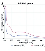
To optimize the synthesis conditions, UV-Vis absorption spectra of nanoparticles produced using silver nitrate concentrations of 0.5 and 1 mM were recorded. Both spectra showed a characteristic surface plasmon resonance (SPR) absorption peak in the visible range, confirming successful Ag-NP biosynthesis. The SPR peak for 1 mM was observed at 400 nm with significantly higher absorption intensity, whereas 0.5 mM showed a peak at 460 nm. The blue shift and increased intensity at 1 mM indicated a higher nanoparticle yield. Therefore, nanoparticles synthesized at 1 mM were selected for further characterization and in vivo experiments (Figure 1).
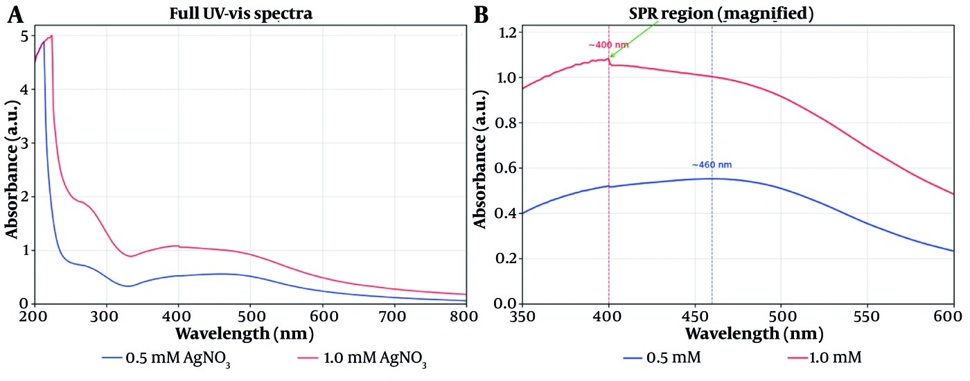
Figure 1.
UV-Vis absorption spectra of silver nanoparticles synthesized with Lavandula stricta aqueous extract at two concentrations of silver nitrate (0.5 and 1 mM); A, Full UV-Vis absorption spectra over 200 - 800 nm; B, Magnified view of the surface plasmon resonance region (350 - 600 nm), with SPR peaks at approximately 460 nm and 400 nm for the 0.5 mM and 1 mM concentrations, respectively. Increasing precursor concentration produced a blue shift and increased absorption intensity, indicating a higher yield of nanoparticle formation.
The XRD diffractogram showed four prominent peaks at 2θ values of 38.1°, 44.3°, 64.5°, and 77°, corresponding to the 111, 200, 220, and 311 crystallographic planes of the face-centered cubic silver structure, consistent with the JCPDS reference card No. 04 - 0783. The average crystallite size, calculated using the Scherrer equation, ranged from 13.8 to 18.7 nm (Figure 2).
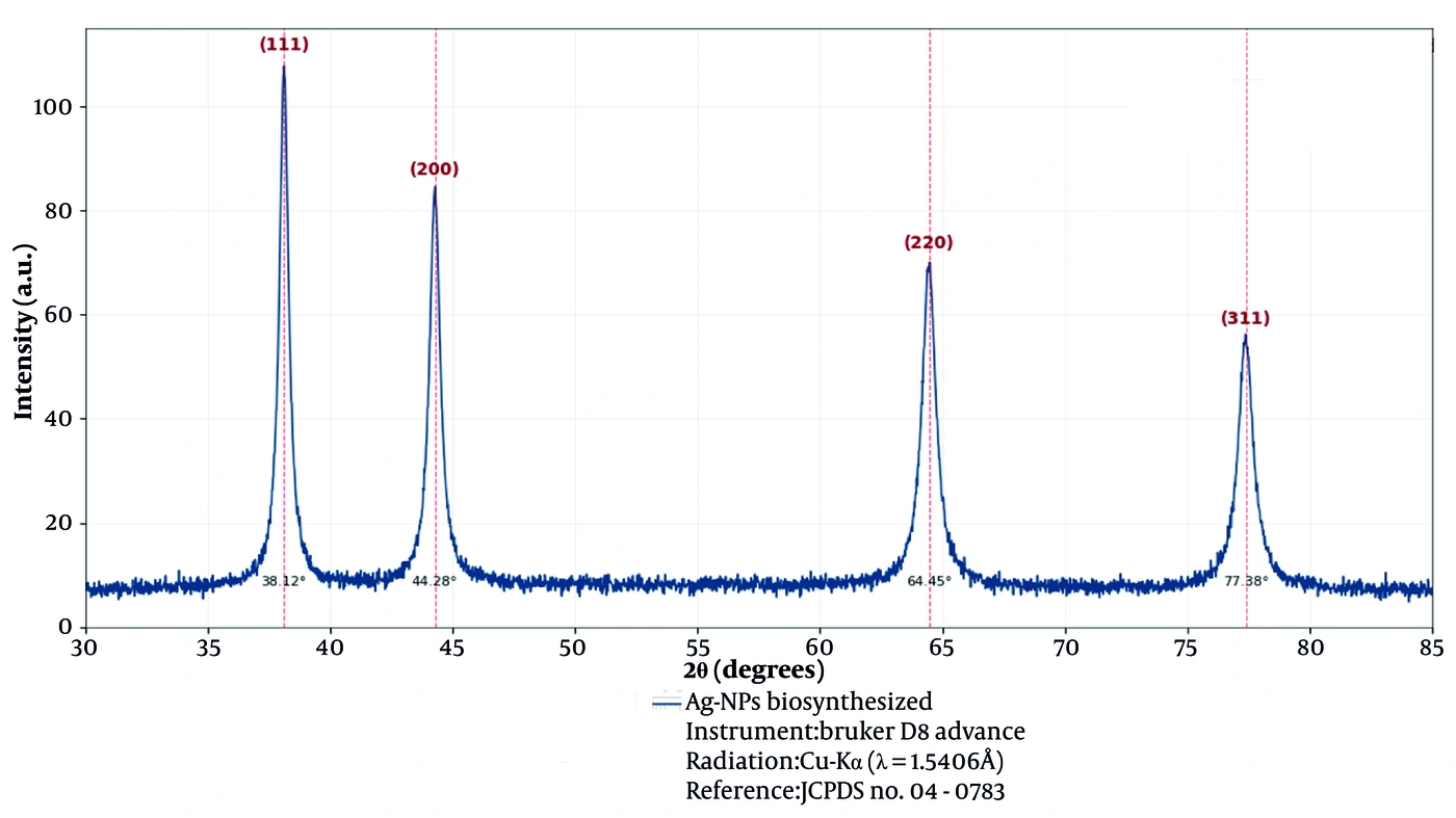
Figure 2.
X-ray diffraction pattern of silver nanoparticles synthesized with Lavandula stricta aqueous extract. Four main peaks were observed at 2θ angles of 38.12°, 44.28°, 64.45°, and 77.38°, corresponding to the 111, 200, 220, and 311 crystal planes of the face-centered cubic structure of silver, respectively, consistent with JCPDS standard card No. 04 - 0783. The average crystallite size calculated using the Scherrer equation was 13.8 - 18.7 nm.
TEM images revealed spherical nanoparticles with a mean particle size of 24 ± 0.71 nm (Figure 3). DLS analysis showed a hydrodynamic size of approximately 135 - 147 nm, with an acceptable PDI and a negative zeta potential of approximately -12 mV. Although this zeta potential is relatively low for stabilization based solely on electrostatic repulsion, the observed colloidal stability is primarily attributable to steric hindrance provided by the phytochemical capping layer surrounding the nanoparticles, as further supported by maintenance of the SPR peak at 400 nm in the UV-Vis spectra. The discrepancy between the TEM and DLS sizes is attributable to the bioactive compound layer surrounding the nanoparticles (Table 1).
Table 1.
Summary of Physicochemical Characterization of Silver Nanoparticles Synthesized with Lavandula stricta Aqueous Extract
| Parameter (Analytical Method) | Result |
|---|---|
| Surface plasmon resonance peak (UV-Vis spectroscopy) | ~400 nm |
| Crystal structure (X-ray diffraction) | Face-centered cubic (planes 111, 200, 220, 311), consistent with JCPDS 04 - 0783 |
| Crystallite size (X-ray diffraction) | 13.8 - 18.7 nm |
| Morphology (transmission electron microscopy) | Spherical |
| Particle size (transmission electron microscopy) | 24 ± 0.71 nm |
| Hydrodynamic size (dynamic light scattering) | Z-Average: 146.2 ± 1.4 nm |
| Zeta potential (laser Doppler velocimetry) | -12.4 ± 0.8 mV |
| Polydispersity index (dynamic light scattering) | 0.30 ± 0.03 |
| Surface functional groups (Fourier transform infrared spectroscopy) | ~3400 cm-1 (O-H), ~1650 cm-1 (C=O), ~550 cm-1 (Ag-O) |
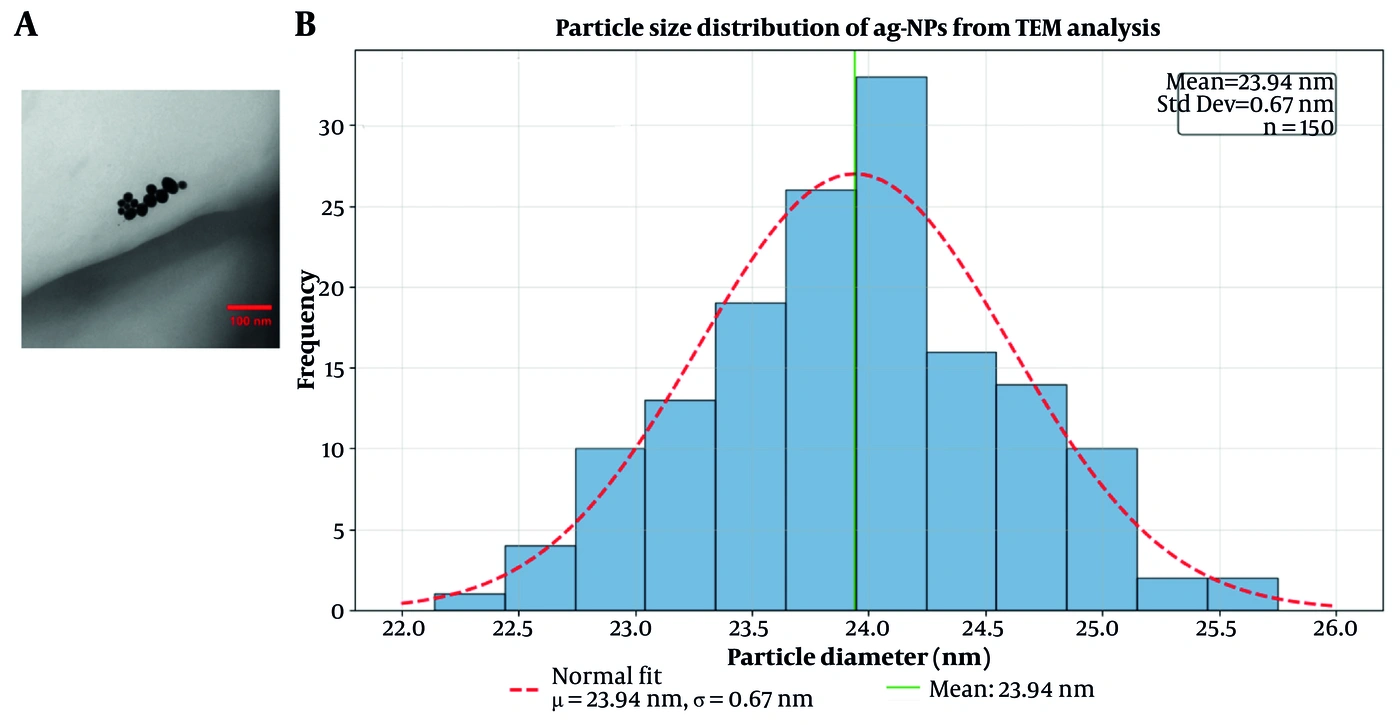
Figure 3.
A, Transmission electron microscopy image of silver nanoparticles synthesized with Lavandula stricta aqueous extract (scale bar, 100 nm); B, Size distribution histogram of nanoparticles based on TEM analysis.
JCPDS: Joint Committee on Powder Diffraction Standards; Z-Average: mean hydrodynamic diameter.
The functional groups involved in Ag+ reduction and nanoparticle stabilization were evaluated by FTIR spectroscopy. As shown in Figure 4 and Table 2, characteristic hydroxyl, carbonyl, and polysaccharide peaks in the extract spectrum showed slight shifts and reduced intensities in the Ag-NP spectrum, suggesting that these phytochemicals participated in Ag+ reduction and surface capping. In addition, a distinct peak at 544 cm-1 was assigned to Ag-O coordination bond vibrations, consistent with previous reports for green-synthesized Ag-NPs (16, 17). FTIR confirms the presence of surface-bound phytochemicals but does not, by itself, prove successful Ag-NP biosynthesis, which was verified by UV-Vis, XRD, and TEM analyses.
Table 2.
Comparison of FTIR Characteristic Peak Positions Between Lavandula stricta Aqueous Extract and the Synthesized Silver Nanoparticles
| Functional Group | Extract (cm-1) | Ag-NPs (cm-1) | Observed Change |
|---|---|---|---|
| O-H stretching | 3452 | 3448 | Slight shift (-4 cm-1), decreased intensity |
| C-H stretching | 2981 | 2980 | No significant shift, decreased intensity |
| C=O stretching | 1684/1654 | 1691 | Shift (+7 to +37 cm-1), decreased intensity |
| C-O stretching | 1044 | 1086 | Shift (+42 cm-1), decreased intensity |
| Ag-O coordination bonds | - | 544 | New peak |
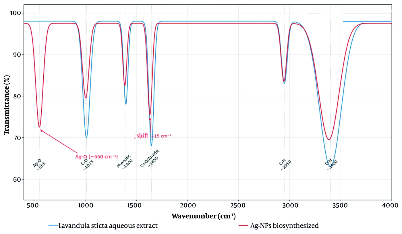
Figure 4.
FTIR spectra of Lavandula stricta aqueous extract (blue) and silver nanoparticles synthesized using this extract (red). Peaks in the extract spectrum include O-H stretching vibrations (2950 cm-1) and C=O (1015 cm-1). In the nanoparticle spectrum, partial shifts and decreased intensity of these peaks, together with a new peak at approximately 550 cm-1 attributed to Ag-O vibration, support the role of bioactive compounds in nanoparticle reduction and surface stabilization.
4.2. Phenolic and Flavonoid Content of the Extract and Ag-NPs
TPC and TFC were measured in the lavender aqueous extract and the synthesized silver nanoparticles (Table 3). The crude lavender extract contained substantial amounts of phenolic and flavonoid compounds; however, these values were markedly reduced in the synthesized silver nanoparticles. The reductions of 64% for TPC and 50% for TFC indicate that a considerable proportion of the phenolic and flavonoid compounds in the extract were consumed during nanoparticle synthesis, serving as reducing agents for conversion of Ag+ ions to Ag0 and as capping agents on the nanoparticle surface.
Table 3.
Total Phenolic and Total Flavonoid Content in Lavender Extract and Synthesized Silver Nanoparticles (N = 3) a
| Samples | TPC (mg GAE/g DW) | TFC (mg RE/g DW) |
|---|---|---|
| Lavender extract | 0.199 ± 0.014 A | 1.046 ± 0.08 A |
| Silver nanoparticles | 0.071 ± 0.006 B | 0.527 ± 0.04 B |
a Values are reported as mean ± SEM. Abbreviations: TPC, total phenolic content; TFC, total flavonoid content; GAE, gallic acid equivalent; RE, rutin equivalent; DW, dry weight. Different capital letters in each column indicate statistically significant differences (P < 0.05) according to Tukey's test.
4.3. In Vitro Radical-Scavenging and Reducing Capacity
The antioxidant activity of the lavender aqueous extract and biosynthesized Ag-NPs was assessed using DPPH radical-scavenging and FRAP reducing-power assays (Table 4). In the DPPH assay, ascorbic acid, used as the standard, showed the strongest radical-scavenging activity, with an IC50 value of 5.8 ± 0.3 µg/mL. The crude extract showed greater activity (IC50 = 120 ± 5.3 µg/mL) than the silver nanoparticles (IC50 = 235 ± 12 µg/mL; P < 0.01). The FRAP assay also showed a higher value for the crude extract (0.432 ± 0.132 mg FeSO4/mg DW) than for the silver nanoparticles (0.277 ± 0.08 mg FeSO4/mg DW); however, this difference was not statistically significant (P = 0.17), possibly because of the relatively small sample size (n = 3). The lower antioxidant activity of the nanoparticles compared with the crude extract is likely attributable to the consumption of some phenolic and flavonoid compounds during silver-ion reduction and particle surface coating. Nevertheless, the nanoparticles retained considerable free-radical-scavenging and iron-reducing capacity, possibly because of residual surface phytochemicals.
Table 4.
Comparison of in Vitro Antioxidant Activity of Lavender Aqueous Extract and Synthesized Silver Nanoparticles (N = 3) a
| Samples | DPPH (IC50, µg/mL) | FRAP (mg FeSO4/g DW) |
|---|---|---|
| Ascorbic acid (standard) | 5.8 ± 0.3 | ND |
| Lavandula stricta aqueous extract | 120 ± 5.3 | 0.432 ± 0.132 |
| Biosynthesized silver nanoparticles | 235 ± 12 | 0.277 ± 0.08 |
a Abbreviations: IC50, half-maximal inhibitory concentration; DW, dry weight; FRAP, ferric reducing antioxidant power; ND, not determined (ascorbic acid was used only as a reference standard for the DPPH assay and was not evaluated in the FRAP assay).
4.4. In Vivo Anti-Inflammatory Effects
Carrageenan administration via subplantar injection resulted in a marked increase in paw thickness in the positive control group compared with the healthy control group (P < 0.0001). Oral pretreatment with indomethacin and the synthesized silver nanoparticles significantly prevented edema formation. As shown in Table 5, edema inhibition was 32.26% in the group receiving silver nanoparticles at 0.6 mg/kg and 31.44% in the group receiving indomethacin at 5 mg/kg. The anti-inflammatory effect of the higher Ag-NP dose (0.6 mg/kg) was comparable to that of indomethacin (P > 0.05), whereas the crude lavender extract showed a milder inhibitory effect of approximately 16% - 17%. The lower Ag-NP dose (0.3 mg/kg) did not significantly inhibit inflammation. These findings indicate the anti-inflammatory potential of Ag-NPs synthesized using lavender aqueous extract, particularly at 0.6 mg/kg.
Table 5.
Effect of Different Treatments on Paw Edema Thickness and Percentage of Inflammation Inhibition in the Carrageenan-Induced Edema Model (N = 6) a
| Treatment Groups | Paw Edema Thickness (mm) | Inflammation Inhibition (%) |
|---|---|---|
| Healthy control | 0.45 ± 0.02 D | - |
| Carrageenan | 1.85 ± 0.22 A | 0.0 |
| Indomethacin (5 mg/kg; standard comparator) | 1.27 ± 0.18 B | 31.44 ± 5.4 B |
| Lavender extract (15 mg/kg) | 1.55 ± 0.15 C | 16.2 ± 4.6 C |
| Lavender extract (30 mg/kg) | 1.53 ± 0.16 C | 17.1 ± 4.7 C |
| Ag-NPs (0.3 mg/kg) | 1.81 ± 0.19 A | 2.08 ± 0.6 A |
| Ag-NPs (0.6 mg/kg) | 1.25 ± 0.14 B | 32.2 ± 5.8 B |
a Values are reported as mean ± SEM. Different letters in each column indicate statistically significant differences (P < 0.05) according to Tukey's test.
4.5. Pro-Inflammatory Cytokine Levels
Carrageenan challenge markedly increased concentrations of the pro-inflammatory cytokines TNF-α and IL-6 in paw tissue. Pretreatment with the synthesized silver nanoparticles significantly suppressed these cytokines (Table 6). Ag-NPs at 0.6 mg/kg reduced TNF-α levels from 134.17 to 20.71 pg/mg tissue, corresponding to 84.6% inhibition, and IL-6 levels from 21.24 to 9.98 pg/mg protein, corresponding to 53% inhibition. Both values were comparable to those in the healthy control group and did not differ significantly (P > 0.05). The crude lavender extract also significantly reduced cytokine levels, although its effect was less pronounced than that of the synthesized nanoparticles.
Table 6.
Effect of Different Treatments on Pro-Inflammatory Cytokine Levels in Paw Tissue (N = 6) a
| Treatment Groups | TNF-α (pg/mg Tissue) | IL-6 (pg/mg Protein) |
|---|---|---|
| Healthy control | 19.49 ± 10.05 A | 7.33 ± 0.65 A |
| Carrageenan | 134.17 ± 2.31 D | 21.24 ± 1.03 D |
| Indomethacin (5 mg/kg) | 18.68 ± 6.16 A | 8.94 ± 0.58 A |
| Lavender extract (15 mg/kg) | 48.89 ± 10.21 B | 15.29 ± 1.59 C |
| Lavender extract (30 mg/kg) | 26.44 ± 10.39 C | 15.41 ± 1.43 C |
| Ag-NPs (0.3 mg/kg) | 48.72 ± 1.95 B | 14.18 ± 0.59 B |
| Ag-NPs (0.6 mg/kg) | 20.71 ± 8.64 A | 9.98 ± 1.26 A |
a Values are reported as mean ± SEM (n = 6). Different capital letters within each column indicate statistically significant differences (P < 0.05) according to Tukey's post hoc test.
4.6. Oxidative Stress Markers
To investigate the in vivo antioxidant mechanism, MDA and TAC were measured (Table 7). Carrageenan-induced inflammation significantly increased MDA levels to 0.201 nmol/mg protein and reduced TAC to 0.456 mmol/L compared with the healthy control group (P < 0.001). Pretreatment with the synthesized silver nanoparticles at 0.6 mg/kg significantly reversed this trend, reducing MDA to 0.075 nmol/mg protein, a 62.7% reduction compared with the carrageenan group, and increasing TAC to 1.13 mmol/L, a 147.5% increase. These values did not differ significantly from those of the healthy control group (P > 0.05).
Table 7.
Effect of Different Treatments on Lipid Peroxidation and Total Antioxidant Capacity (N = 6) a
| Treatment Group | MDA (nmol/mg Protein) | TAC (mmol/L) |
|---|---|---|
| Healthy control | 0.08 ± 0.01 A | 1.16 ± 0.05 A |
| Carrageenan | 0.201 ± 0.02 D | 0.456 ± 0.04 D |
| Indomethacin (5 mg/kg) | 0.09 ± 0.01 A | 0.98 ± 0.06 B |
| Lavender extract (15 mg/kg) | 0.15 ± 0.02 B | 0.65 ± 0.05 C |
| Lavender extract (30 mg/kg) | 0.12 ± 0.01 A | 0.82 ± 0.06 B |
| Ag-NPs (0.3 mg/kg) | 0.14 ± 0.02 B | 0.85 ± 0.07 B |
| Ag-NPs (0.6 mg/kg) | 0.075 ± 0.01 A | 1.13 ± 0.06 A |
a Values are reported as mean ± SEM (n = 6). Different capital letters within each column indicate statistically significant differences (P < 0.05) according to Tukey's post hoc test.
5. Discussion
The present study biosynthesized Ag-NPs using L. stricta aqueous extract and evaluated their in vivo anti-inflammatory and antioxidant activities. The nanoparticles exhibited favorable physicochemical characteristics and moderate colloidal stability and significantly inhibited carrageenan-induced edema. They also exerted anti-inflammatory effects by reducing TNF-α and IL-6 and modulating the oxidative stress markers MDA and TAC. The anti-inflammatory effect of Ag-NPs at 0.6 mg/kg was comparable to that of indomethacin (P > 0.05), whereas the crude plant extract alone showed a weaker effect. These findings suggest that the green synthesis process preserved the therapeutic properties of the extract and may have enhanced biological activity through combined contributions from the silver nanoparticle core and surface-associated phytochemicals (6, 7).
Successful Ag-NP biosynthesis was confirmed by the progressive color change of the reaction solution and characteristic SPR absorption bands at approximately 400 nm for the 1 mM concentration and 460 nm for the 0.5 mM concentration. The XRD pattern revealed a face-centered cubic crystal structure with a crystallite size of 13.8 - 18.7 nm, whereas TEM showed a mean particle size of 24 ± 0.71 nm, comparable to 14.5 nm Ag-NPs synthesized using Grewia tiliaefolia leaf extract (16) and 50 - 60 nm propolis-mediated Ag-NPs (17). The crystallite size reflects the dimensions of coherently diffracting crystalline domains, whereas TEM particle size represents the physical diameter of individual particles. The larger TEM particle size suggests that the nanoparticles may be polycrystalline; however, polycrystallinity cannot be definitively established from these measurements alone because crystallite size calculated from a single diffraction peak may also be influenced by lattice strain. DLS indicated a larger hydrodynamic size of approximately 147 nm. This discrepancy is primarily attributable to the hydrodynamic diameter measured by DLS, which includes the silver core, the surface-associated phytochemical capping layer, and the hydration shell. The PDI was 0.30 ± 0.03, indicating a moderately narrow particle size distribution and suggesting that the larger hydrodynamic size does not reflect massive macroscopic aggregation. DLS is also inherently more sensitive to larger particles, and the moderate zeta potential of -12.4 mV suggests that some loose agglomeration in suspension may contribute to the apparent size. The maintained SPR peak at 400 nm over 24 hours indicates no substantial aggregation during the observation period.
In the in vitro antioxidant assessment, the crude lavender extract showed stronger inhibitory activity than the silver nanoparticles in both DPPH and FRAP assays. This difference likely reflects the consumption of some electron-donating phenolic and flavonoid compounds during the reduction of silver ions (Ag+) to metallic silver (Ag0) and the oxidation of these compounds on the particle surface (4). The reductions in the phenolic and flavonoid contents of the nanoparticles compared with the crude extract, 64% and 50%, respectively, support their participation in silver-ion reduction and particle capping. Nevertheless, the synthesized nanoparticles retained considerable antioxidant activity, likely because of the remaining surface phytochemicals. The relative contribution of the metallic silver core to this activity was not established and warrants further investigation (18, 19).
In the in vivo acute inflammation model, oral pretreatment with silver nanoparticles resulted in more than 30% inhibition of carrageenan-induced paw edema, comparable to indomethacin. These findings are consistent with those of Islam et al. (17), who reported anti-inflammatory activity of propolis-mediated Ag-NPs in a burn wound model. Carrageenan elicits a biphasic inflammatory response, with an early phase at 1 - 2 hours involving histamine and serotonin and a late phase at 3 - 5 hours associated with prostaglandins, bradykinin, and pro-inflammatory cytokines. The significant inhibition of edema at the fourth hour is consistent with an effect on mediators typically associated with the late phase (13).
The higher dose of silver nanoparticles significantly reduced TNF-α and IL-6 concentrations in inflamed tissue. The observed suppression of IL-6 is consistent with the findings of Alfahed et al. (18), who reported marked downregulation of IL-6 expression in diabetic rats treated with resveratrol. TNF-α and IL-6 amplify inflammatory responses and can promote reactive oxygen species generation through the activation of intracellular signaling cascades (8). Their concurrent reduction suggests suppression of key pro-inflammatory mediators. Previous reports suggest that this suppression may involve inflammation-related transcription pathways such as NF-κB; however, this mechanism was not directly examined in the current study and requires further validation (7).
The findings also showed concurrent alleviation of inflammation and oxidative stress. Carrageenan-induced inflammation increased MDA and reduced TAC. Pretreatment with silver nanoparticles reversed these changes, lowering MDA and restoring TAC to levels observed in healthy controls. The observed reduction in MDA is consistent with the findings of Bahadoran et al. (6), who reported reduced hippocampal MDA levels after bavachinin treatment in a seizure model. This dual effect, involving suppression of oxidative stress and augmentation of antioxidant capacity, may partly reflect hydroxyl and carbonyl functional groups on the nanoparticle surface that can contribute to free-radical scavenging and protection of cell membranes from oxidative damage (19, 20).
An apparent discrepancy was observed between the in vitro and in vivo results. Although the crude extract showed stronger in vitro antioxidant activity, Ag-NPs at 0.6 mg/kg produced greater in vivo anti-inflammatory effects, with 32.2% edema inhibition compared with 17.1% for the crude extract at 30 mg/kg. This difference suggests that enhanced in vivo efficacy is not solely attributable to the antioxidant properties of plant compounds and may involve factors such as improved bioavailability and cellular uptake associated with nanoscale dimensions (21, 22). The formation of a protein corona, or persistence of the existing phytochemical corona, may also alter nanoparticle biodistribution, cellular interactions, and pharmacokinetics in biological environments, potentially enhancing therapeutic efficacy despite lower intrinsic antioxidant activity in vitro (23).
The study has several strengths. It provides a comprehensive in vivo evaluation of Ag-NPs biosynthesized using L. stricta extract, includes both a crude extract-only group and a standard drug comparator, and assesses multiple inflammatory and oxidative stress markers in the target tissue.
5.1. Limitations and Future Directions
A dose of 0.6 mg/kg of synthesized silver nanoparticles appeared effective and produced no overt signs of acute toxicity during the observation period. Nevertheless, further studies are required to assess acute and chronic toxicity in vital organs, including the liver and kidneys, before clinical safety can be established. Future research should evaluate molecular signaling pathways such as the mitogen-activated protein kinase and NF-κB pathways and characterize nanoparticle pharmacokinetics. Elemental analyses such as EDX, XPS, elemental mapping, ICP-OES, ICP-MS, or AAS were not available. Consequently, the precise mass of elemental silver within the purified nanoformulation and the overall nanoparticle yield were not determined. The administered doses of 0.3 and 0.6 mg/kg therefore represent the total mass of the purified dried preparation, including the silver core and surface-associated phytochemical capping layer, rather than elemental silver alone. Future studies should quantify elemental composition, silver content, nanoparticle yield, silver release, and the fate of the phytochemical capping layer after administration.
5.2. Translational Implications
The comparable efficacy of Ag-NPs and indomethacin at a much lower administered mass, together with a natural capping layer that may reduce adverse effects associated with conventional nonsteroidal anti-inflammatory drugs, warrants further preclinical investigation. However, comprehensive toxicological, pharmacokinetic, and chronic disease model studies are essential before clinical translation can be considered.
5.3. Conclusions
Silver nanoparticles synthesized using Lavandula stricta aqueous extract possess favorable physicochemical characteristics and exhibit antioxidant and anti-inflammatory activities in a murine model of acute inflammation. These effects were associated with suppression of TNF-α and IL-6 and reduced lipid peroxidation. Because the carrageenan-induced paw edema model represents an acute inflammatory condition, the findings support further preclinical investigation of these biosynthesized nanoparticles for acute inflammatory responses. Comprehensive long-term toxicological evaluation and studies in chronic disease models are required before broader therapeutic potential can be established.