Hidden Unicameral Bone Cyst of the Calcaneus: A Case Report of Military Soldier with Chronic Ankle Pain

Authors

Zaheer Ahmed Gill1, Saeed Bin Ayaz2,*, Nadeem Ahmad2, Ali Raza Qureshi1
1Department of Rehabilitation Medicine, Combined Military Hospital Malir, Karachi 75080, Sindh, Pakistan
2Department of Rehabilitation Medicine, Combined Military Hospital Quetta and Quetta institute of Medical Sciences, Quetta 87300, Baluchistan, Pakistan
*Corresponding Author: Corresponding author: Dr Saeed Bin Ayaz, Department of Rehabilitation Medicine, Combined Military Hospital Quetta and Quetta institute of Medical Sciences, Quetta 87300, Baluchistan, Pakistan. Tel: +92-3335245770, E-mail: Email: [email protected]

Journal of Archives in Military Medicine:Vol. 5, issue 2; e57339
Published online:Jun 14, 2017
Article type:Case Report
Received:Feb 21, 2017
Accepted:Apr 07, 2017
How to Cite:Gill ZA, Bin Ayaz S, Ahmad N, Qureshi AR. Hidden Unicameral Bone Cyst of the Calcaneus: A Case Report of Military Soldier with Chronic Ankle Pain. J Arch Mil Med. 2017;5(2):e57339. doi: https://doi.org/10.5812/jamm.57339

Abstract

Bone cysts are benign bone lesions that are quite uncommon in the calcaneus. Calcaneal cysts generally remain asymptomatic in most patients and are discovered incidentally but in some, they may cause localized bone pain that is progressive in intensity. If not picked on the conventional radiographs, the diagnosis is delayed and the patient remains distressed and dispirited. We, here, report a military soldier with calcaneal cyst who remained misdiagnosed for six months and had been treated as a case of ankle sprain. After correct diagnosis, his problems were finally settled following surgical intervention. The physicians should keep a high degree of suspicion of an alternative diagnosis in all cases where usual management is not beneficial in commonly expected pathologies.

Highlights

1. Introduction

Chronic unremitting pain in the lower limb is sometimes a diagnostic dilemma for the physicians. The primary rare etiology is easily overlooked in the initial evaluation. There have been case reports of people with chronic lower limb (1) and foot pain (2) who were diagnosed late because the initial evaluation did not include the rare causes they suffered from. We, here, report a military soldier with ankle pain who actually had calcaneal cyst and was treated wrongly as a case of ankle sprain for six months.
Calcaneal cysts are benign or unicameral bone cysts (UBC) seen radiographically as single thin-walled lytic bone lesions without surrounding periosteal reaction. UBC are common in humerus, femur, and tibia but quite uncommon in the calcaneus (3). Calcaneal cysts generally remain asymptomatic in most patients and are discovered incidentally but in some, they may cause localized bone pain that is progressive in intensity. If not picked on the conventional radiographs, the diagnosis is delayed and the patient remains distressed and dispirited especially if he or she is a walking worker. This report highlights the importance of a detailed evaluation in a case of chronic unrelieved ankle pain and the importance of advanced imaging techniques if conventional imaging fails to pick the etiology.

2. Case Presentation

A 22-year-old otherwise healthy male soldier was referred to an orthopedic surgeon for the evaluation of chronic unremitting pain in right ankle that had been troubling him for the past six months. The patient graded the pain as 6/10 on Numeric Rating Scale. The pain aggravated on walking and running and was relieved on taking rest. There was no history of trauma or any joint disorder in the past. His family history was negative for any inflammatory joint disorder. The orthopedic surgeon ordered radiographs of the affected ankle and foot that were read as negative for any fracture, dislocation, or a space occupying lesion by the radiologist (Figure 1A). His lab investigations including complete blood count, erythrocyte sedimentation rate, C-reactive protein, and serum uric acid levels were all within normal limits. He was labelled as having ankle sprain and was suspected a malingerer. Thereafter, he was referred to our department for physiotherapy.
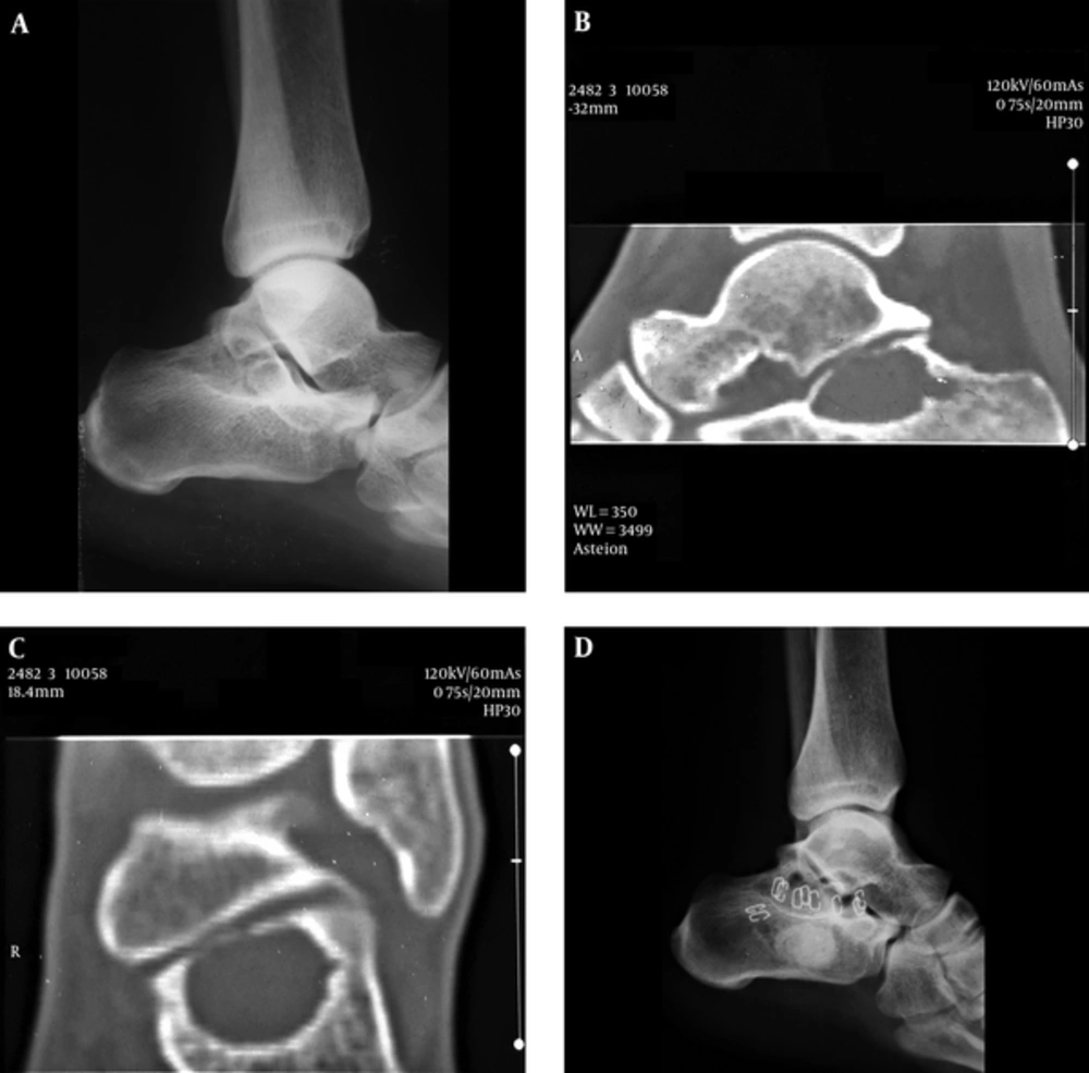
A, Normal Appearing X-Ray of the Left Ankle Joint; B and C, Computerized Tomography Scan of the Left Ankle Showing Single Lytic Bone Lesion in the Calcaneus Without Surrounding Periosteal Reaction; D, X-Ray of the Left Ankle Joint Following Bone Cement Filling
Figure 1.
A, Normal Appearing X-Ray of the Left Ankle Joint; B and C, Computerized Tomography Scan of the Left Ankle Showing Single Lytic Bone Lesion in the Calcaneus Without Surrounding Periosteal Reaction; D, X-Ray of the Left Ankle Joint Following Bone Cement Filling
In our department, we examined him again. He had no swelling but tenderness over the calcaneus with reduced ankle inversion to 20° and eversion to 5°. Ankle plantarflexion and dorsiflexion were normal and there was no neurological deficit.
Keeping high sympathy for the patient, a computerized tomography scan of the ankle was requested that demonstrated a calcaneal cyst (Figure 1B and C). The patient was then referred back to the orthopedic surgeon for surgical intervention.
The orthopedic surgeon did surgical curettage followed by bone cement filling (Figure 1D). The range of motion exercises were continued and at 10 months’ post-surgical follow up, he got rid of pain, however, the ankle eversion was minimally restricted (10°). Written informed consent was taken from the patient for the publication of this case report.

3. Discussion

Several theories have been postulated to explain the origin of UBC, including trauma and inflammation, however, none has been conclusive (4). Calcaneal cysts are usually asymptomatic, but may present as progressively mounting bony pain with increased local skin temperature (5). A bony swelling may be palpable and movement in the adjacent joint may be affected (5). Active treatment of calcaneal cysts is recommended because the outcome is more favorable than the conservative management. There are chances of calcaneal fracture and collapse (6). Among active options, the healing rate was found to be similar with steroids or bone marrow injection but a higher rate of healing was reported when demineralized bone matrix was added (7). The surgical options are flexible intramedullary nails without curettage (7) and curettage followed by utilization of allograft, autograft, or bone cement producing healing rate approaching 90% (7, 8). Healing rate is also high with the utilization of intramedullary nails (7). The emerging treatment methods that are used as adjuvants or alternative therapies with varying success rates are Argon beam coagulation, sclerotherapy, radiotherapy, arterial embolization, and medical management with bisphosphonates or denosumab (9, 10). The alternative therapies are primarily reserved for inoperable lesions or lesion at anatomic locations where surgery may cause significant morbidity (9).
In conclusion, this report endorses the long-heard aphorism “the patient is mostly right”. Thinking out of the box and raising a high suspicion of an alternative diagnosis is recommended in all cases where usual management is not beneficial in commonly expected pathologies.
Acknowledgments

Footnotes

  • Authors’ Contribution:Zaheer Ahmed Gill conceived the report and recorded the important findings. Saeed Bin Ayaz did literature search and drafted the manuscript. Nadeem Ahmad drafted and reviewed the manuscript. Ali Raza Qureshi recorded the important findings

  • Financial Disclosure:Nil.

  • Ethical Approval:Written consent was taken from the patient for this case report.

  • Funding/Support:Nil.

References

Copyright

Copyright © 2017, Journal of Archives in Military Medicine. This is an open-access article distributed under the terms of the Creative Commons Attribution-NonCommercial 4.0 International License (http://creativecommons.org/licenses/by-nc/4.0/) which permits copy and redistribute the material just in noncommercial usages, provided the original work is properly cited.

Similar Articles

20
Oct
2014

Chondroblastoma of the Navicular Bone

XiaoNa Li,
ZhiGang Peng,
JingPin Zhao,
ZeKun Zhang

Li X, Peng Z, Zhao J, Zhang Z. Chondroblastoma of the Navicular Bone. I J Radiol. 2014;11(4):e10848. doi: https://doi.org/10.5812/iranjradiol.10848

19
Jun
2016
Simple Bone Cyst with Mesenchymal Proliferation Involving Epiphysis of the Proximal Fibula: A Case Report

Simple Bone Cyst with Mesenchymal Proliferation Involving Epiphysis of the Proximal Fibula: A Case Report

Hye Young Choi,
So Young Park,
Wook Jin,
Ji Seon Park,
Kyung Nam Ryu,
Yong Koo Park

Choi HY, Park SY, Jin W, Park JS, Ryu KN, et al. Simple Bone Cyst with Mesenchymal Proliferation Involving Epiphysis of the Proximal Fibula: A Case Report. I J Radiol. 2017;14(4):e34183. doi: https://doi.org/10.5812/iranjradiol.34183

30
Mar
2011

Simple Bone Cyst of the Mandible: Report of Two Cases

M. Imanimoghaddam,
Adineh Javadian Langaroodi,
S. Nemati,
S. Ataei Azimi

Imanimoghaddam M, Javadian Langaroodi A, Nemati S, Ataei Azimi S. Simple Bone Cyst of the Mandible: Report of Two Cases. I J Radiol. 2011;8(1):e78757. doi:

31
Mar
2026
Anterior Mandibular Mural Unicystic Ameloblastoma Crossing the Midline: A Rare Case with Histopathological Diagnostic Challenges

Anterior Mandibular Mural Unicystic Ameloblastoma Crossing the Midline: A Rare Case with Histopathological Diagnostic Challenges

Fatemeh Salehi,
Seyed Amirhossein Rahimizadeh Hosseini,
Sahar Seifollahi

Salehi F, Rahimizadeh Hosseini SA, Seifollahi S. Anterior Mandibular Mural Unicystic Ameloblastoma Crossing the Midline: A Rare Case with Histopathological Diagnostic Challenges. J Kermanshah Univ Med Sci. 2026;30(1):e171344. doi: https://doi.org/10.5812/jkums-171344

30
Apr
2017

A Simple Approach for Diagnosis of Chronic Tuberculous Osteomyelitis: A Case Report

Mohammadreza Salehi,
Seyed Ali Dehghan Manshadi,
Zeynab Yassin,
Malihe Hassannejad

Salehi M, Dehghan Manshadi SA, Yassin Z, Hassannejad M. A Simple Approach for Diagnosis of Chronic Tuberculous Osteomyelitis: A Case Report. Arch Clin Infect Dis. 2017;12(2):e57281. doi: https://doi.org/10.5812/archcid.57281

More by these authors

Zaheer Ahmed GillPubMedScholar
Saeed Bin AyazPubMedScholar
Nadeem AhmadPubMedScholar
Ali Raza QureshiPubMedScholar
Share
Cited by
Metrics