
1. Introduction
Ameloblastoma is among the most common odontogenic tumors (1). The World Health Organization (WHO) defines it as a locally invasive, polymorphic neoplasm characterized by follicular or plexiform patterns within a fibrous stroma (2, 3). Unicystic ameloblastoma, a variant of ameloblastoma, exhibits relatively less aggressive biological behavior (4). Approximately 80% of cases occur exclusively in the mandibular third molar region, often in association with an unerupted tooth, and are most frequently observed in young patients during the second decade of life (8-13). These tumors typically demonstrate slow growth but can exhibit substantial local invasion (5). This case highlights the essential role of comprehensive histopathological assessment in the evaluation of all jaw lesions, even when a lesion appears clinically benign.
2. Case Presentation
A 42-year-old woman was referred to the Department of Oral and Maxillofacial Surgery at Isfahan University of Medical Sciences with a chief complaint of painless swelling in the anterior mandible that had developed over the preceding two months. The swelling was described as entirely painless, with no associated pus discharge or paresthesia. Her medical history was significant for asthma for the previous five years, which was well controlled with medication.
Clinical examination revealed a solitary, hard, painless swelling measuring approximately 3 × 5 cm in the anterior mandible. Palpation indicated a bony consistency, and the mass was fixed to the underlying structures (Figure 1). Intraoral assessment confirmed the swelling, without evidence of pus drainage, inflammation, or regional lymphadenopathy. The overlying mucosa was intact, and the patient denied numbness of the lower lip or adjacent teeth. Aspiration yielded no fluid.
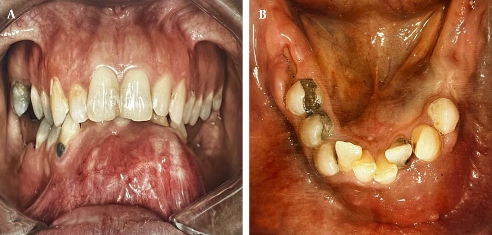
Figure 1.
Intraoral clinical photograph demonstrating swelling in the anterior mandibular
Radiographic evaluation with panoramic radiography revealed a well-defined, corticated lesion with a mixed radiolucent–radiopaque appearance in the anterior mandible, extending from the distal aspect of the right first premolar to the distal aspect of the left second premolar. The lesion caused root displacement of teeth 31, 32, 33, and 43 and root resorption of teeth 31, 33, 34, and 42. Disappearance of the periodontal ligament space was noted around the affected teeth, except for the lower left canine (Figure 2A). Computed tomography (CT) scans disclosed a mixed-density lesion with areas of dystrophic calcification, accompanied by thinning and expansion of the mandibular buccal and lingual cortical plates (Figure 2B). The lesion extended toward the inferior border of the mandible.
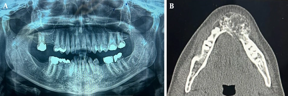
Figure 2.
A, Panoramic radiograph illustrating a well-defined, corticated mixed radiolucent-radiopaque lesion, extending from tooth 44 to tooth 35; B, Axial computed tomography (CT) scan revealing thinning and expansion of the buccal and lingual cortical plates, with internal high-density areas suggestive of dystrophic calcifications.
Based on the clinical and radiographic findings, a provisional diagnosis of a benign odontogenic tumor was made. The differential diagnosis included central ossifying fibroma, desmoplastic ameloblastoma, and fibro-osseous lesions, including fibrous and cemento-osseous dysplasia.
An incisional biopsy was performed, yielding four soft, tan fragments; the largest measured 1.1 × 0.4 × 0.3 cm, and the smallest measured 0.6 × 0.3 × 0.1 cm. Histopathological examination initially diagnosed the lesion as an ameloblastic fibroma.
Subsequently, under general anesthesia, bilateral submandibular incisions were made, with careful dissection to preserve the marginal mandibular branch of the facial nerve. A patient-specific cutting guide derived from CT data was used to facilitate a precise osteotomy. Segmental mandibular resection was performed with a 1.5-cm oncologic safety margin, and the resected specimen was submitted for histopathological analysis. Detailed evaluation by the Department of Oral Pathology at Isfahan University of Medical Sciences revealed islands of odontogenic epithelium within fibrous connective tissue, with peripheral palisading and stellate reticulum-like features. The final diagnosis was mural unicystic ameloblastoma (Figure 3). No clinical or radiographic evidence of recurrence was observed during the short six-month follow-up period. The chronological sequence of the patient’s clinical course, diagnostic procedures, and treatment is summarized in Table 1.
Table 1.
Chronological Timeline of the Patient's Clinical Presentation, Diagnostic Interventions, Surgical Treatment, and Follow-Up
| Date | Clinical Events, Diagnostic Testing, and Interventions |
|---|---|
| May 2025 | The patient noticed gradual, painless swelling in the anterior mandible. |
| July 2025 | The patient was referred to the Oral and Maxillofacial Surgery Department at Isfahan University, and clinical examination was performed. |
| July 2025 | Radiographic evaluation, including panoramic radiography and CT, showed a well-defined mixed radiolucent-radiopaque lesion. |
| August 2025 | Incisional biopsy was performed using 4 pieces, with a maximum size of 1.1 cm. The preliminary diagnosis was ameloblastic fibroma. |
| August 2025 | Segmental mandibular resection with a 1.5-cm safety margin was performed under general anesthesia. |
| September 2025 | Final histopathological examination was performed. The final diagnosis was mural unicystic ameloblastoma. |
| November 2025 | No clinical or radiographic evidence of recurrence was observed during the short six-month follow-up period. |
| Future Plan | Long-term follow-up is planned every 6 months for 2 years and then annually for 5 - 10 years. |
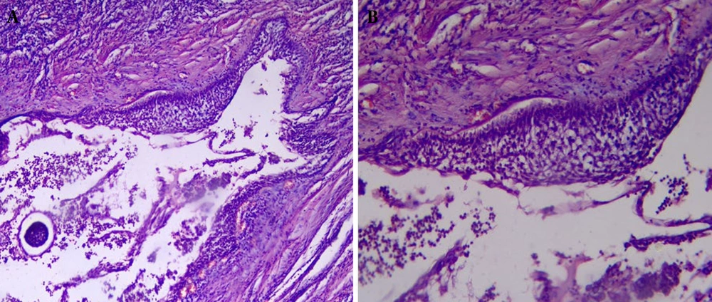
Figure 3.
A, Histopathological photomicrograph showing the cystic lining composed of ameloblastic epithelium with a hyperchromatic, polarized basal layer; B, the suprabasal cells are loosely arranged, resembling the stellate reticulum.
3. Discussion
Ameloblastoma is an odontogenic tumor that is typically benign, characterized by locally aggressive growth and a rare potential for malignant transformation and metastasis (6). It accounts for approximately 1% of all oral tumors and 13% to 78% of odontogenic tumors (7, 8). It can originate from the dental lamina or enamel organ, odontogenic cyst linings (predominantly dentigerous cysts), or basal epithelial cells of the oral epithelium (8). According to the 2022 WHO classification of odontogenic tumors, ameloblastomas are categorized into conventional, unicystic, extraosseous/peripheral, adenoid, and metastasizing subtypes (9). The unicystic variant comprises 5% to 15% of all ameloblastomas, with more than 90% occurring in the mandible. These lesions predominantly occur in the posterior mandible, with approximately 80% associated with an impacted mandibular third molar. Anterior mandibular involvement remains rare and atypical. In the present case, the lesion arose in the anterior mandible, representing an uncommon anatomical site (10).
Unicystic ameloblastomas are typically observed in the second and third decades of life and show a slight male predominance (11). Presentation in the fifth decade is infrequent; however, the present case involved a 42-year-old woman, representing a deviation from typical demographic patterns.
Patients with unicystic ameloblastoma often present with painless swelling, facial asymmetry, impacted or displaced teeth, tooth mobility, root changes such as resorption or divergence, occlusal irregularities, and tooth extrusion (12). The patient in the present report presented with painless intraoral swelling, which is consistent with these manifestations.
Radiographically, unicystic ameloblastomas typically appear as well-defined, unilocular low-density lesions, are infrequently multilocular, and are most often located in the posterior mandible near third molars (13). In contrast, the lesion in this case had an atypical anterior mandibular location, with mixed radiolucent-radiopaque features, root resorption, and cortical expansion, contributing to diagnostic complexity.
Ackermann's classification divides unicystic ameloblastomas into luminal (Group 1), intraluminal/plexiform (Group 2), and mural (Group 3) subtypes. Groups 1 and 2 may be managed conservatively through enucleation, whereas Group 3 requires aggressive intervention because of its higher recurrence risk. Histopathological examination is pivotal for accurate subtyping and treatment guidance. Management strategies range from conservative approaches, such as marsupialization, enucleation, and curettage with or without adjunctive therapies, to radical methods such as marginal or segmental resection (14). Treatment selection depends on lesion size, location, patient age, surgical expertise, and patient preferences (13). Given the diagnosis of the mural subtype in this case, segmental mandibular resection with a 1.5-cm safety margin was performed; this radical approach is associated with reduced recurrence rates (15, 16).
In this case, the anterior location and mixed radiographic pattern with root resorption initially suggested desmoplastic ameloblastoma, whereas the incisional biopsy indicated ameloblastic fibroma. Only after complete resection and thorough histopathological review was mural unicystic ameloblastoma confirmed. This finding underscores that definitive diagnosis requires comprehensive histological analysis of the entire lesion, because preoperative clinical and radiographic assessments alone are insufficient. Moreover, the epithelial lining in unicystic ameloblastomas may vary and may occasionally mimic dentigerous cyst epithelium in nonspecific areas. The true nature of the lesion often becomes evident after enucleation, when serial sectioning of the entire resected specimen is performed during histopathological evaluation to identify mural invasion and confirm the final diagnosis (17).
An important aspect of the present case was the discrepancy between the initial incisional biopsy diagnosis and the final histopathological diagnosis after surgical resection. The incisional biopsy suggested ameloblastic fibroma; however, evaluation of the entire resected specimen established the diagnosis of mural unicystic ameloblastoma. This difference can be attributed to sampling limitations and lesion heterogeneity, because an incisional biopsy represents only a limited portion of the lesion and may fail to capture areas of mural invasion in heterogeneous odontogenic tumors. Furthermore, histopathological overlap between ameloblastic fibroma and early or mural variants of unicystic ameloblastoma may complicate the initial interpretation. In the present case, examination of the resected specimen demonstrated epithelial islands and cords within a fibrous connective tissue stroma, with peripheral palisading and stellate reticulum-like cells. These features, particularly the mural epithelial proliferation illustrated in Figure 3, supported the final diagnosis of mural unicystic ameloblastoma.
The diagnostic pathway for complex jaw lesions such as unicystic ameloblastoma may involve considerable clinical uncertainty, particularly when clinical, radiographic, and initial histopathological findings are discordant. Similar challenges have been reported in other bone pathologies, in which differentiating aggressive lesions from inflammatory or benign conditions requires careful multidisciplinary assessment and correlation of imaging, histopathology, and clinical findings (18). In addition, managing diagnostically challenging oral lesions may impose substantial cognitive and professional demands on dental practitioners. Previous investigations have suggested that complex clinical decision-making and diagnostic ambiguity can contribute to professional stress and burnout among dentists, highlighting the importance of effective stress management and emotional intelligence in clinical practice (19). Furthermore, although ameloblastoma is considered a benign odontogenic tumor, certain variants, particularly those demonstrating mural invasion, exhibit biological behavior resembling neoplastic aggressiveness, including local invasion and recurrence potential. Consequently, continued research into tumor biology and the exploration of novel therapeutic agents in oncology remain relevant to understanding the broader biological behavior of such lesions (20).
Clinically, this case underscores that odontogenic tumors should be routinely considered in the differential diagnosis of anterior mandibular lesions, even when radiographic features mimic fibro-osseous lesions or cysts. Because incisional biopsies may underestimate biological behavior, especially in mural variants, serial sectioning of the entire surgical specimen is essential for accurate subtyping. Management must be subtype-specific: conservative approaches may be suitable for luminal or intraluminal variants, whereas mural lesions require radical resection with adequate safety margins to minimize recurrence risks, which are comparable to those of conventional ameloblastoma. Long-term surveillance for 5 - 10 years and multidisciplinary collaboration among radiologists, pathologists, and surgeons remain imperative for optimal outcomes. Reporting such cases continues to refine prognostic indicators and standardized management protocols for unicystic ameloblastoma.
3.1. Conclusions
Unicystic ameloblastoma is an uncommon odontogenic tumor that can exhibit locally invasive behavior and a risk of recurrence. Accurate histopathological diagnosis is important for selecting an appropriate treatment approach. Lesions displaying aggressive characteristics may benefit from more comprehensive surgical intervention. Regular long-term radiographic follow-up is recommended to detect potential recurrence, especially in subtypes such as the mural variant. Additional case reports could contribute to a better understanding of its pathogenesis and biological characteristics. Further studies may help clarify prognostic factors and outcomes.
3.2. Patient Perspective
The patient reported that the gradual swelling in the anterior mandible initially caused concern despite the absence of pain. After receiving detailed explanations regarding the diagnosis and surgical management, she felt reassured and agreed to the proposed treatment plan. After surgery, she expressed satisfaction with the treatment outcome, particularly with the preservation of facial symmetry and functional recovery. At the six-month follow-up visit, the patient reported no discomfort and expressed willingness to continue long-term follow-up to monitor for possible recurrence.