Solitary SBC occurs most frequently in long tubular bone. They arise in the metaphysis adjacent to the physeal plate. With skeletal growth, the lesion grows away from the physeal plate, so the cyst may appear to migrate into the diaphysis. SBC in adults can usually be seen in the diaphysis. Crossing the growth plate and epiphyseal involvement is considered rare.
Cappana et al. reviewed 607 SBC patients and found that only 12 patients were confirmed SBC with epiphyseal involvement (
1). Jaffee first reported SBC with epiphyseal involvement, and other authors have reported that lesions in the metaphysis can cross the physeal plate and result in involvement of the epiphyseal area (
2-
6).
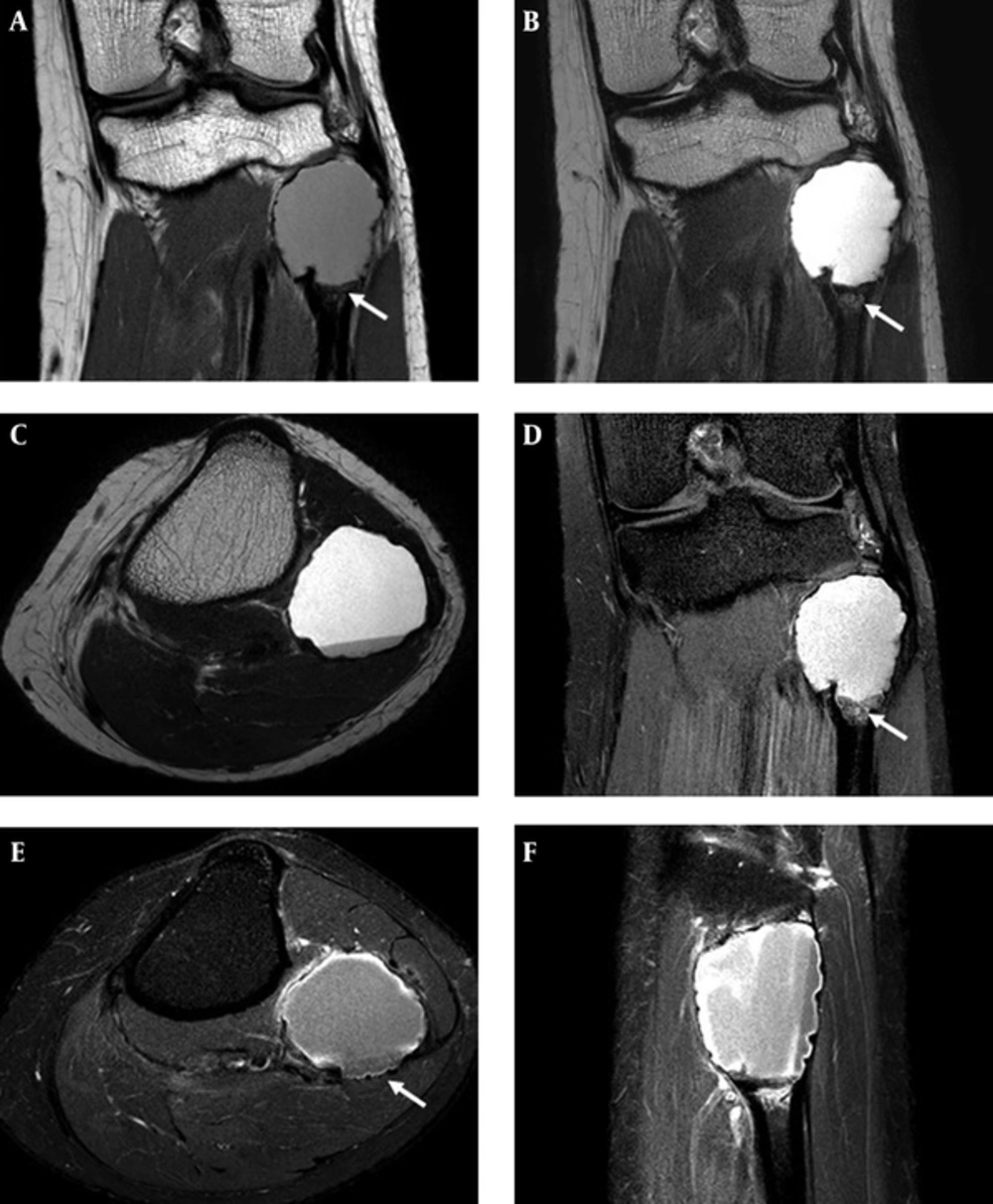
Our case showed a cystic lesion that involved the entire epiphysis as well as within the soft tissue component. Therefore, our initial differential diagnosis did not include SBC. This is unusual due to the mesenchymal tissue presenting as a soft tissue component on the MR image.
Our initial differential diagnosis was giant cell tumor (GCT) or chondroblastoma with secondary aneurysmal bone cyst. Secondary aneurysmal bone cyst formation of giant cell tumor occurs in up to 14% of cases (
7,
8). It may be present in a portion of GCT and fluid-fluid levels are seen throughout lesion on MRI (
9). Typically, GCT originates in the metaphysis and extends into the epiphysis, often to near the articular surface that is eccentric in location (
9). Plain radiography reveals a lytic lesion with a well-defined and non-sclerotic margin. The majority of cases show a geographic appearance with a narrow transition zone. Chondroblastoma is an uncommon benign cartilage tumor. Typically, it occurs in the epiphysis and often extends into the metaphysis. Radiographic findings include a well-defined, lytic geographic lesion eccentrically located. The majority of cases show a thin sclerotic margin. Mild cortical expansion and endosteal scalloping may occur. Periosteal reaction along adjacent metaphysis and internal chondroid matrix can be seen in up to 50% of cases. It may also contain intralesional fluid-fluid levels of secondary aneurysmal bone cyst, in reportedly 10% - 15% of cases (
9,
10).
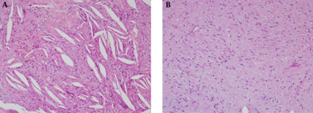
Pathologically, it demonstrated cholesterol clefts, scattered giant cells and fibrous depositions compatible with a SBC. Within the lesion, mesenchymal tissue proliferation consisting of storiform spindle cells was seen. Mesenchymal tissue is embryonic connective tissue of the lymphatic and circulatory systems, and connective tissue of the body such as bone and cartilage. This finding is unusual and its etiology is unclear.
Growth arrest or shortening as a result of a SBC is relatively uncommon, and most such cases are possibly related to surgical curettage of the epiphysis or previous fractures (
4,
6). Epiphyseal involvement of SBC seems to have a greater association with growth arrest or deformity than their metaphyseal lesions (
4,
6). Our patient had no evidence of growth arrest or shortening.
In conclusion, epiphyseal involvement of a SBC is generally rare. Furthermore, coexisting soft tissue component is even more unusual. Less than 50% of proven simple bone cyst cases meet all criteria. Therefore, SBC should be considered in the diagnosis of cystic lesions even though it is not a common entity.