Cover image of the article: Reproductive Toxicity of Aflatoxins: A Brief Report with Implications for Public Health Policy

Image Credit:J Health Rep Technol

Reproductive Toxicity of Aflatoxins: A Brief Report with Implications for Public Health Policy

Authors

Cyrus JaliliCyrus Jalili ORCID1, Mehdi KianMehdi Kian ORCID2, Ardeshir AbbasiArdeshir Abbasi ORCID3, Touraj Zamir NastaTouraj Zamir Nasta ORCID2,*
1Medical Biology Research Center, Health Technology Institute, Kermanshah University of Medical Sciences, Kermanshah, Iran
2Medicinal Plant Research Center, Ahvaz Jundishapur University of Medical Sciences, Ahvaz, Iran
3Department of Immunology, Faculty of Medical Sciences, Tarbiat Modares University, Tehran, Iran
*Corresponding Author: Medicinal Plant Research Center, Ahvaz Jundishapur University of Medical Sciences, Ahvaz, Iran. Email: toraj.nasta@gmail.com

Journal of Health Reports and Technology:Vol. 12, issue 3; e172876
Published online:Jul 31, 2026
Article type:Brief Report
Received:Jun 16, 2026
Accepted:Jul 19, 2026
How to Cite:Jalili C, Kian M, Abbasi A, Zamir Nasta T. Reproductive Toxicity of Aflatoxins: A Brief Report with Implications for Public Health Policy. J Health Rep Technol. 2026;12(3):e172876. doi: https://doi.org/10.5812/jhrt-172876

Abstract

Context:

Aflatoxins (AFs) are toxic metabolites produced by Aspergillus species and pose a serious threat to human and animal health. Exposure to AFs may lead to reproductive disorders and reduced fertility.

Objectives:

This health policy brief highlights the potential reproductive health risks associated with chronic AF exposure and emphasizes the need for continuous monitoring of food quality and storage conditions to reduce contamination.

Evidence Acquisition:

This health policy brief synthesizes evidence from our previously published studies and other relevant literature to assess the effects of AF exposure on fertility in in vitro and in vivo models. Evidence was identified from several databases.

Results:

Aflatoxin B1 (AFB1) and aflatoxin G1 (AFG1) disrupt spermatogenesis by interfering with genes involved in cell-cycle progression, including cyclins. They also induce the expression of apoptosis-related genes in vivo, activate the intrinsic apoptotic pathway, and exert cytotoxicity in TM3 mouse Leydig cells.

Conclusions:

AFs may affect fundamental molecular mechanisms involved in spermatogenesis, potentially reducing fertility. Reducing chronic exposure through improved food storage and monitoring is important for protecting reproductive health.

1. Background

Aflatoxins (AFs) are toxic metabolites produced by certain Aspergillus species and are commonly found in foods such as peanuts, corn, nuts, oilseeds, cereals, dried fruits, milk, dairy products, and animal feed (1). Studies in animal models have shown that aflatoxin exposure can induce reproductive toxicity, resulting in testicular damage, altered estrous cycles, and reduced fertility (2, 3). AFs can cause testicular tissue atrophy by disrupting molecular pathways involved in spermatogenesis, thereby reducing fertility (4).
Our recent study investigating the association between aflatoxin B1 (AFB1) and the intrinsic and extrinsic pathways of apoptosis in TM3 cells confirmed that AF exposure suppresses TM3 cell viability, triggers apoptotic pathways, and ultimately damages male reproductive cells (5).
Reducing aflatoxin exposure is important for protecting reproductive health through proper food storage, improved agricultural practices, and routine food monitoring. This health policy brief highlights the potential reproductive risks of chronic aflatoxin exposure and emphasizes the need for continuous food-safety surveillance.

2. Objectives

This health policy brief highlights that the chronic consumption of AFs poses a serious threat to reproductive health. Evidence was synthesized from our previously published studies and the relevant literature to summarize the reproductive toxicity of AFs and discuss their public health implications. Continuous monitoring of food quality and storage conditions is essential to minimize aflatoxin contamination in food products.

3. Methods

This health policy brief synthesized evidence from previously published studies on the reproductive health effects of AFB1 and aflatoxin G1 (AFG1) in vivo and in vitro. The primary emphasis was on original studies reporting the molecular and histopathological effects of AFs on the reproductive system. Relevant evidence was identified through searches of Google Scholar, ScienceDirect, Scopus, and PubMed. The retrieved studies were critically reviewed, and findings were narratively synthesized according to AF type, experimental model, exposure conditions, and reported molecular and histopathological outcomes.

4. Results

In an animal study, 24 albino mice were exposed to AFG1 at a daily dose of 20 µg/kg body weight via intraperitoneal (i.p.) injection for 35 days. AFG1 significantly altered Cyclin D1, p21, and ERα expression (P < 0.05) and increased the proportion of seminiferous tubules with negative TDI, RI, and SPI values, suggesting disruption of testicular cell-cycle regulation and spermatogenesis (3).
Another animal study reported that AFG1 exposure (20 µg/kg for 28 days) increased MDA and NO levels and decreased SOD levels in mouse testicular tissue, accompanied by intercellular edema and seminiferous tubular atrophy (4).
In another animal study, mice were exposed to AFB1 at a daily dose of 20 µg/kg body weight via intraperitoneal (i.p.) administration for 7, 14, and 21 days. AFB1 reduced Cyclin D1, CDK4, and ERα expression and increased p21 and p53 expression in mouse testicular tissue (P < 0.05), suggesting cell-cycle dysregulation and activation of p53-associated apoptotic signaling (2).
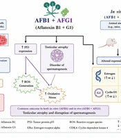
A recent review by Jalili et al. (1) highlighted oxidative stress, DNA damage, cell-cycle dysregulation, and apoptosis as potential mechanisms underlying AFB1-induced reproductive toxicity. Finally, an in vitro study in TM3 cells exposed to 3.6 µM AFB1 for 24 h showed increased caspase-3 and caspase-9 levels, indicating activation of apoptotic pathways (5). The main molecular and reproductive effects of AFB1 and AFG1 reported in these studies are summarized in Figure 1.
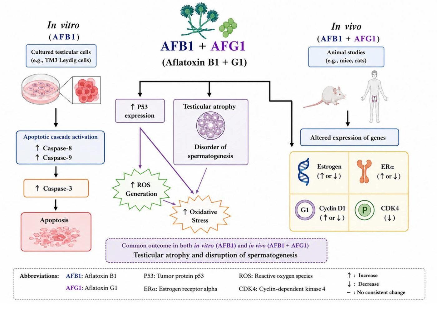
Schematic representation of the effects of AFB1 and AFG1 on testicular cells in vitro and in vivo. In vivo exposure altered the expression of estrogen, ERα, Cyclin D1, and CDK4, while in vitro exposure increased caspase production and activated the apoptotic cascade. Both exposures to AFB1 and AFG1 were associated with testicular atrophy, disrupted spermatogenesis, increased p53 expression, oxidative stress, and reactive oxygen species generation.
Figure 1.
Schematic representation of the effects of AFB1 and AFG1 on testicular cells in vitro and in vivo. In vivo exposure altered the expression of estrogen, ERα, Cyclin D1, and CDK4, while in vitro exposure increased caspase production and activated the apoptotic cascade. Both exposures to AFB1 and AFG1 were associated with testicular atrophy, disrupted spermatogenesis, increased p53 expression, oxidative stress, and reactive oxygen species generation.

5. Discussion

AF exposure through contaminated food is a public health concern. Preclinical evidence indicates that AFs may disrupt spermatogenesis through altered cell-cycle regulation, estrogen signaling, apoptosis, and oxidative stress (1), although their effects on human fertility remain uncertain. Zamir-Nasta et al. (3) reported that AFG1 exposure in albino mice increased Cyclin D1 and p21 expression and reduced ERα expression, with alterations in TDI, RI, and SPI.
Another study by Zamir-Nasta et al. (4) indicated that AFG1-related changes in Cyclin D1, SOD, MDA, and NO were associated with testicular damage, tubular atrophy, and intercellular edema. Zamir-Nasta et al. (2) showed that AFB1 reduced Cyclin D1, CDK4, and ERα expression and increased p21 and p53 expression, suggesting p53-associated apoptotic signaling.
Although Cyclin D1 showed opposite changes following AFG1 and AFB1 exposure, both increased and decreased expression may disrupt cell-cycle regulation and spermatogenesis. Jalili et al. (5) further showed that AFB1 activated intrinsic apoptosis in TM3 cells. These findings provide biological plausibility for aflatoxin-associated reproductive toxicity but do not establish direct effects on human fertility.
Regarding human evidence, Francis et al. (6) detected AFs more frequently in semen from infertile men than in fertile controls. Similarly, Suhail et al. (7) reported significantly higher seminal AFB1 levels in infertile Iraqi men than in fertile controls. However, these observational findings do not establish causality.
AF contamination is an important food-safety concern, particularly in cereals, maize, and nuts. National surveillance and food-safety standards may help reduce dietary AF exposure, although their effects on reproductive health remain uncertain (8, 9).

5.1. Limitations

The evidence is predominantly derived from animal and in vitro studies. Species differences, experimental conditions, and exposure doses may limit translation to humans. Further epidemiological studies are needed to clarify the relationship between chronic AF exposure and male reproductive health.

5.2. Conclusions

Monitoring high-risk foods, including cereals, maize, and nuts, through routine food-safety surveillance is warranted as a precautionary measure. Available evidence suggests a potential association between AF exposure and adverse male reproductive outcomes, but its impact on human fertility remains uncertain. Improved food storage and routine monitoring of high-risk foods are practical measures that may help reduce chronic AF exposure and protect reproductive health. These are precautionary policy proposals, and further human studies are needed to clarify reproductive risks.
Footnotes

  • AI Use Disclosure:The authors declare that no generative AI tools were used in the creation of this article.

  • Authors' Contribution:Writing – Original Draft, Project Administration, Methodology, Data Curation, and Conceptualization: C. J.; Supervision, Software, and Investigation: M. K., A. A., and T. Z. N.; Writing – Review & Editing, Writing – Original Draft, Supervision, and Conceptualization: T. Z. N.

  • Conflict of Interests Statement:No potential conflict of interest reported by the authors.

  • Data Availability:The dataset presented in the study is available on request from the corresponding author during submission or after publication.

  • Funding/Support:The authors gratefully acknowledge the Research Council of Kermanshah University of Medical Sciences for their financial support.

References

  • 1.
    Jalili C, Ranjbar Shamsi R, Amiri B, Kakebaraie S, Jalili F, Nasta TZ. Genotoxic and cytotoxic effects of aflatoxin on the reproductive system: Focus on cell cycle dynamics and apoptosis in testicular tissue. Toxicology. 2024;504. 153773. [PubMed ID: 38484789]. https://doi.org/10.1016/j.tox.2024.153773.
  • 2.
    Zamir-Nasta T, Razi M, Shapour H, Malekinejad H. Roles of p21, p53, cyclin D1, CDK‐4, estrogen receptor α in aflatoxin B1‐induced cytotoxicity in testicular tissue of mice. Environmental Toxicology. 2018;33(4):385-395. [PubMed ID: 29274131]. https://doi.org/10.1002/tox.22524.
  • 3.
    Zamir-Nasta T, Pazhouhi M, Ghanbari A, Abdolmaleki A, Jalili C. Expression of cyclin D1, p21, and estrogen receptor alpha in aflatoxin G1-induced disturbance in testicular tissue of albino mice. Research in Pharmaceutical Sciences. 2021;16(2):182-192. [PubMed ID: 34084205]. [PubMed Central ID: PMC8102931]. https://doi.org/10.4103/1735-5362.310525.
  • 4.
    Zamir-Nasta T, Kakebaraei S, Pazhouhi M, Jalili C. Investigating the Erα and Cyclin D1 gene expression levels, changes in SOD, NO, and MDA levels, and histopathology of mice testicular tissue treated with AFG1. J Mazandaran Univ Med Sci. 2021;31(198):1-12.
  • 5.
    Jalili C, Abbasi A, Rahmani-Kukia N, Andarzi S, Kakebaraie S, Zamir Nasta T. The relationship between aflatoxin B1 with the induction of extrinsic/intrinsic pathways of apoptosis and the protective role of taraxasterol in TM3 leydig cell line. Ecotoxicology and Environmental Safety. 2024;276. 116316. [PubMed ID: 38615640]. https://doi.org/10.1016/j.ecoenv.2024.116316.
  • 6.
    Francis S, Kortei NK, Sackey M, Richard SA. Aflatoxin B1 induces infertility, fetal deformities, and potential therapies. Open Medicine. 2024;19(1). 20240907. [PubMed ID: 38283584]. [PubMed Central ID: PMC10818061]. https://doi.org/10.1515/med-2024-0907.
  • 7.
    Suhail M, Al-Musawi MT, Al-obaidy LHA. STUDY THE CORRELATION BETWEEN AFLATOXIN B1 AND MALE INFERTILITY IN IRAQI PATIENTS. Iraqi Journal of market research and consumer protection. 2026;18(2):290-297. https://doi.org/10.28936/jmracpc18.2.2026.(22).
  • 8.
    Sajjad Y, Dib J, Soliman N, Alhmoudi M, Sajjad SG, Kandil H, et al. The Role of Mycotoxins in Reproductive health: Mechanisms, Evidence, and Clinical Implications. Journal of IVF-Worldwide. 2025;3(1). https://doi.org/10.46989/001c.132398.
  • 9.
    Yohannis E, Urugo MM, Teka TA, Getachew P, Tola YB, Forsido SF, et al. Aflatoxin Contamination in Agri‐Food Systems: A Comprehensive Review of Toxicity, Food Security, Economic Impacts, and Sustainable Mitigation Across the Value Chain. Food Science & Nutrition. 2025;13(10). e71104. [PubMed ID: 41111890]. [PubMed Central ID: PMC12529245]. https://doi.org/10.1002/fsn3.71104.

Copyright

Copyright © 2026, Journal of Health Reports and Technology. This open-access article is available under the Creative Commons Attribution 4.0 (CC BY 4.0) International License (https://creativecommons.org/licenses/by/4.0/), which allows for the copying and redistribution of the material only for noncommercial purposes, provided that the original work is properly cited.

Similar Articles

11
Oct
2012
The Role of Aflatoxins in Hepatocellular Carcinoma

The Role of Aflatoxins in Hepatocellular Carcinoma

Hui-Chen Wu,
Regina Santella

Wu H, Santella R. The Role of Aflatoxins in Hepatocellular Carcinoma. Hepat Mon. 2012;12(10):7238. doi: https://doi.org/10.5812/hepatmon.7238

1
Mar
2019
Relationship between synthesis of P21 and Cyclin D1 proteins and histomorphometric parameters of mice testicular tissue in presence of Aflatoxin G1

Relationship between synthesis of P21 and Cyclin D1 proteins and histomorphometric parameters of mice testicular tissue in presence of Aflatoxin G1

Cyrus Jalili,
Toraj Zamir-Nasta,
laya ghahari,
Seyran Kakeh baraei

Jalili C, Zamir-Nasta T, ghahari L, Kakeh baraei S. Relationship between synthesis of P21 and Cyclin D1 proteins and histomorphometric parameters of mice testicular tissue in presence of Aflatoxin G1. koomesh. 2019;21(1):e153057. doi:

22
Jun
2016
Aflatoxins in Food Products in Iran: a Review of the Literature

Aflatoxins in Food Products in Iran: a Review of the Literature

Mohammad Taghi Hedayati,
Saeid Mahdavi Omran,
Abbas Soleymani,
Mojtaba Taghizadeh Armaki

Hedayati MT, Mahdavi Omran S, Soleymani A, Taghizadeh Armaki M. Aflatoxins in Food Products in Iran: a Review of the Literature. Jundishapur J Microbiol. 2016;9(7):e33235. doi: https://doi.org/10.5812/jjm.33235

31
Jul
2021
Occurrence of Aflatoxins in Commercial Cereal-based Baby Foods in Iran: A Probabilistic Risk Assessment to Health

Occurrence of Aflatoxins in Commercial Cereal-based Baby Foods in Iran: A Probabilistic Risk Assessment to Health

Moein Bashiry,
Hassan Yazdanpanah,
Ehsan Sadeghi,
Sajad shokri,
Leila Mirmoghtadaie,
Amir Mohammad Mortazavian
,et al.

Bashiry M, Yazdanpanah H, Sadeghi E, shokri S, Mirmoghtadaie L, et al. Occurrence of Aflatoxins in Commercial Cereal-based Baby Foods in Iran: A Probabilistic Risk Assessment to Health. Iran J Pharm Res. 2021;20(3):e124499. doi: https://doi.org/10.22037/ijpr.2021.114631.14961

31
Dec
2014
Aflatoxin M1 and B1 contaminations in products of animal origin in Iran

Aflatoxin M1 and B1 contaminations in products of animal origin in Iran

R Mahmoudi,
A Golchin,
N Hoosseinzadeh,
P Ghajarbeygi

Mahmoudi R, Golchin A, Hoosseinzadeh N, Ghajarbeygi P. Aflatoxin M1 and B1 contaminations in products of animal origin in Iran. J Inflamm Dis. 2024;18(4):e155852. doi:

More by these authors

Cyrus JaliliPubMedScholar
Mehdi KianPubMedScholar
Ardeshir AbbasiPubMedScholar
Touraj Zamir NastaPubMedScholar
Share
Cited by
Metrics