Cover image of the article: Prevalence of the luxS Gene and Biofilm Formation in Urinary Tract Klebsiella pneumoniae Isolates

Image Credit:Jundishapur J Microbiol

Prevalence of the luxS Gene and Biofilm Formation in Urinary Tract Klebsiella pneumoniae Isolates

Authors

Sara KootiSara Kooti ORCID1, Mohammad Rahmanizadeh2, Amirhooshang AlvandiAmirhooshang Alvandi ORCID3,*, Touraj Esmailzadeh2, Ehsan Moradi-JooEhsan Moradi-Joo ORCID4, Zahra Nikkhooy5, Raham Armand6, Hadi HossainpourHadi Hossainpour ORCID6,**
1Infectious Ophthalmologic Research Center, Ahvaz Jundishapur University of Medical Sciences, Ahvaz, Iran
2Department of Microbiology, School of Medicine, Kermanshah University of Medical Sciences, Kermanshah, Iran
3Department of Microbiology, Medical Technology Research Center, Research Institute for Health Technology, Kermanshah University of Medical Sciences, Kermanshah, Iran
4Public Health, School of Health, Abadan University of Medical Sciences, Abadan, Iran
5Department of Biology, Faculty of Sciences, Shahid Chamran University, Ahvaz, Iran
6Behbahan Faculty of Medical Sciences, Behbahan, Iran
Corresponding Authors:
*Corresponding Author: Department of Microbiology, School of Medicine, Medical Technology Research Center, Research Institute for Health Technology, Kermanshah University of Medical Sciences, Kermanshah, Iran. Email: ah_alvandi@kums.ac.ir
**Corresponding Author: Behbahan Faculty of Medical Sciences, Behbahan, Iran. Email: hadi.hossinpour@yahoo.com

Jundishapur Journal of Microbiology:Vol. 19, issue 8; e172198
Published online:Aug 31, 2026
Article type:Research Article
Received:May 23, 2026
Accepted:Jul 26, 2026
How to Cite:Kooti S, Rahmanizadeh M, Alvandi A, Esmailzadeh T, Moradi-Joo E, et al. Prevalence of the luxS Gene and Biofilm Formation in Urinary Tract Klebsiella pneumoniae Isolates. Jundishapur J Microbiol. 2026;19(8):e172198. doi: https://doi.org/10.5812/jjm-172198

Abstract

Background:

Klebsiella pneumoniae is an important opportunistic pathogen associated with urinary tract infections (UTIs), and its ability to form biofilms contributes to persistence and antimicrobial resistance.

Objectives:

This study aimed to evaluate the prevalence of Klebsiella isolates from urinary tract infections, their biofilm-forming ability, and the presence of the luxS gene, using molecular methods.

Methods:

In this cross-sectional study, 51 Klebsiella isolates obtained from urine samples were examined. Species identification was performed using standard microbiological methods, including culture on MacConkey and blood agar, followed by biochemical identification using the IMViC tests. Antimicrobial susceptibility was assessed using the disk diffusion method, biofilm formation was evaluated using the microtiter plate assay, and molecular analysis was performed using polymerase chain reaction (PCR) with specific primers targeting the luxS gene. Data were analyzed using SPSS.

Results:

Among the 51 samples, 32 isolates were identified as K. pneumoniae and 19 as K. oxytoca. Ampicillin exhibited the highest resistance rate among the Klebsiella isolates. All isolates were susceptible to gentamicin, and 74.5% were susceptible to amikacin. In addition, the luxS gene was detected in 32 of the 51 isolates.

Conclusions:

The isolates showed high levels of antimicrobial resistance. Biofilm formation may contribute to the persistence of these bacteria and their reduced susceptibility to antimicrobial agents.

1. Background

Klebsiella pneumoniae is a ubiquitous encapsulated bacterium found in diverse environments, including soil, water, and medical surfaces. It commonly colonizes human mucosal sites, particularly the oropharynx and gastrointestinal tract, from which it can disseminate to other tissues (1). Klebsiella pneumoniae is a significant opportunistic pathogen responsible for serious clinical conditions, such as liver abscesses, meningitis, and urinary tract infections (UTIs) (2). Although newborns, older adults, and immunocompromised individuals are at the highest risk, community-acquired infections are increasingly reported (3). Klebsiella pneumoniae is recognized as a major cause of Gram-negative infections, including septicemia, endocarditis, pneumonia, and surgical site infections, and is characterized by increasing rates of antimicrobial resistance (4-7). Biofilm formation by K. pneumoniae was first reported in the late 1980s and was later confirmed in vivo. Approximately 40% of isolates form biofilms, increasing to 63% in catheterized patients, and many ventilator-associated pneumonia strains also demonstrate this ability. Biofilms increase stress tolerance and antibiotic resistance, thereby enhancing bacterial virulence (8, 9).
Bacteria communicate through quorum sensing (QS), a regulatory system based on the production and detection of extracellular signaling molecules known as autoinducers (AIs) (10). The LuxS enzyme facilitates the synthesis of AI-2 from S-ribosylhomocysteine (11). AI-2 signaling regulates the expression of numerous genes and influences key physiological processes, such as virulence, protein secretion, extracellular polysaccharide production, iron uptake, motility, and biofilm formation (12). LuxS strongly affects the bacterial phenotype and contributes to K. pneumoniae pathogenicity through several mechanisms (13). LuxS has been implicated in the modulation of host immune responses, biofilm formation, and the regulation of capsular polysaccharide (14). Previous studies have demonstrated that LuxS-mediated AI-2 quorum sensing influences biofilm architecture and the expression of outer-membrane-associated genes in K. pneumoniae. Subsequent evidence indicates that LuxS-dependent virulence effects are strain-specific and growth phase-dependent, particularly among isolates with different mucoid phenotypes (15).
Although the molecular role of LuxS in quorum sensing, virulence modulation, and biofilm formation in K. pneumoniae has been extensively studied, few studies have reported the distribution of the luxS gene among clinical urinary isolates and its possible association with biofilm-forming ability. Furthermore, epidemiological evidence on this topic in Iran remains limited. Therefore, this study was conducted to determine the prevalence of the luxS gene among urinary Klebsiella isolates and to assess its association with biofilm formation and antimicrobial resistance patterns, thereby providing clinically relevant information on the role of LuxS in UTIs.

2. Objectives

The present study aimed to determine the prevalence of the luxS gene in clinical urinary isolates of K. pneumoniae and to assess the association of the luxS gene with biofilm formation and antimicrobial resistance patterns, thereby providing clinically relevant insights into the molecular determinants of K. pneumoniae UTIs.

3. Methods

3.1. Bacterial Isolates

This cross-sectional study was conducted from September 2022 to March 2023 at the reference laboratory of Kermanshah University of Medical Sciences, Iran. The study used anonymized urinary isolates routinely received by the microbiology laboratory for diagnostic purposes. Clinical samples were cultured on MacConkey and blood agar and incubated at 37 °C for 24 hours. Colonies were identified by microscopy, biochemical tests, and IMViC. Confirmed isolates were stored at -70 °C in trypticase soy broth containing 18% glycerol for further analysis. All eligible Klebsiella isolates recovered from urine specimens during the study period were consecutively included. Accordingly, all available isolates meeting the inclusion criteria were included in the study population, and the sample size was determined by the total number of eligible isolates collected rather than by a predefined target sample size.

3.2. Antimicrobial Susceptibility Testing

Antibiotic susceptibility was assessed using the disk diffusion method on Mueller-Hinton agar. The antibiotics tested were amikacin, ampicillin, sulfamethoxazole-trimethoprim, ceftazidime, cefazolin, gentamicin, nalidixic acid, and nitrofurantoin. Results were interpreted according to CLSI guidelines (16). Escherichia coli ATCC 25922 and K. pneumoniae ATCC 9997 were used as quality-control strains.

3.3. Biofilm Assay

Biofilm formation was evaluated using the microtiter plate assay. Briefly, bacterial suspensions were inoculated into sterile 96-well microplates and incubated under appropriate conditions for biofilm development. The wells were gently washed to remove planktonic cells, and the adherent biofilm was fixed and stained with crystal violet. The bound dye was then solubilized, and optical density (OD) was measured using a microplate reader. Based on the OD values, isolates were classified as non-biofilm producers or as weak, moderate, or strong biofilm producers according to established criteria described in previous studies (17-19).

3.4. Genomic DNA Preparation

DNA was extracted using the boiling method (20). DNA absorbance and concentration at 260/280 nm were measured using a NanoDrop spectrophotometer (NanoDrop Technologies, Inc., Wilmington, DE, USA). The DNA templates were stored at -20 °C until further analysis.

3.5. PCR Amplification of the luxS Gene

Polymerase chain reaction (PCR) using gene-specific primers (Table 1) was performed to detect the luxS gene. The PCR was performed in a final volume of 25 µL containing 15 µL Taq premix, 2 µL template DNA, 1 µL forward primer, 1 µL reverse primer, and 6 µL sterile distilled water. Amplification began with initial denaturation at 95 °C for 3 minutes, followed by 35 cycles of denaturation, annealing at 53 °C for 1 minute, and extension. A final extension at 72 °C for 5 minutes was performed to complete DNA synthesis. The amplified PCR products were separated by electrophoresis on a 1% agarose gel and visualized under UV illumination. Klebsiella pneumoniae ATCC 9997 was included as a control strain to verify amplification accuracy.
Table 1.
Primer Sequences and PCR Parameters Used for luxS Gene Amplification
ReferenceLengthTm (°C)Primer Sequence (5'-3')DirectionGene Name
(21)475 bp53GCCGTTGTTAGATAGTTTCACAGFluxS
(21)475 bp53CAGTTCGTCGTTGCTGTTGATGRluxS

3.6. Statistical Analysis

Statistical analysis was performed using SPSS version 19 (Chicago, IL, USA). Chi-square tests were used to evaluate associations between categorical variables, such as luxS gene presence and biofilm intensity. A P value < 0.05 was considered statistically significant.

4. Results

4.1. Bacterial Identification and Demographic Features

A total of 51 urinary isolates were obtained and examined. Of these, 6 (11.8%) isolates were recovered from male patients and 45 (88.2%) from female patients. The mean age of the patients was 40.12 years. Using conventional culture methods and IMViC biochemical testing, 32 isolates (62.75%) were identified as K. pneumoniae, whereas 19 isolates (37.25%) were identified as K. oxytoca. These results indicate that K. pneumoniae was the predominant species among the urinary isolates.

4.2. Antimicrobial Susceptibility Pattern

Antimicrobial susceptibility testing showed that the isolates exhibited distinct resistance profiles. The highest resistance rates were observed for ampicillin (88.2%) and gentamicin (86.2%). In contrast, the highest susceptibility rate was observed for amikacin (74.5%) (Table 2).
Table 2.
Antimicrobial Susceptibility Profile of Klebsiella pneumoniae Isolates Obtained from Urine Samples a
AntibioticsSusceptibilityIntermediateResistant
AM6 (11.8)-45 (88.2)
CAZ16 (31.4)22 (43.1)13 (25.5)
NA16 (31.4)23 (45)12 (23.5)
GM44 (86.2)4 (7.8)3 (6)
FM32 (62.74)7 (13.72)12 (23.5)
AN38 (74.5)8 (15.68)5 (9.8)
SXT35 (68.6)4 (7.8)12 (23.5)
CZ15 (29.4)7 (13.7)29 (56.9)
a Values are expressed as No. (%). Abbreviations: AM, ampicillin; CAZ, ceftazidime; NA, nalidixic acid; GM, gentamicin; FM, nitrofurantoin; AN, amikacin; SXT, sulfamethoxazole/trimethoprim; CZ, cefazolin.

4.3. Biofilm Formation

Biofilm-forming capacity was assessed for all isolates. A considerable proportion of the isolates was able to produce biofilm. The proportions of strong, moderate, and weak biofilm producers were significantly associated with one another (chi-square = 10.83, df = 2, P = 0.0044), indicating variation in biofilm-forming potential among the isolates. Biofilm formation indicates an increased ability of the isolates to persist in the urinary tract and to withstand antimicrobial therapy (Table 3).
Table 3.
Association Between Biofilm-Forming Capacity and Presence of the luxS Gene Among Urinary Klebsiella pneumoniae Isolates a
Biofilm strengthluxS+luxS-Total
Weak156
Moderate909
Strong221436
Total321951
a Values are expressed as number. Chi-square test = 10.83; df = 2; P = 0.0044.

4.4. Detection of the luxS Gene

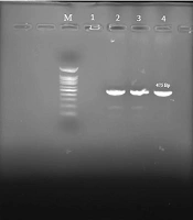
To determine the distribution of the luxS gene, PCR was performed on 51 Klebsiella strains isolated from urine cultures. The results showed that 32 isolates (62.7%) harbored the gene (Figure 1), indicating a substantial frequency within the tested population. Furthermore, the data demonstrated a clear correlation between the presence of luxS and biofilm-forming capacity. Overall, these clinical isolates exhibited substantial luxS carriage, marked biofilm development, and antibiotic resistance.
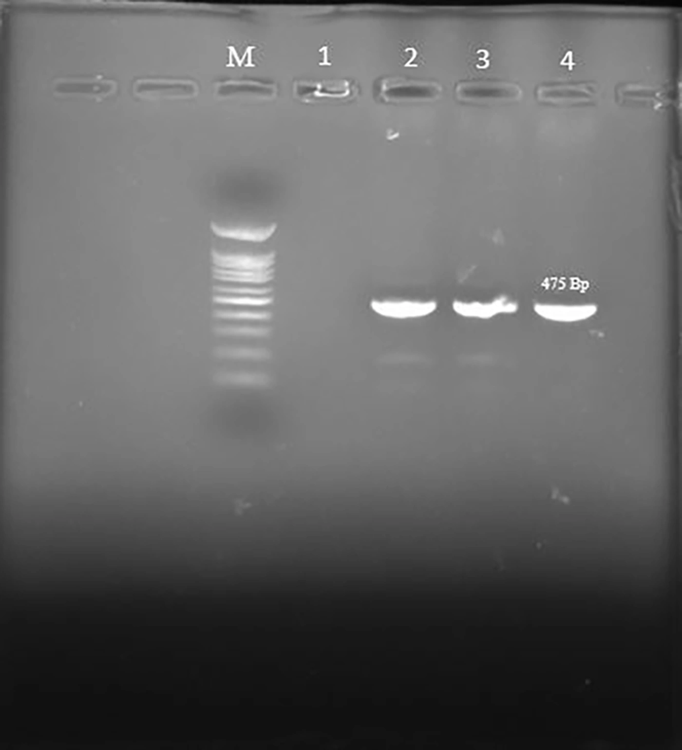
Agarose gel electrophoresis of PCR products obtained for detection of the luxS gene in <i>Klebsiella pneumoniae</i> isolates. Lane M: 100-bp DNA ladder; lane 1: negative control; lane 2: positive control; lanes 3 and 4: positive isolates (475 bp).
Figure 1.
Agarose gel electrophoresis of PCR products obtained for detection of the luxS gene in Klebsiella pneumoniae isolates. Lane M: 100-bp DNA ladder; lane 1: negative control; lane 2: positive control; lanes 3 and 4: positive isolates (475 bp).

5. Discussion

The increasing incidence of antibiotic-resistant Klebsiella species worldwide represents a serious public health threat (22). Biofilm formation further increases the pathogenic potential of these organisms by protecting bacterial cells from host immune responses and reducing antimicrobial penetration. In the present study, we evaluated antimicrobial resistance patterns, biofilm-forming ability, and the presence of the luxS gene among urinary Klebsiella isolates. Among the 51 isolates examined, most were obtained from female patients (88.2%), consistent with previous reports indicating a higher prevalence of UTIs among women (23). This increased susceptibility is attributed to the shorter female urethra and its close anatomical relationship with the perianal area, which facilitates colonization of the urinary tract by Gram-negative bacteria (24). Female sex remains a major epidemiological risk factor for UTIs.
Antimicrobial susceptibility testing revealed high resistance rates to ampicillin (88.2%) and cefazolin (56.9%). Resistance to the other tested antibiotics varied but remained clinically significant. These findings are consistent with previous reports indicating high resistance levels among Klebsiella isolates from UTIs (23, 25-26). Differences among studies are likely related to geographic location, clinical specimen type, antibiotic-prescribing guidelines, and local hygiene practices. In the current study, most isolates were strong biofilm producers (70.5%), whereas fewer were moderate (17.6%) or weak (11.7%) biofilm producers. These results are similar to the findings of Vuotto et al., who reported a predominance of strong biofilm-producing urinary K. pneumoniae isolates (22). Other studies have confirmed the ability of clinical Klebsiella strains to form biofilms, although prevalence rates vary by methodology and sample type (25, 27). Biofilm formation is a key factor in catheter- and device-related infections and a major contributor to chronicity and treatment failure (28).
In addition to its general role as a virulence factor, biofilm formation in K. pneumoniae is a complex, highly regulated process influenced by multiple regulatory systems, including quorum sensing. Quorum sensing enables bacterial populations to coordinate gene expression in response to cell density through the production and detection of signaling molecules. Among these systems, the LuxS-mediated signaling pathway is particularly important because it is responsible for the synthesis of autoinducer-2 (AI-2), a universal signaling molecule involved in interspecies bacterial communication (21, 29). In the present study, the luxS gene was detected in 62.7% of the isolates, and the results suggested an association between luxS gene carriage and biofilm-forming capacity. These findings support the hypothesis that LuxS-mediated quorum sensing may contribute to the regulation of biofilm formation in clinical Klebsiella isolates. Previous studies have shown that disruption of the luxS gene can significantly affect biofilm architecture, bacterial adhesion, and community behavior, indicating that the LuxS/AI-2 system plays an important role in biofilm development (30, 31).
The AI-2 signaling molecule produced by LuxS participates in the regulation of genes involved in adhesion, extracellular matrix production, motility, and stress responses, all of which are critical for the initiation and maturation of bacterial biofilms (32, 33). In K. pneumoniae, quorum sensing has also been associated with increased virulence, enhanced persistence in host tissues, and improved adaptation to hostile environmental conditions. Recent investigations focusing specifically on K. pneumoniae have further highlighted the importance of the LuxS/AI-2 system in biofilm regulation and pathogenicity. Experimental studies have demonstrated that alteration or deletion of luxS affects biofilm biomass, outer membrane protein expression, and the structural stability of biofilm communities (34, 35). Moreover, the LuxS/AI-2 signaling pathway has been implicated in facilitating horizontal gene transfer and plasmid dissemination, potentially contributing to the spread of antimicrobial resistance among clinical isolates (15, 36). Additional reports indicate that quorum sensing may modulate virulence-associated traits, including capsule production and stress adaptation, thereby enhancing bacterial persistence in host tissues (37, 38).
Overall, the findings of the present study indicate that biofilm formation and the presence of the luxS gene are closely associated virulence characteristics in urinary Klebsiella isolates. This relationship may partly explain infection persistence and the reduced effectiveness of antimicrobial therapy in biofilm-associated UTIs. Therefore, understanding the molecular mechanisms underlying quorum sensing and biofilm regulation may contribute to the development of novel therapeutic strategies targeting bacterial communication systems in addition to conventional antibiotic therapy.

5.1. Conclusions

The present study demonstrated frequent strong biofilm-forming ability and a high prevalence of the luxS gene in urinary Klebsiella isolates, especially K. pneumoniae. The observed association between luxS carriage and biofilm formation suggests that quorum sensing may play an important role in the pathogenicity and persistence of these isolates in UTIs. Furthermore, the high level of antimicrobial resistance observed, particularly to ampicillin and cefazolin, underscores the therapeutic challenges posed by these organisms. Overall, the results highlight the clinical importance of biofilm formation and luxS-dependent quorum sensing in Klebsiella infections. Improved understanding of these mechanisms could lead to new strategies for preventing and treating UTIs associated with biofilms and drug resistance.
Acknowledgments

Footnotes

  • AI Use Disclosure:The authors declare that no generative AI tools were used in the creation of this article.

  • Authors' Contribution:Study concept and design: H. H. and Ah. A.; Analysis and interpretation of data: E. M. and M. R.; Statistical analysis: R. A. and T. E.; Drafting of the manuscript: H. H. and S. K.; Critical revision of the manuscript for important intellectual content: Ah. A., S. K., and Z. N.

  • Conflict of Interests Statement:The authors declare no competing interests.

  • Data Availability:The dataset presented in the study is available on request from the corresponding author during submission or after publication.

  • Ethical Approval:The study was approved by the Ethics Committee of Kermanshah University of Medical Sciences (IR.KUMS.REC.1397.861).

  • Funding/Support:No funding was received for this study.

References

  • 1.
    Dao TT, Liebenthal D, Tran TK, Ngoc Thi Vu B, Ngoc Thi Nguyen D, Thi Tran HK, et al. Klebsiella pneumoniae oropharyngeal carriage in rural and urban Vietnam and the effect of alcohol consumption. PLoS One. 2014;9(3). e91999. [PubMed ID: 24667800]. [PubMed Central ID: PMC3965401]. https://doi.org/10.1371/journal.pone.0091999.
  • 2.
    Maleki NS, Babazadeh F, Arzanlou M, Teimourpour R, Dogaheh HP. Serotyping and molecular profiles of virulence-associated genes among Klebsiella pneumoniae isolates from teaching hospitals of Ardabil, Iran: A cross-sectional study. Health Science Reports. 2023;6(9). e1557. [PubMed ID: 37706015]. [PubMed Central ID: PMC10496611]. https://doi.org/10.1002/hsr2.1557.
  • 3.
    Chang D, Sharma L, Dela Cruz CS, Zhang D. Clinical epidemiology, risk factors, and control strategies of Klebsiella pneumoniae infection. Frontiers in Microbiology. 2021;12. 750662. [PubMed ID: 34992583]. [PubMed Central ID: PMC8724557]. https://doi.org/10.3389/fmicb.2021.750662.
  • 4.
    Khodadadian R, Rahdar HA, Javadi A, Safari M, Khorshidi A. Detection of VIM-1 and IMP-1 genes in Klebsiella pneumoniae and relationship with biofilm formation. Microbial Pathogenesis. 2018;115:25-30. [PubMed ID: 29248513]. https://doi.org/10.1016/j.micpath.2017.12.036.
  • 5.
    Kooti S, Zamani K, Sisakht MT, Mansury D, Motamedifar M. Phenotypic and genotypic detection of antibiotic resistance among metallo-beta-lactamases producing Klebsiella pneumoniae strains isolated from patients in intensive care units in Shiraz, Iran. Gene Reports. 2019;17. 100522. https://doi.org/10.1016/j.genrep.2019.100522.
  • 6.
    Akintoyese TO, Alao JO, Oladipo EK, Oyedemi OT, Oyawoye OM. Antimicrobial resistance and virulence in Klebsiella pneumoniae: a four-month study in Osogbo, Nigeria. Antimicrobial Stewardship & Healthcare Epidemiology. 2025;5(1). e64. [PubMed ID: 40026760]. [PubMed Central ID: PMC11869054]. https://doi.org/10.1017/ash.2025.21.
  • 7.
    Rapp RP, Urban C. Klebsiella pneumoniae carbapenemases in Enterobacteriaceae: history, evolution, and microbiology concerns. Pharmacotherapy. 2012;32(5):399-407. [PubMed ID: 22488420]. https://doi.org/10.1002/j.1875-9114.2012.01035.x.
  • 8.
    Vuotto C, Longo F, Balice M, Donelli G, Varaldo P. Antibiotic resistance related to biofilm formation in Klebsiella pneumoniae. Pathogens. 2014;3(3):743-758. [PubMed ID: 25438022]. [PubMed Central ID: PMC4243439]. https://doi.org/10.3390/pathogens3030743.
  • 9.
    Ribeiro SM, Cardoso MH, Cândido EDS, Franco OL. Understanding, preventing and eradicating Klebsiella pneumoniae biofilms. Future Microbiology. 2016;11(4):527-538. [PubMed ID: 27064296]. https://doi.org/10.2217/fmb.16.7.
  • 10.
    Zhang J, Feng T, Wang J, Wang Y, Zhang XH. The mechanisms and applications of quorum sensing (QS) and quorum quenching (QQ). Journal of Ocean University of China. 2019;18(6):1427-1442. https://doi.org/10.1007/s11802-019-4073-5.
  • 11.
    Dong YH, Zhang LH. Quorum sensing and quorum-quenching enzymes. J Microbiol. 2005;43:101-9. [PubMed ID: 15765063].
  • 12.
    Kalia VC. Quorum sensing inhibitors: an overview. Biotechnology Advances. 2013;31(2):224-245. [PubMed ID: 23142623]. https://doi.org/10.1016/j.biotechadv.2012.10.004.
  • 13.
    Taşkın Kafa AH, Aslan R, Durna Daştan S, Çeli̇k C, Hasbek M, Emi̇noğlu A. Molecular diversity of Klebsiella pneumoniae clinical isolates: antimicrobial resistance, virulence, and biofilm formation. Nucleosides Nucleotides Nucleic Acids. 2025;44(5):361-377. [PubMed ID: 38718417]. https://doi.org/10.1080/15257770.2024.2344741.
  • 14.
    Niazy AA. LuxS quorum sensing system and biofilm formation of oral microflora: A short review article. Saudi Dent J. 2021;33(3):116-123. [PubMed ID: 33679103]. [PubMed Central ID: PMC7910685]. https://doi.org/10.1016/j.sdentj.2020.12.007.
  • 15.
    Tang M, Zhao D, Zhang Y, Qian C, Chen H, Chen L, et al. Impact of LuxS on virulence and pathogenicity in Klebsiella pneumoniae exhibiting varied mucoid phenotypes. Infect Immun. 2024;92(3). e0001224. [PubMed ID: 38358274]. [PubMed Central ID: PMC10929404]. https://doi.org/10.1128/iai.00012-24.
  • 16.
    Li Y, Kumar S, Zhang L, Wu H. Klebsiella pneumonia and its antibiotic resistance: A bibliometric analysis. Biomed Res Int. 2022;2022(1). 1668789. [PubMed ID: 35707374]. [PubMed Central ID: PMC9192197]. https://doi.org/10.1155/2022/1668789.
  • 17.
    Briega I, Garde S, Sánchez C, Rodríguez-Mínguez E, Picon A, Ávila M. Evaluation of biofilm production and antibiotic resistance/susceptibility profiles of Pseudomonas spp. isolated from milk and dairy products. Foods. 2025;14(7):1105. [PubMed ID: 40238238]. [PubMed Central ID: PMC11989221]. https://doi.org/10.3390/foods14071105.
  • 18.
    Guéneau V, Jiménez G, Castex M, Briandet R. Insights into the genomic and phenotypic characteristics of Bacillus spp. strains isolated from biofilms in broiler farms. Appl Environ Microbiol. 2024;90(9). e0066324. [PubMed ID: 39158314]. [PubMed Central ID: PMC11409695]. https://doi.org/10.1128/aem.00663-24.
  • 19.
    Stepanović S, Vuković D, Dakić I, Savić B, Švabić-Vlahović M. A modified microtiter-plate test for quantification of staphylococcal biofilm formation. J Microbiol Methods. 2000;40(2):175-179. [PubMed ID: 10699673]. https://doi.org/10.1016/S0167-7012(00)00122-6.
  • 20.
    Mahmoudi H, Hossainpour H, Moradi M, Alikhani MY. Distribution of iron uptake systems encoding genes among the clinical isolates of Escherichia coli compared to foodstuffs isolates. Infect Disord Drug Targets. 2020;20(4):517-522. [PubMed ID: 30659552]. https://doi.org/10.2174/1871526519666190119112542.
  • 21.
    Balestrino D, Haagensen JAJ, Rich C, Forestier C. Characterization of type 2 quorum sensing in Klebsiella pneumoniae and relationship with biofilm formation. J Bacteriol. 2005;187(8):2870-2880. [PubMed ID: 15805533]. [PubMed Central ID: PMC1070389]. https://doi.org/10.1128/JB.187.8.2870-2880.2005.
  • 22.
    Vuotto C, Longo F, Pascolini C, Donelli G, Balice MP, Libori MF, et al. Biofilm formation and antibiotic resistance in Klebsiella pneumoniae urinary strains. J Appl Microbiol. 2017;123(4):1003-1018. [PubMed ID: 28731269]. https://doi.org/10.1111/jam.13533.
  • 23.
    Shakya P, Shrestha D, Maharjan E, Sharma VK, Paudyal R. ESBL production among E. coli and Klebsiella spp. causing urinary tract infection: a hospital based study. Open Microbiol J. 2017;11(1):23-30. [PubMed ID: 28553414]. [PubMed Central ID: PMC5427687]. https://doi.org/10.2174/1874285801711010023.
  • 24.
    Magliano E, Grazioli V, Deflorio L, Leuci AI, Mattina R, Romano P, et al. Gender and age-dependent etiology of community-acquired urinary tract infections. ScientificWorldJournal. 2012;2012:349597-6. [PubMed ID: 22629135]. [PubMed Central ID: PMC3351074]. https://doi.org/10.1100/2012/349597.
  • 25.
    Karimi K, Zarei O, Sedighi P, Taheri M, Doosti-Irani A, Shokoohizadeh L. Investigation of antibiotic resistance and biofilm formation in clinical isolates of Klebsiella pneumoniae. Int J Microbiol. 2021;2021:5573388-6. [PubMed ID: 34221021]. [PubMed Central ID: PMC8219462]. https://doi.org/10.1155/2021/5573388.
  • 26.
    Parsaie Mehr V, Shokoohizadeh L, Mirzaee M, Savari M. Molecular typing of Klebsiella pneumoniae isolates by enterobacterial repetitive intergenic consensus (ERIC)-PCR. Infect Epidemiol Microbiol. 2017;3(4):112-116. https://doi.org/10.18869/modares.iem.3.4.112.
  • 27.
    Seifi K, Kazemian H, Heidari H, Rezagholizadeh F, Saee Y, Shirvani F, et al. Evaluation of biofilm formation among Klebsiella pneumoniae isolates and molecular characterization by ERIC-PCR. Jundishapur J Microbiol. 2016;9(1). [PubMed ID: 27099694]. [PubMed Central ID: PMC4834130]. https://doi.org/10.5812/jjm.30682.
  • 28.
    Zarei O, Arabestan MR, Majlesi A, Mohammadi Y, Alikhani MY. Determination of virulence determinants of Escherichia coli strains isolated from patients with colorectal cancer compared to the healthy subjects. Gastroenterol Hepatol Bed Bench. 2019;12(1):52-59. [PubMed ID: 30949320]. [PubMed Central ID: PMC6441484].
  • 29.
    Pereira CS, Thompson JA, Xavier KB. AI-2-mediated signalling in bacteria. FEMS Microbiol Rev. 2013;37(2):156-181. [PubMed ID: 22712853]. https://doi.org/10.1111/j.1574-6976.2012.00345.x.
  • 30.
    Chen X, Schauder S, Potier N, Van Dorsselaer A, Pelczer I, Bassler BL, et al. Structural identification of a bacterial quorum-sensing signal containing boron. Nature. 2002;415(6871):545-549. [PubMed ID: 11823863]. https://doi.org/10.1038/415545a.
  • 31.
    Reading NC, Sperandio V. Quorum sensing: the many languages of bacteria. FEMS Microbiol Lett. 2006;254(1):1-11. [PubMed ID: 16451172]. https://doi.org/10.1111/j.1574-6968.2005.00001.x.
  • 32.
    Li YH, Tian X. Quorum sensing and bacterial social interactions in biofilms. Sensors. 2012;12(3):2519-2538. [PubMed ID: 22736963]. [PubMed Central ID: PMC3376616]. https://doi.org/10.3390/s120302519.
  • 33.
    Antunes LCM, Ferreira RBR, Buckner MMC, Finlay BB. Quorum sensing in bacterial virulence. Microbiology. 2010;156(8):2271-2282. [PubMed ID: 20488878]. https://doi.org/10.1099/mic.0.038794-0.
  • 34.
    Chen L, Wilksch JJ, Liu H, Zhang X, Torres VVL, Bi W, et al. Investigation of LuxS-mediated quorum sensing in Klebsiella pneumoniae. J Med Microbiol. 2020;69(3):402-413. [PubMed ID: 32223838]. [PubMed Central ID: PMC7377169]. https://doi.org/10.1099/jmm.0.001148.
  • 35.
    Zhou Y, Yang Y, Wu C, Zhou P, Gao H, Wang B, et al. LuxS/AI-2 system facilitates the dissemination of antibiotic-resistant plasmids in Klebsiella pneumoniae. Int J Antimicrob Agents. 2024;64(6). 107361. [PubMed ID: 39455017]. https://doi.org/10.1016/j.ijantimicag.2024.107361.
  • 36.
    Al-Maeni MARM, Al-Khazraji SFR, Hasan HM. Study the relation between luxS gene and forming biofilm and non-forming isolates of Klebsiella pneumonia. Iraqi J Sci. 2025:4137-4147. https://doi.org/10.24996/ijs.2025.66.10.10.
  • 37.
    Li Y, Ni M. Regulation of biofilm formation in Klebsiella pneumoniae. Front Microbiol. 2023;14. 1238482. [PubMed ID: 37744914]. [PubMed Central ID: PMC10513181]. https://doi.org/10.3389/fmicb.2023.1238482.
  • 38.
    Bin Mohammad Muzaki MZ, Subramoni S, Summers S, Kjelleberg S, Rice SA. Klebsiella pneumoniae AI-2 transporters mediate interspecies interactions and composition in a three-species biofilm community. NPJ Biofilms Microbiomes. 2024;10(1). 91. [PubMed ID: 39341797]. [PubMed Central ID: PMC11439081]. https://doi.org/10.1038/s41522-024-00546-0.

Copyright

Copyright © 2026, Kooti et al. This open-access article is available under the Creative Commons Attribution 4.0 (CC BY 4.0) International License (https://creativecommons.org/licenses/by/4.0/), which allows for unrestricted use, distribution, and reproduction in any medium, provided that the original work is properly cited.

Similar Articles

17
Oct
2023
Quinolone Resistance in Biofilm-Forming Klebsiella pneumoniae-Related Catheter-Associated Urinary Tract Infections: A Neglected Problem

Quinolone Resistance in Biofilm-Forming Klebsiella pneumoniae-Related Catheter-Associated Urinary Tract Infections: A Neglected Problem

Mahdi Dadashi Firouzjaei,
Peyman Hendizadeh,
Mehrdad Halaji,
Sajad Yaghoubi,
Mohammad Teimourian,
Akramossadat Hosseini
,et al.

Dadashi Firouzjaei M, Hendizadeh P, Halaji M, Yaghoubi S, Teimourian M, et al. Quinolone Resistance in Biofilm-Forming Klebsiella pneumoniae-Related Catheter-Associated Urinary Tract Infections: A Neglected Problem. Jundishapur J Microbiol. 2023;16(8):e136170. doi: https://doi.org/10.5812/jjm-136170

26
Oct
2020
Detection of OqxAB and QepA Efflux Pumps and Their Association with Antibiotic Resistance in Klebsiella pneumoniae Isolated From Urinary Tract Infection

Detection of OqxAB and QepA Efflux Pumps and Their Association with Antibiotic Resistance in Klebsiella pneumoniae Isolated From Urinary Tract Infection

Manijeh Dehnamaki,
Maryam Ghane,
Laleh Babaeekhou

Dehnamaki M, Ghane M, Babaeekhou L. Detection of OqxAB and QepA Efflux Pumps and Their Association with Antibiotic Resistance in Klebsiella pneumoniae Isolated From Urinary Tract Infection. Int J Infect. 2020;7(4):e107397. doi: https://doi.org/10.5812/iji.107397

2
Jan
2016
Evaluation of Biofilm Formation Among Klebsiella pneumoniae Isolates and Molecular Characterization by ERIC-PCR

Evaluation of Biofilm Formation Among Klebsiella pneumoniae Isolates and Molecular Characterization by ERIC-PCR

Kimia Seifi,
Hossein Kazemian,
Hamid Heidari,
Fereshteh Rezagholizadeh,
Yasaman Saee,
Fariba Shirvani
,et al.

Seifi K, Kazemian H, Heidari H, Rezagholizadeh F, Saee Y, et al. Evaluation of Biofilm Formation Among Klebsiella pneumoniae Isolates and Molecular Characterization by ERIC-PCR. Jundishapur J Microbiol. 2016;9(1):e30682. doi: https://doi.org/10.5812/jjm.30682

26
Aug
2024
Characterization of Clonal Groups of Antibiotic-Resistant Biofilm-Forming Staphylococcus aureus Strains Isolated from Patients with Urinary Tract Infections in Isfahan, Iran

Characterization of Clonal Groups of Antibiotic-Resistant Biofilm-Forming Staphylococcus aureus Strains Isolated from Patients with Urinary Tract Infections in Isfahan, Iran

Narges Sadat Mostafavi,
Fateh Rahimi,
Sanaz Khashei

Mostafavi NS, Rahimi F, Khashei S. Characterization of Clonal Groups of Antibiotic-Resistant Biofilm-Forming Staphylococcus aureus Strains Isolated from Patients with Urinary Tract Infections in Isfahan, Iran. Arch Clin Infect Dis. 2024;19(4):e143383. doi: https://doi.org/10.5812/archcid-143383

16
Feb
2025
Hypervirulent Klebsiella pneumoniae: Biofilm Formation, Capsular Type, Virulence Genes, and Genotyping in Amol Hospitals, Northern Iran

Hypervirulent Klebsiella pneumoniae: Biofilm Formation, Capsular Type, Virulence Genes, and Genotyping in Amol Hospitals, Northern Iran

Nassim Tavassoli,
Rahem Khoshbakht,
Fatemeh Zaboli,
Mojtaba Khosravi

Tavassoli N, Khoshbakht R, Zaboli F, Khosravi M. Hypervirulent Klebsiella pneumoniae: Biofilm Formation, Capsular Type, Virulence Genes, and Genotyping in Amol Hospitals, Northern Iran. Jundishapur J Microbiol. 2025;18(2):e157119. doi: https://doi.org/10.5812/jjm-157119

More by these authors

Sara KootiPubMedScholar
Mohammad RahmanizadehPubMedScholar
Amirhooshang AlvandiPubMedScholar
Touraj EsmailzadehPubMedScholar
Ehsan Moradi-JooPubMedScholar
Zahra NikkhooyPubMedScholar
Raham ArmandPubMedScholar
Hadi HossainpourPubMedScholar
Share
Cited by
Metrics
Indexed in