
1. Background
Diarrhea is a common cause of hospitalization and contributes substantially to the health care burden (1, 2). In patients with cancer, it may delay chemotherapy and lead to dehydration, impaired absorption of oral medications, bacterial translocation, and even sepsis. It can also prolong hospitalization, increase costs, and, in severe cases, result in death (3). Causes may be infectious or noninfectious and include chemotherapy-associated epithelial cell loss, hyperosmotic enteral nutrition, graft-versus-host disease, mucositis, and radiation injury (4). Identifying the underlying cause of diarrhea in these patients is crucial for effective treatment but is often challenging (5). Routine fecal bacterial cultures may miss a substantial proportion of the etiologic agents of gastroenteritis, including pathogenic subtypes of E. coli and common viral and parasitic causes. Multiplex PCR-based syndromic panels enable clinicians to test for a broad range of pathogens (6, 7), including gastrointestinal pathogens.
2. Objectives
Diarrhea is common among patients with cancer; however, studies examining its detailed epidemiology in this population remain limited. Similarly, data on the causes of gastroenteritis identified by syndromic panels in patients with hematologic or oncologic disease remain limited (8). This retrospective cohort study aimed to investigate the causes of gastroenteritis in hematology/oncology patients in our setting, focusing on those who developed hospital-acquired gastroenteritis.
3. Methods
This retrospective cohort study was conducted at the Bahrain Oncology Center, Kingdom of Bahrain, the national referral center for comprehensive oncology and hematology services. The facility includes inpatient units for medical oncology, hematology, and bone marrow transplantation. The study period was March 1, 2020, through November 30, 2022. The findings are reported in accordance with the Strengthening the Reporting of Observational Studies in Epidemiology (STROBE) guidelines (9). The primary outcome was the distribution of gastrointestinal pathogens identified by the multiplex GI PCR panel among patients with cancer and hospital-acquired gastroenteritis.
Secondary outcomes included: 1) comparison of pathogen distribution between neutropenic and non-neutropenic hematology/oncology patients; 2) assessment of agreement and diagnostic performance of the GI panel compared with conventional stool culture; and 3) all-cause 30-day mortality following GI panel positivity. Initially, all stool FilmArray GI Panel test results from the hematology/oncology clinics were extracted from hospital electronic medical records. Demographic data, underlying diseases, diarrhea severity, stool analysis, stool culture, and other relevant data were then recorded for patients who fulfilled the study inclusion criteria.
3.1. Study Inclusion Criteria
This study retrospectively evaluated the last 50 consecutive patients with a positive GI panel result who met the inclusion criteria. Each patient was counted only once, and relapses were excluded. Patients were included if they met all 3 of the following criteria: 1) no diarrhea was present on admission, but diarrhea developed > 72 hours after admission and fulfilled the gastroenteritis criterion of 3 or more episodes of diarrhea per day; 2) a gastrointestinal panel was performed; and 3) the gastrointestinal panel result was positive. Patients meeting these criteria were considered to have hospital-acquired gastroenteritis. Exclusion criteria were: 1) diarrhea not fulfilling the gastroenteritis definition described above; 2) age < 18 years; 3) diarrhea for which a GI panel was not performed; and 4) diarrhea present on admission and/or hospitalization because of diarrhea.
3.1.1. Case Identification Flow
All patients for whom a GI pathogen panel was requested during hospitalization were screened. Among these, only patients with a positive GI panel result who fulfilled the study inclusion criteria were further evaluated. From this group, 50 consecutive eligible patients meeting all clinical criteria were included in the final analytic cohort. The study was designed to characterize the epidemiology and short-term outcomes of microbiologically confirmed infectious causes of hospital-acquired gastroenteritis. Therefore, the analysis was limited to GI panel-positive cases to ensure that all included episodes had a defined etiologic agent.
3.2. Microbiology Testing
In our setting, routine stool culture detected conventional bacterial enteropathogens such as Salmonella spp., Shigella spp., Vibrio cholerae, and Plesiomonas shigelloides. In contrast, the GI multiplex PCR panel detected a broader range of bacterial, viral, and parasitic pathogens, including diarrheagenic E. coli pathotypes that were not recoverable by routine culture (10-12). Stool culture was routinely performed on Salmonella-Shigella (SS) agar in addition to blood agar, with examination primarily for Salmonella and Shigella within 48 hours of culture and, when present, for less commonly isolated organisms such as V. cholerae and P. shigelloides. Suspected colonies were identified using Bruker MALDI-TOF, and suspected Shigella spp. colonies were identified using BD Phoenix.
The BioFire FilmArray GI Panel is a 2-stage multiplex PCR test designed to detect 22 targets. The pathogens covered were Campylobacter (C. jejuni/C. coli/C. upsaliensis), Clostridioides (previously Clostridium) difficile (toxin A/B), P. shigelloides, Salmonella spp., Yersinia enterocolitica, Vibrio spp. (V. parahaemolyticus/V. vulnificus), V. cholerae, diarrheagenic E. coli/Shigella [enteroaggregative E. coli (EAEC), enteropathogenic E. coli (EPEC), enterotoxigenic E. coli (ETEC) lt/st, Shiga-like toxin-producing E. coli (STEC) stx1/stx2, E. coli O157, and Shigella/enteroinvasive E. coli (EIEC)], Cryptosporidium, Cyclospora cayetanensis, Entamoeba histolytica, Giardia lamblia, adenovirus F40/41, astrovirus, norovirus GI/GII, rotavirus A, and sapovirus (I, II, IV, and V).
The GI Panel uses a 2-step multiplex amplification system. During the first stage, multiplex PCR is performed with a reverse-transcription step that converts viral RNA into complementary DNA (cDNA) before amplification, thereby increasing the amount of detectable target nucleic acid in the specimen. In the second stage, the amplified products from the first reaction are diluted and combined with a new PCR master mix containing an intercalating fluorescent dye (LCGreen® Plus, BioFire Diagnostics). This mixture is distributed across a second-stage PCR array composed of multiple wells. Each well contains triplicate primers designed to detect specific nucleic acid sequences corresponding to the targeted pathogens. These primers are nested within the amplicons generated during the first-stage multiplex reaction, enhancing analytical specificity. Following the second amplification step, the system gradually increases the temperature while continuously measuring fluorescence in each well. The resulting melting curves are analyzed using dedicated software to determine the presence of specific pathogens.
3.2.1. Concordance and Sensitivity Calculations
For concordance and sensitivity calculations, only cases in which concomitant stool culture and GI panel testing were performed were analyzed. As noted above, stool cultures could detect only Salmonella spp., Shigella spp., Vibrio spp., or P. shigelloides. Therefore, for sensitivity and agreement analyses of the GI panel and stool culture, organisms not routinely recovered by culture were excluded, and only cases in which stool culture and/or the GI panel identified Salmonella spp., Shigella spp., Vibrio spp., or P. shigelloides were included.
3.3. Statistical Analysis
SPSS version 25.0 (IBM, New York, USA) was used for statistical analysis. Categorical variables were expressed as frequencies and percentages, whereas continuous variables were summarized as mean ± standard deviation (SD). Demographic, clinical, and laboratory characteristics, as well as outcomes, are presented using descriptive statistics. The chi-square test and Fisher exact test were used to compare categorical values between 2 groups. A 2-sided P value < 0.05 was considered statistically significant. The dataset was complete, with no missing values for the primary and secondary outcomes; therefore, no data imputation was necessary. Given the exploratory nature of subgroup analyses (eg, neutropenic vs non-neutropenic), no formal adjustment for multiple comparisons, such as the Bonferroni correction, was applied.
4. Results
4.1. General Characteristics of the Study Cohort
A total of 148 GI panel tests were performed on patients who developed diarrhea > 72 hours after hospital admission during the study period, of which 98 (66.2%) were negative. The remaining 50 cases (mean age, 57.9 ± 15 years; 29 female) that fulfilled the study inclusion criteria comprised the study cohort. Twenty-five cases had a hematologic diagnosis, and 25 had an oncologic diagnosis (Table 1). Chemotherapy/immunotherapy was administered to 34 patients (68%). In addition, five patients had received chemotherapy/immunotherapy 15 - 30 days before the onset of diarrhea. C. difficile (34%), EPEC (30%), and EAEC (24%) were the most common etiologic agents. Diarrheagenic E. coli was detected in 27 patients (54%) (12). Only 1 microorganism was detected in 32 samples (64%), whereas 2 or more microorganisms were detected in 18 samples (36%). Overall etiologic agents are summarized in Table 2 and Figure 1. No pathogen overlap or temporal clustering was observed within ± 48-hour periods. Although this finding may suggest the absence of a common-source hospital outbreak, it does not definitively indicate an external source. No relapse or reinfection occurred within 30 days after the described gastroenteritis episode. Among the 22 pathogens detectable by the GI Panel, 14 different pathogens were identified in the study cohort.
Table 1.
Demographics a
| Variables | Values (n = 50) |
|---|---|
| Age (y) | 57.9 ± 15.4 |
| Gender (female) | 29 (58) |
| Underlying diagnosis | |
| Hematologic diseases | 25 (50) |
| Oncologic diseases | 25 (50) |
| Hematological | |
| Multiple myeloma | 4 (8) |
| Non-Hodgkin lymphoma | 12 (24) |
| Autoimmune hemolytic anemia | 1 (2) |
| AML | 3 (6) |
| CML | 0 (0) |
| Hodgkin lymphoma | 3 (6) |
| ALL | 1 (2) |
| Leukemia (other) | 1 (2) |
| Oncological | |
| Breast cancer | 5 (10) |
| Colon cancer | 0 (0) |
| Rectal cancer | 3 (6) |
| Lung cancer | 1 (2) |
| Ovarian cancer | 3 (6) |
| Bladder cancer | 1 (2) |
| Endometrial cancer | 2 (4) |
| Adenocarcinoma of unknown origin | 1 (2) |
| Cervical cancer | 1 (2) |
| Liver cancer | 1 (2) |
| Maxillary squamous cell carcinoma | 1 (2) |
| Nasopharyngeal cancer | 1 (2) |
| Prostate cancer | 1 (2) |
| Renal cancer | 1 (2) |
| Testicular cancer | 1 (2) |
| Thyroid cancer | 1 (2) |
| Tonsillar cancer | 1 (2) |
| Best supportive care | 4 (8) |
| ANC level (109/L) | 4.00 ± 5.40 (0.02 - 27.30) |
| Patients with neutropenia (ANC < 0.5 × 10⁹/L) | 17 (34) |
| Total number of microorganisms detected in GI panel | 72 |
a Values are expressed as mean ± SD, No. (%) or range. Abbreviations: ANC, Absolute neutrophil count; ALL, acute lymphoblastic leukemia; AML, acute myeloid leukemia; CML, chronic myeloid leukemia; SD, standard deviation.
Table 2.
Etiologic Distribution a
| Etiologic Agent | Hematology Cases (n = 25) | Oncology Cases (n = 25) | Total (n = 50) | P-Value |
|---|---|---|---|---|
| C. difficile | 9 (36) | 8 (32) | 17 (34) | 1 |
| EPEC | 6 (24) | 9 (36) | 15 (30) | 0.538 |
| EAEC | 6 (24) | 6 (24) | 12 (24) | 1 |
| Salmonella spp. | 6 (24) | 0 (0) | 6 (12) | 0.022 |
| Shiga-like toxin-producing E. coli | 3 (12) | 1 (4) | 4 (8) | 0.609 |
| Vibrio spp. | 2 (8) | 2 (8) | 4 (8) | 1 |
| ETEC | 3 (12) | 0 (0) | 3 (6) | 0.234 |
| Norovirus | 2 (8) | 1 (4) | 3 (6) | 1 |
| E. coli O157 | 0 (0) | 2 (8) | 2 (4) | 0.489 |
| Cryptosporidium | 2 (8) | 0 (0) | 2 (4) | 0.489 |
| Campylobacter | 0 (0) | 1 (4) | 1 (2) | 1 |
| Giardia | 1 (4) | 0 (0) | 1 (2) | 1 |
| Rotavirus | 1 (4) | 0 (0) | 1 (2) | 1 |
| Sapovirus | 0 (0) | 1 (4) | 1 (2) | 1 |
| Total number of microorganisms detected in GI panel | 41 | 31 | 72 |
a Values are expressed as No. (%). Abbreviations: EPEC, Enteropathogenic E. coli; EAEC, enteroaggregative E. coli; ETEC, enterotoxigenic E. coli; EIEC, enteroinvasive E. coli; GI, gastrointestinal.
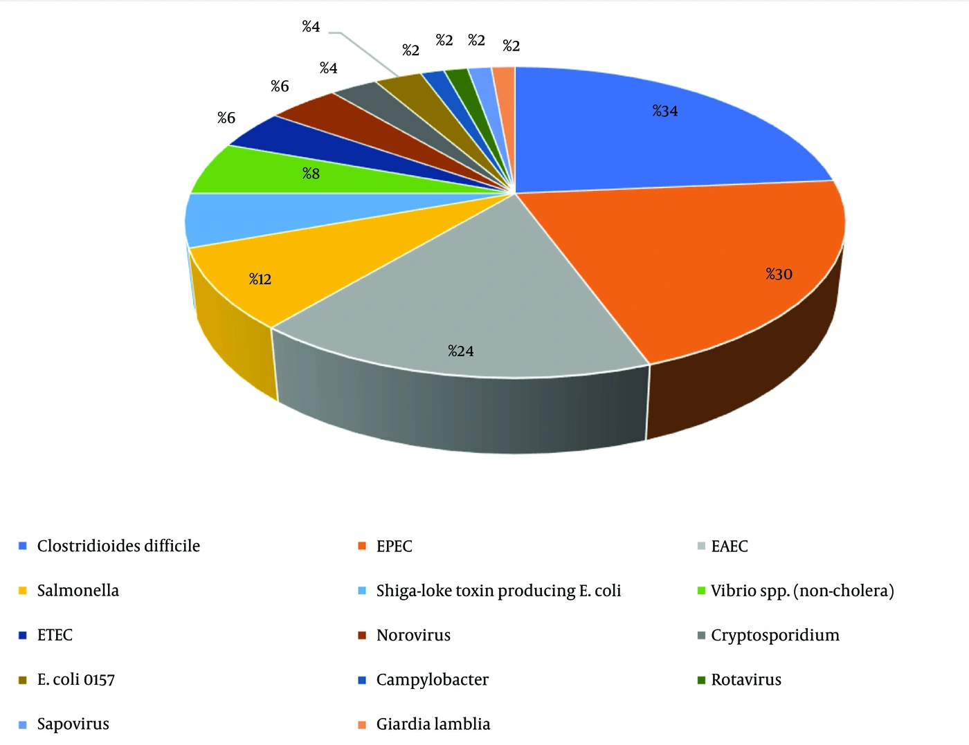
Figure 1.
Overall etiology of hospital-acquired gastroenteritis in patients with cancer. EPEC: Enteropathogenic E. coli; EAEC: enteroaggregative E. coli; ETEC: enterotoxigenic E. coli.
4.2. Etiologic Distribution in Neutropenic and Non-Neutropenic Patients
A separate subgroup analysis was performed to assess differences in etiology. The predominant pathogens identified in non-neutropenic cases were EAEC and EPEC (16 cases), followed by C. difficile (11 cases). In neutropenic cases, the order differed slightly, with C. difficile (6 cases), EAEC (5 cases), EPEC (3 cases), and Salmonella (3 cases) being the most prevalent. The distribution of these pathogens did not differ significantly between neutropenic and non-neutropenic cases (Table 3).
Table 3.
Distribution and Comparison of Etiologic Agents by Neutropenic (< 500/mm3) and Non-Neutropenic Status a
| Etiologic Agent | Non-neutropenic (n = 33) | Neutropenic (n = 17) | Total | P-Value |
|---|---|---|---|---|
| C. difficile | 11 (33.3) | 6 (35.3) | 17 | 0.890 |
| EPEC | 10 (30.3) | 5 (29.4) | 15 | 0.948 |
| EAEC | 9 (27.3) | 3 (17.6) | 12 | 0.450 |
| Salmonella spp. | 3 (9.1) | 3 (17.6) | 6 | 0.378 |
| ETEC | 3 (9.1) | 0 (0) | 3 | 0.200 |
| Shiga-like toxin-producing E. coli | 1 (3.0) | 3 (17.6) | 4 | 0.071 |
| Vibrio spp. | 3 (9.1) | 1 (5.9) | 4 | 0.692 |
| E. coli O157 | 1 (3.0) | 1 (5.9) | 2 | 0.626 |
| Norovirus | 1 (3.0) | 2 (11.8) | 3 | 0.218 |
| Cryptosporidium | 1 (3.0) | 1 (5.9) | 2 | 0.626 |
| Campylobacter | 1 (3.0) | 0 (0) | 1 | 0.468 |
| Giardia | 1 (3.0) | 0 (0) | 1 | 0.468 |
| Rotavirus | 0 (0) | 1 (5.9) | 1 | 0.159 |
| Sapovirus | 1 (3.0) | 0 (0) | 1 | 0.468 |
| Total number of microorganisms detected in GI Panel | 46 | 26 | 72 | 0.810 |
a Values are expressed as No. (%) or number. Abbreviations: EPEC, Enteropathogenic E. coli; EAEC, enteroaggregative E. coli; ETEC, enterotoxigenic E. coli; GI, gastrointestinal.
4.3. Etiologic Distribution in Best Supportive Care Patients
Best supportive care (BSC) in patients with cancer refers to high-quality symptom and adverse-effect management intended to enhance quality of life. This includes pain management, nutritional support, and palliative care services (13). When no cancer treatment options remain, the course of care may be classified as BSC, provided there is mutual agreement among the treating physician, patient, and family. Four cases were classified as BSC either on the day of GI panel positivity or within 30 days. The detected pathogens were C. difficile (n = 2), sapovirus (n = 1), and EAEC (n = 1).
4.4. Concordance of Stool Culture and the Gastrointestinal Panel
Concomitant stool cultures were obtained in 30 of 50 cases (60%). Stool cultures were obtained concurrently in 9 cases that tested positive for Salmonella, Shigella, or Vibrio spp. on the GI Panel, which are etiologic agents assessed by conventional stool culture. Of these, 5 (55.5%) also had positive stool-culture results for Salmonella spp. or Vibrio spp. All 5 stool culture-positive cases were also positive by the GI panel. Therefore, when stool culture was considered the gold standard, the GI panel had a sensitivity of 100%. Conversely, when the GI panel was considered the gold standard, stool culture had a sensitivity of 55.5%. The agreement rate between stool culture and the GI panel was 55.5% (5 of 9). Because the GI panel was positive for other pathogens in 25 cases in which concomitant GI panel testing and conventional stool culture were performed, the GI panel delineated the etiology of diarrhea in an additional 83.3% (25/30) of cases.
4.5. Day-30 Mortality Following GI Panel Positivity
The all-cause day-30 mortality rate following GI panel positivity was 14% (7/50). Three patients had ongoing diarrhea on the day of death. In addition, one patient had persistent Klebsiella pneumoniae bloodstream infection, and another had candidemia on the day of death. We evaluated whether BSC status was associated with higher mortality in the study population. Four patients were classified as BSC on the day of GI panel positivity and 46 as non-BSC. In the BSC group, 2 patients died (50%) during the 30-day follow-up period after GI panel positivity, whereas 2 survived. In the non-BSC group, 5 patients died (10.9%) during the 30-day follow-up, and 41 survived (Figure 2). There was no statistically significant association between BSC status and mortality among patients who developed diarrhea (P = 0.089, Fisher exact test), neutropenic status on the day of GI positivity, or hematologic vs oncologic diagnosis (Table 4).
Table 4.
One-Month Mortality Versus Neutropenic Status, Best Supportive Care Status, and Underlying Malignancy Type a
| Variables | One-Month Mortality | P-Value |
|---|---|---|
| Neutropenia (ANC < 500/mm3) | 3/17 (17.6%) | 0.6765 |
| No neutropenia (ANC < 500/mm3) | 4/33 (12.1%) | |
| Best supportive care status positive | 2/4 (50%) | 0.089 |
| Best supportive care status negative | 5/46 (10.9%) | |
| Hematology case | 3/25 (12%) | 1 |
| Oncological malignancy | 4/25 (16%) |
a Abbreviation: ANC, Absolute neutrophil count.
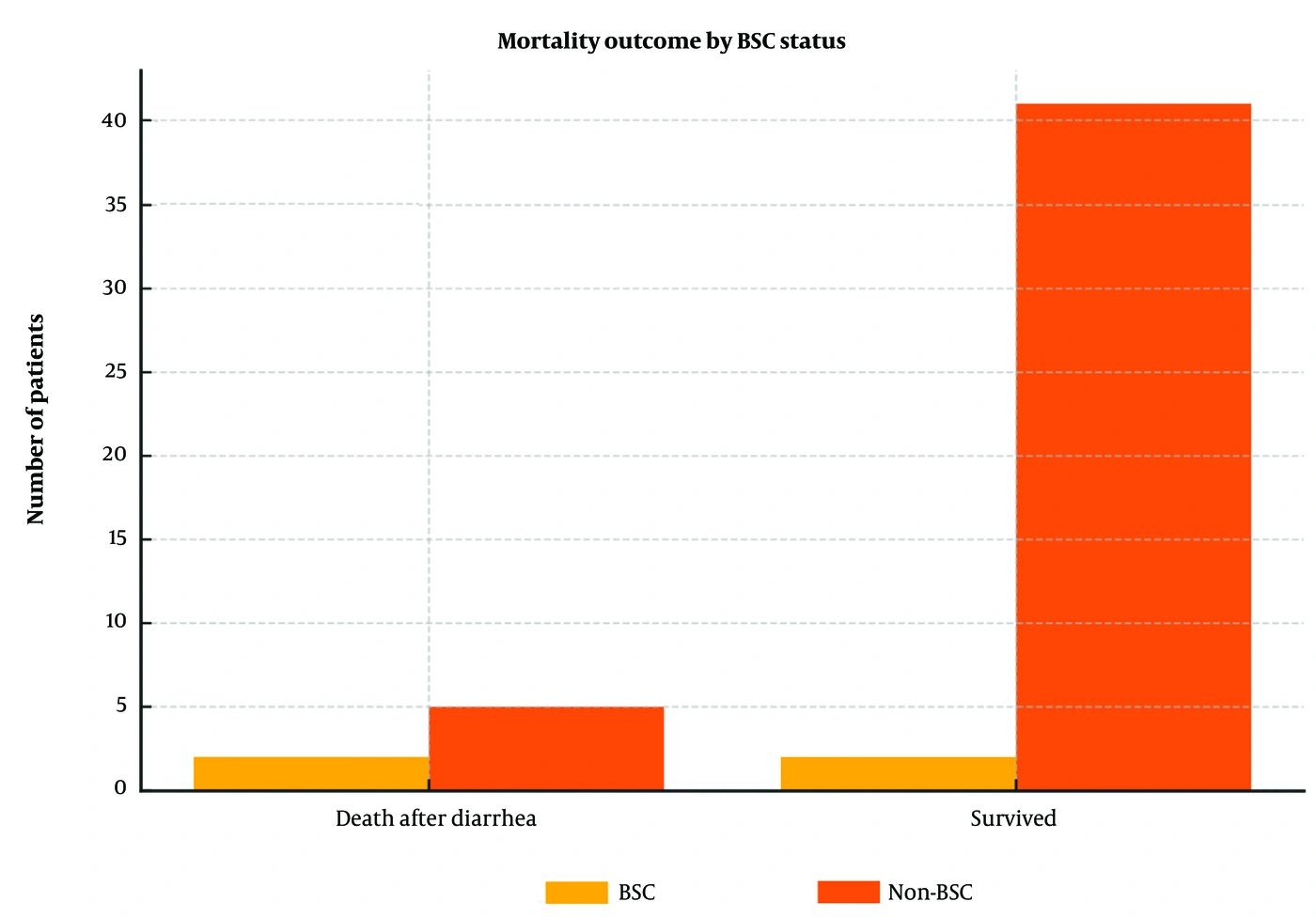
Figure 2.
Outcome of the study population by best supportive care status.
5. Discussion
Diarrhea in patients with hematologic malignancies or solid tumors may arise from infectious and noninfectious causes. Infectious episodes may be associated with pathogens acquired in the community or hospital environment (14). Although acute gastroenteritis is generally self-limiting and antimicrobial therapy is not routinely indicated, the approach differs in immunocompromised individuals. In this population, infectious diarrhea may warrant antimicrobial treatment because of the higher risk of complications (15). Effective management and hospital infection control largely depend on timely and accurate identification of the underlying pathogen (16). Traditional diagnostic approaches, including stool culture, microscopic examination, and antigen-based assays, are often time-consuming and technically demanding, and their diagnostic scope remains limited. These methods may fail to detect several clinically relevant organisms, particularly diarrheagenic E. coli strains and many viral pathogens. In light of these limitations, multiplex molecular diagnostic panels that simultaneously screen for a broad spectrum of bacterial, viral, and parasitic agents offer a more rapid and sensitive alternative. Although such assays have been assessed in different clinical contexts, data on their diagnostic performance specifically in oncology patients remain limited (17, 18).
The implementation of molecular diagnostic techniques has been shown to substantially increase detection rates of enteric pathogens compared with conventional laboratory methods (19). The FilmArray GI Panel (BioFire-bioMérieux, France) is an FDA-cleared multiplex assay capable of identifying 22 gastrointestinal pathogens directly from stool samples using nested multiplex PCR technology. This system provides results within approximately 2 hours and is designed to be user-friendly, facilitating earlier clinical decision-making and the initiation of targeted therapy (15). Reported performance characteristics of the panel include a sensitivity of 95%, a specificity of 100%, a positive predictive value of 100%, and a negative predictive value of 99%, whereas corresponding values for routine methods were 75%, 99%, 94%, and 94%, respectively (8). The high negative predictive value is particularly valuable in hemato-oncology patients because a negative result may prompt clinicians to investigate noninfectious causes of diarrhea, such as chemotherapy-related toxicity or graft-versus-host disease, which are common in this group (8). In our institution, the FilmArray GI Panel is routinely used to evaluate infectious causes of acute diarrhea in patients with cancer. In our cohort, 14/22 (63.6%) detectable pathogens were identified, suggesting that the assay covers the predominant regional etiologic agents. Moreover, compared with stool culture alone, the GI panel identified an infectious etiology in an additional 25 patients (83.3%), highlighting its added diagnostic value in this population.
A strong association has been reported between diarrhea caused by diarrheagenic E. coli and immunosuppression after recent chemotherapy (20). Similarly, our analysis showed that more than half of the cases (54%) were infected with diarrheagenic E. coli. Although adenovirus and rotavirus constitute one-third of causative agents (21), particularly in pediatric gastroenteritis, this rate was lower in our cohort because only adults were included. C. difficile remains an important cause of morbidity and mortality, particularly in older hospitalized patients, and is commonly associated with antibiotic exposure (22). Consistent with this, our series showed that C. difficile was among the leading pathogens responsible for infectious diarrhea in patients with malignancy. Because organisms other than C. difficile are more likely to be associated with improperly cooked food, including undercooked or restaurant-prepared food, our Infection Prevention and Control (IPC) Unit placed greater emphasis on strict food safety at home for discharged patients after these results. Given that no pathogen overlap or clustering was observed within a +/- 48-hour period, patients may have been infected through food brought from home or other outsourced food sources, such as hospital cafes or vendors outside the hospital. Similarly, in a study evaluating only diarrheagenic E. coli in patients with cancer, one-third of cases developed diarrhea after 48 hours of hospitalization without clusters (20). Based on the presented analysis, the IPC Unit suggested: 1) enhancing food-safety measures for both inpatient and discharged hemato-oncology patients and 2) introducing further regulations to reduce the potential use of outsourced food, including home-cooked meals, for inpatients.
Our results showed that non-Hodgkin lymphoma was prevalent in the study population, although breast and colon cancers are the 2 most common cancers in our country (23). We propose that the increased frequency of non-Hodgkin lymphoma may be linked to a higher likelihood of neutropenia; 8 of 12 patients with non-Hodgkin lymphoma had neutropenia < 500/mm3 on the day of GI panel positivity (data not shown). Although most cases of infectious diarrhea are acute and self-limited, they may still lead to serious complications and death (24). In our study, the overall all-cause 30-day mortality rate was 14%. One-month mortality was 50% (2/4) in BSC cases versus 10.9% (5/46) in other cases. Previous studies have indicated that BSC status is often associated with higher mortality, regardless of cancer etiology (25). Although the difference was not statistically significant (P = 0.089), our findings were consistent with these reports, as mortality was also higher among BSC cases, that is, patients for whom active cancer treatment was unavailable because of advanced disease. The lack of temporal clusters led us to hypothesize that individual exposures, such as food brought from home or outsourced sources, may contribute to hospital-acquired gastroenteritis in this cohort. However, the study did not include direct exposure measurements, dietary logs, or molecular strain typing (eg, pulsed-field gel electrophoresis [PFGE] or whole-genome sequencing [WGS]) to link clinical isolates to specific food vehicles. Consequently, prospective multicenter studies are required to validate these hypotheses and provide more definitive data regarding infection sources.
5.1. Study Limitations
This study has several limitations. The epidemiologic profile of nosocomial gastroenteritis observed here reflects the clinical environment of a national referral center in Bahrain. Although these results provide important insights into pathogens affecting immunocompromised patients in this region, their generalizability to other geographic areas or community-based hospitals may be limited. Therefore, the findings of this single-center retrospective cohort study should be verified in larger multicenter studies. Because the study was restricted to hospital-acquired gastroenteritis episodes identified through GI multiplex PCR panel testing, not all diarrhea or gastroenteritis episodes occurring during the study period were included. Consequently, the full denominator of diarrhea episodes could not be determined, and selection bias in the representation of etiologic pathogens may exist. None of the included patients underwent autopsy; therefore, a direct causal relationship between 30-day mortality and gastroenteritis could not be established.
In addition, detection of these pathogens by molecular techniques may not necessarily indicate that the detected agent was responsible for the diarrhea because some patients may have been asymptomatic carriers. Nevertheless, all patients in the cohort had clinically significant diarrhea meeting the gastroenteritis criteria and had positive GI panel results. Thus, although a causal relationship between diarrhea and GI panel results could not be proven, the etiologic agents reported here were likely contributors to the diarrhea. Patients admitted with non-hemato-oncologic diseases could have similar or different pathogen profiles; however, because our center is an oncology hospital, such patients could not be included. The sample size could not be increased because of relatively long periods of GI Panel unavailability after November 2022. Pathogens causing infectious diarrhea can substantially affect health outcomes in patients with cancer, leading to prolonged illness, disruption of cancer treatment, extended hospital stays, and, in severe cases, increased mortality. At this stage, only the 30-day mortality rate was assessed, but we aim to explore other variables in future studies. To the best of our knowledge, this is the first study to evaluate GI panel results and provide a detailed description of the etiology of hospital-acquired gastroenteritis in the hematology/oncology patient subgroup.
5.2. Conclusions
Our results suggest that, despite its cost, the GI panel can substantially aid in identifying the probable cause of gastroenteritis. Routine stool cultures appear to have limited ability to identify the etiology. EAEC, EPEC, Salmonella, and C. difficile appear to be important etiologic agents of diarrhea in hematology/oncology patients in our region. We recommend enhancing food safety in this patient population and providing structured home-health education on food handling, preparation, and safety specifically for immunocompromised patients.