
1. Introduction
Mitochondria are lipid-bilayer membrane organelles commonly referred to as the “powerhouse” of the cell owing to their pivotal role in producing ATP through oxidative phosphorylation (1). Beyond this function, mitochondria play several other crucial roles, including programmed cell death, redox balance, ion homeostasis, and innate immune signaling (2). The importance of mitochondrial health and function in progenitor cells becomes even more pronounced during proliferation (3, 4). During this state, mitochondrial number increases, and cellular metabolism shifts from glycolysis to oxidative phosphorylation, a transformation that increases mitochondrial sensitivity to free radicals (3, 5).
Pharmacological approaches to restoring mitochondrial structure and function are broadly categorized as targeted or untargeted mitochondrial drugs (6). Untargeted strategies include inducing mitochondrial biogenesis with PPAR agonists (e.g., pioglitazone), Sirt1 activators (e.g., resveratrol), and AMPK activators (e.g., PXL770), and modulating the redox state with antioxidants such as sonlicromanol (6-9). Targeted approaches include direct modulation of specific mitochondrial pathways, such as the use of mitochondrial pyruvate-carrier inhibitors (for example, thiazolidinediones) and enhancement of oxidative phosphorylation with coenzyme Q10 (Q10) (10). Mitochondria-targeted delivery enables higher local drug concentrations while minimizing systemic exposure, thereby improving the therapeutic index and reducing off-target toxicity (11).
Q10 (ubiquinone) is a lipid-soluble cofactor located in the inner mitochondrial membrane (12). It transfers electrons from complexes I and II, and from fatty-acid and amino-acid oxidation, to complex III. Its reduced form, ubiquinol, functions as a potent antioxidant that protects against lipid peroxidation and oxidative damage to proteins and DNA (13). Consistent with this cytoprotective role, recent in vivo evidence indicates that coenzyme Q10 supplementation attenuates oxidative-stress–mediated tissue damage, reinforcing the rationale for developing efficient Q10 delivery systems (14). Owing to its essential role in mitochondrial bioenergetics and antioxidant defense, together with its low toxicity, Q10 is widely used in mitochondrial disorders (15). However, crystalline Q10 has poor water solubility and low bioavailability, necessitating solubilized formulations or mitochondria-targeted analogs (16) to enhance cellular and mitochondrial delivery (17).
Various strategies and systems are used to deliver drugs to mitochondria, with the choice depending on the therapeutic goal. The principal nanocarrier classes designed for mitochondria-targeted delivery include liposomes, polymeric nanoparticles, and inorganic nanoparticles (18). Cationic liposomes are commonly prepared by combining cationic lipids with zwitterionic or neutral helper lipids, whereas mitochondria-targeted liposomes can be obtained by functionalizing the lipid bilayer with lipophilic cations such as triphenylphosphonium (TPP) (19). Direct conjugation of TPP to drug molecules can reduce intrinsic potency through steric hindrance and unfavorable physicochemical changes (20); anchoring TPP to lipid carriers such as liposomes is therefore a promising strategy to enhance mitochondrial targeting while maintaining drug activity (21, 22).
Liposomes are phospholipid vesicles and versatile carriers capable of incorporating both hydrophobic and hydrophilic agents. Compared with non-liposomal mitochondrial carriers (polymeric dendrimers, metal nanoparticles, and micelles), cationic liposomes typically enable the use of endogenous or biomimetic lipids, improving biocompatibility; allow easier control over size, charge, and surface targeting ligands; and interact closely with mitochondrial membranes to promote direct drug transfer (23, 24). Direct TPP conjugation to liposomes can disrupt bilayer packing, induce curvature stress, and reduce colloidal stability owing to the cationic and hydrophobic nature of TPP, potentially causing leakage, fusion, and aggregation (25, 26). Nevertheless, stearyl-TPP (STPP) anchoring provides stable and predictable membrane insertion without requiring complex lipid modifications (27).
Research gap and aim. Most TPP strategies conjugate TPP directly to the drug (risking loss of potency) or rely on stearyl-TPP for a narrow payload range; systematic design-of-experiments optimization and physicochemical characterization of a purpose-synthesized, lipid-anchored TPP conjugate remain underexplored. The present study addresses this gap. TPP was conjugated to octadecylamine (ODA) through a mild synthetic strategy to generate a lipid-compatible targeting moiety, and the resulting TPP-anchored liposomes were formulated and systematically optimized using a factorial design, leading to selection of the most suitable formulation based on predefined physicochemical criteria (Figure 1). Cellular and in vivo evaluation is deferred to the subsequent phase of the project.
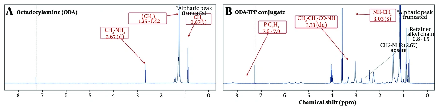
Figure 1.
optimized TPP-functionalized cationic liposomal coenzyme Q10 for mitochondrial targeting. Created with BioRender.com.
2. Methods
2.1. Materials
(2-Carboxyethyl)triphenylphosphonium bromide was purchased from Merck (China). Octadecylamine was obtained from Merck (Germany). 1-Ethyl-3-(3-dimethylaminopropyl)carbodiimide (EDC) was purchased from Alfa Aesar (USA), and N-hydroxysuccinimide (NHS) was obtained from Merck (Germany). Pharmaceutical-grade coenzyme Q10 (Q10) was sourced from Dana Pharmaceutical Company (Iran). L-α-phosphatidylcholine (soybean lecithin) was purchased from Acros (Belgium). Cholesterol was obtained from Solarbio (China). Sodium chloride (NaCl) was purchased from Merck (Germany).
2.2. Preparation of the Targeting Agent
The synthesis of the octadecylamine–triphenylphosphonium conjugate (ODA-TPP) was performed in several steps. Reflux conditions were maintained at 50 - 60 °C. (2-Carboxyethyl)triphenylphosphonium (820 mg) was dissolved in 45 mL ethanol; EDC (620 mg) was added, and the mixture was stirred for 1 h. After NHS (460 mg) was added and stirring was continued, ODA (540 mg) was introduced dropwise while maintaining stirring at 500 rpm for a further 24 h. The mixture was transferred to a flask, half of the solvent was evaporated at 50 °C, and the residue was stored in a refrigerator. Product separation was initiated by salting-out (NaCl solution), followed by chloroform extraction; after separation of the organic layer, the solvent was removed to yield a semi-solid, light-yellow product.
The conjugate was obtained with an 84.6% yield. Reaction progress and product purity were monitored by thin-layer chromatography (Rf = 0.42; chloroform:methanol 9:1 v/v), and the purity of the isolated ODA-TPP was 96.8% as determined by HPLC.
2.3. FTIR and NMR
Fourier-transform infrared (FTIR) and nuclear magnetic resonance (NMR) spectroscopy were used to confirm chemical-bond formation. FTIR analysis was performed using a Thermo Avatar FTIR spectrometer to identify characteristic functional groups and bonding interactions. Structural confirmation was further performed by 1H-NMR spectroscopy. All analyses were conducted under standard operating conditions (28).
2.4. Preparation of the Liposomal Formulation
Liposomes were prepared by thin-film hydration (29). ODA-TPP, lecithin, cholesterol, ODA, and Q10 were dissolved in 5 mL chloroform and sonicated for 5 min. The solvent was removed using a rotary evaporator (50 °C, 120 rpm), and the film was vacuum-dried overnight. The dried film was hydrated with preheated phosphate buffer (pH 7.0 - 7.4, 50 °C) to generate a liposomal dispersion, which was downsized by high-shear homogenization (up to 15,000 rpm, 10 min) with additional probe sonication (100 W, 5 min) to obtain a uniform nanosized dispersion (30). During homogenization, the temperature was maintained below 40 °C to minimize Q10 oxidation and lipid degradation. The final dispersion was transferred to amber glass vials, stored at 4 °C, and further characterized.
2.5. Optimization Using a Full Factorial Design
A 23 randomized full factorial design was used to evaluate three independent factors, each at two levels (Table 1) (31); all eight combinations were prepared and evaluated. Preliminary experiments defined the critical variables and their ranges. Each factor was coded at two levels, −1 (low) and +1 (high): Chol:PC (0.2/0.5), PC (10/25 mg), and drug amount (2.5/5.0 mg). Uncoded (natural-unit) regression equations were derived by back-substitution of the coding transformation. Multi-response optimization used a desirability-function approach, minimizing particle size and maximizing entrapment efficiency (composite desirability 0.786 at the optimum).
Table 1.
Liposome Formulation Components
| Formulation | Factorial state | Chol:PC (w/w) | PC (mg) | Drug (mg) | TPP (mg) | ODA (mg) |
|---|---|---|---|---|---|---|
| Lipo-Q10 - 1 | – – – | 0.2 | 10 | 2.5 | 3 | 5 |
| Lipo-Q10 - 2 | + – – | 0.5 | 10 | 2.5 | 3 | 5 |
| Lipo-Q10 - 3 | – + – | 0.2 | 25 | 2.5 | 3 | 5 |
| Lipo-Q10 - 4 | + + – | 0.5 | 25 | 2.5 | 3 | 5 |
| Lipo-Q10 - 5 | – – + | 0.2 | 10 | 5.0 | 3 | 5 |
| Lipo-Q10 - 6 | + – + | 0.5 | 10 | 5.0 | 3 | 5 |
| Lipo-Q10 - 7 | – + + | 0.2 | 25 | 5.0 | 3 | 5 |
| Lipo-Q10 - 8 | + + + | 0.5 | 25 | 5.0 | 3 | 5 |
2.6. Particle Size and Zeta Potential
Particle size was evaluated at 25 °C by photon-correlation spectroscopy using a Scatter Scope 1 QUIDIX analyzer (South Korea) (32). Surface charge (zeta potential) was assessed using a Malvern Zetasizer Nano (Malvern Instruments Ltd., Malvern, UK). All samples were measured in triplicate. Particle size (Z-average) and PDI were derived from intensity-weighted cumulants analysis, and representative intensity size-distribution profiles were recorded.
2.7. Q10 Quantification and HPLC Method Validation
Q10 was quantified by high-performance liquid chromatography (HPLC) using a LiChrospher C8 column with a methanol/acetonitrile mobile phase (70:30, v/v) at 1 mL min-1, UV detection at 275 nm, and a retention time of ≈9 - 10 min. Quantification used a six-point calibration curve over 1 - 30 ppm, performed in triplicate (33).
The method was validated according to ICH Q2(R1). Linearity was established over 1 - 30 µg/mL (Area = 796.8·C − 376; R2 = 0.9996). The limits of detection and quantification (3.3σ/S and 10σ/S) were 0.37 and 1.12 µg/mL, respectively. Precision was intra-day relative standard deviation (RSD) 1.2% and inter-day RSD 1.8%, and accuracy (mean recovery) was 98.4%. Robustness, solution-stability, and forced-degradation (stability-indicating) evaluations under ICH Q2(R1) were not undertaken; therefore, the method was validated for quantification only and is not established as stability-indicating (see Study limitations).
2.8. Entrapment Efficiency
Free (unencapsulated) drug was separated from the vesicles by high-speed centrifugation (12,000 rpm, 20 min), followed by secondary centrifugation of the supernatant; the free drug in the supernatant was quantified to obtain the entrapment efficiency (EE%) indirectly, and the pellet was additionally dissolved in methanol to enable direct quantification of the entrapped drug as a cross-check. Ultrafiltration and size-exclusion chromatography are valid alternatives; however, double centrifugation with direct/indirect cross-validation yielded internally consistent EE values (RSD 0.5 - 2.6%). EE% was calculated as:
where Wtotal is the total drug added and Wfree is the drug recovered in the supernatant.
2.9. In Vitro Release and Membrane-Diffusion Study
Release from the optimized liposomal formulation and from the free-drug suspension was studied using Franz diffusion cells fitted with a pre-hydrated regenerated-cellulose membrane (MWCO 12,000 - 14,000 Da). The receptor compartment contained 30 mL phosphate-buffered saline (pH 7.4) at 37 ± 0.5 °C under continuous magnetic stirring at 300 rpm. Aliquots were withdrawn at predetermined intervals and replaced with fresh pre-warmed medium to maintain sink conditions. Q10 was quantified by HPLC and expressed as the cumulative percentage released over time; release data were fitted to standard kinetic models, and the model with the highest R2 was selected (34). This Franz-cell configuration, which used a synthetic regenerated-cellulose membrane, characterizes membrane-controlled diffusion of coenzyme Q10 under sink conditions rather than true release from the vesicles; it was selected for its defined, constant diffusion area and well-mixed receptor compartment. Sample-and-separate and dialysis approaches, which more directly report vesicle-release kinetics, are designated as future work.
2.10. Stability Studies
The stability of the optimized formulation was evaluated over three months of storage at room temperature. At predetermined intervals, samples were analyzed for changes in particle size, zeta potential, and encapsulation efficiency as indicators of physical and chemical stability (32).
2.11. Transmission Electron Microscopy
The morphology and size of the liposomes were examined by TEM (Philips EM 208S, Netherlands) at 80 kV. Samples were negatively stained with 2% phosphotungstic acid after adsorption onto carbon-coated copper grids, enabling visualization of vesicle shape, lamellarity, and nanostructure to corroborate the DLS measurements (35).
2.12. Data Analysis
All experiments were performed in triplicate, and data are reported as mean ± standard deviation. Each formulation was prepared as three independent batches (n = 24 for the factorial analysis), each measured in analytical triplicate. The effects of the independent variables on particle size and entrapment efficiency were evaluated by two-way ANOVA. Residual normality was confirmed by the Shapiro–Wilk test (EE p = 0.34; size P = 0.43); because the saturated 23 model estimates all main and interaction terms over replicated runs, the residual error equals pure error and a separate lack-of-fit term is not estimable. Release profiles were compared by two-way repeated-measures ANOVA (formulation × time) and the similarity factor f2.
3. Results
3.1. FTIR
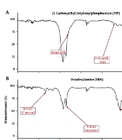
The FTIR spectrum of ODA showed two broad N–H stretching bands at 3300 - 3400 cm-1 (symmetric and asymmetric; primary amine) and a bending peak at ≈1600 - 1650 cm-1, together with strong C–H stretching bands at 2850 - 2950 cm-1 from the octadecyl chain (spectrum b). The spectrum of (2-carboxyethyl) triphenyl phosphonium bromide (TPP) displayed the carboxylic-acid C=O stretching band at ≈1710 - 1730 cm-1, with aromatic-ring vibrations at 1430 - 1600 cm-1 (C=C) and 690 - 750 cm-1 (out-of-plane C–H) (spectrum a). After EDC/NHS coupling, the product spectrum showed loss of the broad carboxylic O–H band (2500 - 3500 cm-1) and a new amide I (C=O) peak at ≈1650 - 1710 cm-1, a sharp amide N–H stretch at 3300 - 3350 cm-1, and the absence of the primary-amine bend at 1600 - 1650 cm-1; the aromatic phosphonium and alkyl C–H bands remained intact (spectrum c). These observations confirm the formation of a secondary amide bond between the amine group of ODA and the carboxyl group of TPP (Figure 2 and Tables 2 and 3).
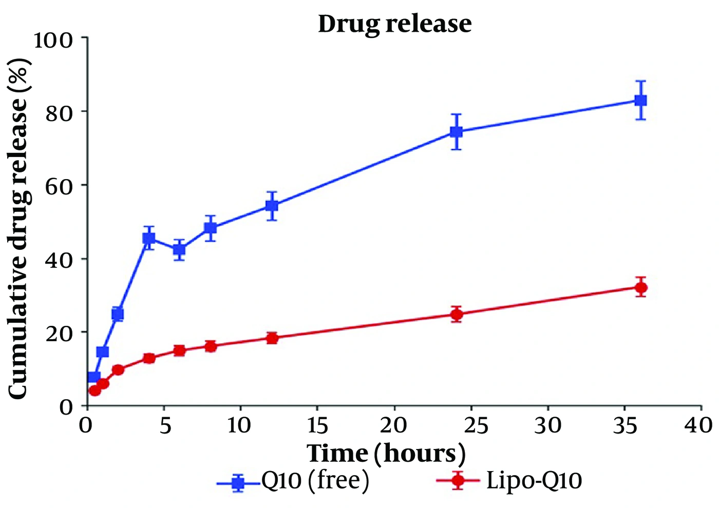
Figure 2.
FTIR spectra of (a) (2-carboxyethyl)triphenylphosphonium bromide (TPP), (b) octadecylamine (ODA), and (c) the synthesized ODA–TPP conjugate. Formation of the amide linkage was confirmed by the disappearance or substantial reduction of spectral features associated with the carboxylic acid and primary amine functionalities, accompanied by the appearance of characteristic amide I and amide II absorption bands. The preservation of alkyl-chain and aromatic phosphonium-related vibrations further indicates that the structural integrity of both precursor moieties was maintained during synthesis.
Table 2.
Characteristic FTIR Absorption Bands
| Species | Band (cm-1) | Assignment |
|---|---|---|
| ODA | 3300 - 3400 / 1600 - 1650 | N–H stretch / bend (primary amine) |
| ODA | 2850 - 2950 | C–H stretch (octadecyl) |
| TPP | 1710 - 1730 / 2500 - 3500 | C=O / O–H (carboxylic acid) |
| TPP | 1430 - 1600 / 690 - 750 | Aromatic C=C / C–H |
| ODA-TPP | 1650 - 1710 | Amide I (C=O); O–H and amine bend absent |
| ODA-TPP | 3300 - 3350 | Amide N–H; aromatic + alkyl retained |
Table 3.
1H-NMR Structural Assignments
| Species | δ (ppm) | Multiplicity | Assignment |
|---|---|---|---|
| ODA | 0.87 / 1.25 - 1.42 | t / m | CH3 / (CH2)n chain |
| ODA | 2.67 | d (J = 14.1 Hz) | CH2–NH2 |
| ODA-TPP | 3.03 / 3.33 | s / dq | NH–CH2 / CH2–CH2–CO–NH |
| ODA-TPP | ~7.6 - 7.9 | m (15H) | P–C6H5 (triphenylphosphonium) |
| ODA-TPP | 2.67 | Absent | Loss of CH2–NH2 (coupling confirmed) |
3.2. 1H NMR
The 1H-NMR spectrum of ODA showed a doublet at 2.67 ppm (J = 14.1 Hz) corresponding to the methylene protons adjacent to the primary amine (CH2–NH2), a terminal methyl triplet at 0.87 ppm, and long-chain methylene signals at 1.25 - 1.42 ppm (spectrum a). After EDC/NHS coupling, the 2.67 ppm doublet disappeared, confirming consumption of the primary amine. A new doublet of quartets appeared at 3.33 ppm (J = 6.4, 12.6 Hz, 2H; CH2 – CH2 – CO – NH) and a new singlet at 3.03 ppm (4H; NH – CH2), while the alkyl-chain methylene signals were retained at 0.77 - 1.5 ppm (spectrum b). Collectively, the loss of the CH2–NH2 signal, the emergence of the 3.33 and 3.03 ppm resonances, and the preservation of the alkyl signals confirm the formation of the secondary amide bond between ODA and TPP (Figure 3).
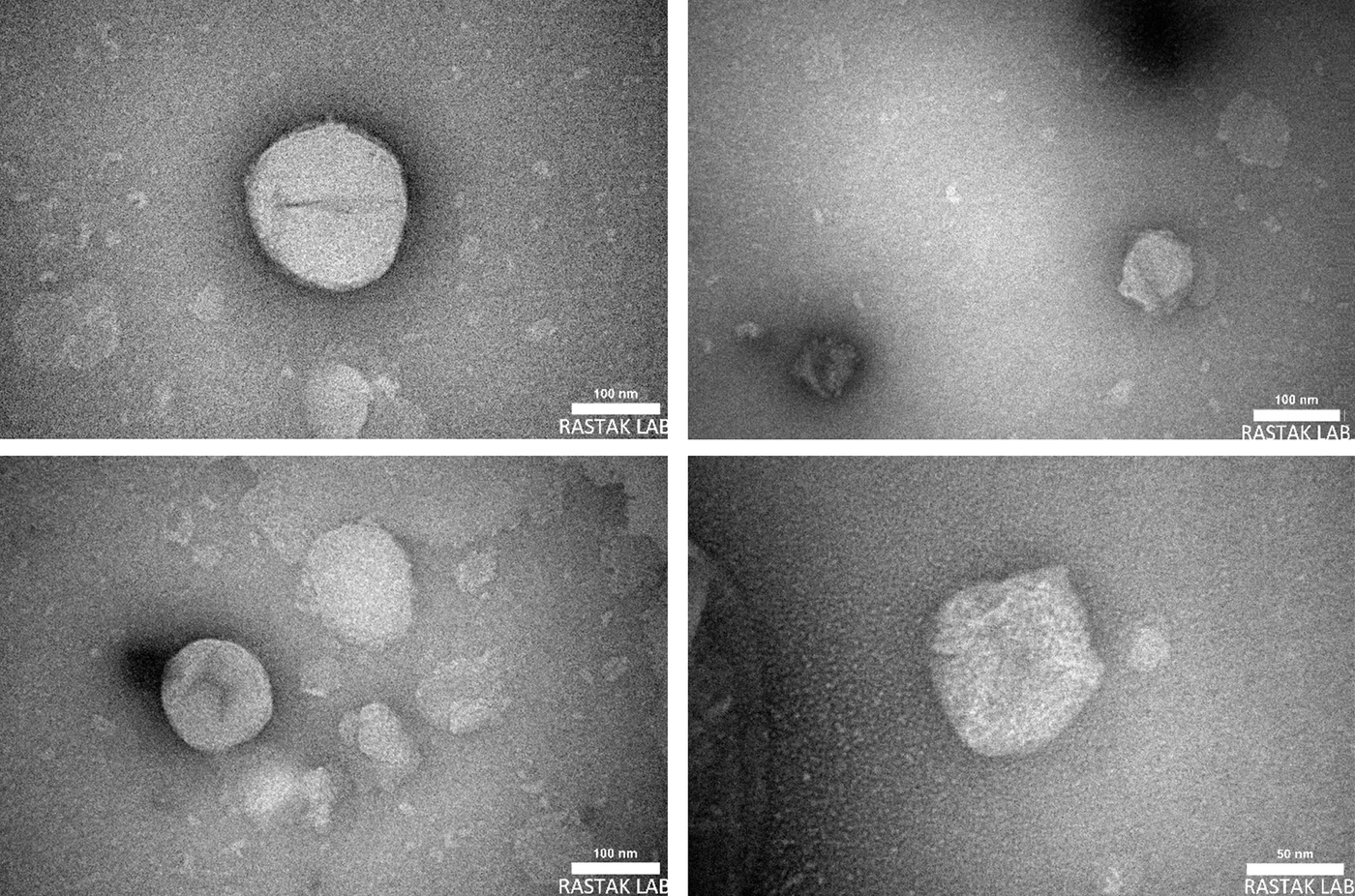
Figure 3.
1H NMR spectra of (a) octadecylamine (ODA) and (b) ODA–TPP conjugate synthesized via EDC/NHS-mediated amidation. Successful conjugation was evidenced by the disappearance of the methylene resonance adjacent to the terminal primary amine of ODA and the emergence of signals attributable to the newly formed amide linkage and triphenylphosphonium-containing structure. The persistence of the characteristic alkyl-chain resonances indicates that the lipophilic tail is structurally preserved after coupling.
3.3. Optimization Based on Particle Size and Entrapment Efficiency
A 23 full factorial design with three replicates per run (n = 24) was used to optimize the liposomes by evaluating the effects of the cholesterol-to-phosphatidylcholine ratio (Chol:PC, A: 0.2 - 0.5), PC amount (B: 10 - 25 mg), and drug amount (C: 2.5 - 5.0 mg) on particle size (Y1) and entrapment efficiency (EE, Y2). The mean particle size was 139.46 nm and the mean EE was 88.35%, with low coefficients of variation (4.31% and 5.72%), indicating a reproducible process.
Factorial regression revealed a significant model for particle size (F = 3.82, P = 0.013). Increasing Chol:PC increased particle size (effect +5.25; coefficient +2.63; p = 0.023), whereas increasing PC decreased it (effect −7.08; coefficient −3.54; p = 0.004); the drug amount showed a non-significant inverse trend (p = 0.415). The PC × drug interaction was marginal (p = 0.080), and the three-way interaction approached significance (P = 0.092). The uncoded regression equation was:
For EE, the model was robust (F = 9.41, P < 0.001; adjusted R2 = 71.91%). PC significantly enhanced EE (effect +8.217; coefficient +4.108; P < 0.001), whereas the drug amount reduced it (effect −2.533; coefficient −1.267; P = 0.033); the Chol:PC effect was non-significant (P = 0.181), and interactions were negligible (P > 0.347). The uncoded equation was:
Model diagnostics. EE model: F = 9.41, p < 0.001 (R2 = 80.46%, adjusted R2 = 71.91%, predicted R2 = 56.04%). Particle-size model: F = 3.82, p = 0.013 (R2 = 62.57%, adjusted R2 = 46.20%, predicted R2 = 15.79%). All variance-inflation factors were 1.00 (orthogonal design), and residuals were normally distributed (Shapiro–Wilk p = 0.34 and 0.43). The desirability-based optimum (Chol:PC 0.2, PC 25 mg, drug 2.5 mg; desirability 0.786) predicted a size of 129.3 nm and an EE of 95.0%.
Responses for the experimental matrix are summarized in Table 4. Particle sizes ranged from 129.3 to 146.7 nm (all < 150 nm, favoring passive targeting), and EE ranged from 82.6% to 94.9%. The optimal formulation balancing minimal size and maximal EE was Chol:PC = 0.2, PC = 25 mg, drug = 2.5 mg, with predicted values of 129.3 nm and 94.9%.
Table 4.
Physicochemical Characteristics of the Factorial Formulations a
| Formulation | Factorial state | Particle size (nm) | PDI | Zeta potential (mV) | EE (%) |
|---|---|---|---|---|---|
| Lipo-Q10 - 1 | – – – | 146.7 ± 5.5 | 0.240 ± 0.046 | 8.5 ± 0.6 | 86.8 ± 2.3 |
| Lipo-Q10 - 2 | + – – | 145.0 ± 2.6 | 0.343 ± 0.026 | 7.5 ± 0.7 | 84.2 ± 1.8 |
| Lipo-Q10 - 3 | – + – | 129.3 ± 3.8 | 0.187 ± 0.021 | 11.7 ± 0.3 | 95.0 ± 2.3 |
| Lipo-Q10 - 4 | + + – | 140.3 ± 8.0 | 0.307 ± 0.021 | 8.2 ± 0.4 | 92.5 ± 2.3 |
| Lipo-Q10 - 5 | – – + | 136.7 ± 6.1 | 0.327 ± 0.031 | 10.8 ± 0.6 | 83.3 ± 2.9 |
| Lipo-Q10 - 6 | + – + | 143.7 ± 3.2 | 0.313 ± 0.021 | 8.0 ± 0.6 | 82.7 ± 2.8 |
| Lipo-Q10 - 7 | – + + | 134.7 ± 1.5 | 0.297 ± 0.031 | 10.2 ± 0.4 | 91.3 ± 4.0 |
| Lipo-Q10 - 8 | + + + | 139.3 ± 6.7 | 0.277 ± 0.015 | 9.6 ± 0.5 | 91.0 ± 2.2 |
a Values are expressed as mean ± SD.
3.4. In Vitro Release and Membrane Diffusion
In vitro release from the selected formulation and the free-drug suspension was assessed in triplicate over 36 h under sink conditions (PBS, pH 7.4, 37 °C; Figure 4). Cumulative release is summarized in Table 5 (mean ± SD, n = 3).
Table 5.
Cumulative Drug Release (%) from the Liposomal Formulation and the Free-Drug Suspension a
| Time (h) | Lipo-Q10 (%) | Free Q10 (%) |
|---|---|---|
| 0.5 | 4.1 ± 0.4 | 7.8 ± 0.8 |
| 1 | 6.0 ± 0.6 | 14.6 ± 1.2 |
| 2 | 9.8 ± 0.9 | 24.9 ± 1.9 |
| 4 | 12.9 ± 1.1 | 45.5 ± 3.1 |
| 6 | 15.0 ± 1.3 | 42.4 ± 2.8 |
| 8 | 16.2 ± 1.4 | 48.2 ± 3.4 |
| 12 | 18.4 ± 1.5 | 54.3 ± 3.9 |
| 24 | 24.8 ± 2.1 | 74.4 ± 4.8 |
| 36 | 32.3 ± 2.6 | 83.0 ± 5.2 |
a Values are expressed as mean ± SD.
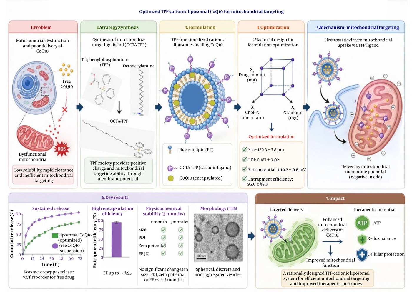
Figure 4.
Cumulative drug release profiles of the selected liposomal formulation (red squares) and free drug suspension (blue circles) over 36 hours. Data points represent mean values ± SD (n = 3). The liposomal formulation exhibits sustained release compared to the rapid release from the suspension.
A two-way repeated-measures ANOVA (formulation × time) confirmed significant differences in cumulative release between the formulation and the free drug. The similarity factor was f2 = 24.4 (< 50), confirming dissimilar profiles and a sustained-release effect. To elucidate the mechanism, the mean data were fitted to zero-order, first-order, Higuchi, and Korsmeyer–Peppas models (Tables 6 and 7).
Table 6.
Kinetic Modeling of Drug Release
| Models and Parameters | Lipo-Q10 | Free Q10 |
|---|---|---|
| Zero-order | ||
| R2 | 0.924 | 0.820 |
| k (%/h) | 0.71 | 1.90 |
| First-order | ||
| R2 | 0.948 | 0.960 |
| k (h-1) | 0.009 | 0.045 |
| Higuchi | ||
| R2 | 0.987 | 0.944 |
| k | 4.98 | 13.80 |
| Korsmeyer–Peppas | ||
| R2 | 0.986 | 0.942 |
| n | 0.458 | 0.616 |
| k | 6.26 | 14.28 |
Table 7.
Physicochemical Stability of the Optimized Formulation Over Three Months a
| Time Points | Particle Size (nm) | PDI | Zeta Potential (mV) | EE (%) |
|---|---|---|---|---|
| First day | 129.3 ± 3.8 | 0.187 ± 0.021 | 11.70 ± 0.312 | 95.0 ± 2.3 |
| 1st month | 130.0 ± 3.6 | 0.262 ± 0.070 | 11.93 ± 0.251 | 93.8 ± 2.5 |
| 2nd month | 132.3 ± 1.2 | 0.284 ± 0.040 | 11.06 ± 0.776 | 92.7 ± 2.5 |
| 3rd month | 134.0 ± 1.0 | 0.287 ± 0.039 | 11.20 ± 0.458 | 92.5 ± 1.9 |
a Values are expressed as mean ± SD.
For the coenzyme Q10 data, the formulation best fit the Higuchi model (R2 = 0.987), with a Korsmeyer–Peppas exponent n = 0.458, indicating diffusion-controlled (Fickian) release; the free drug best fit first-order kinetics (R2 = 0.960), consistent with concentration-dependent dissolution.
3.5. Stability of the Optimized Liposomes
Over three months at room temperature, particle size (129.3 → 134 nm, p = 0.158), zeta potential (11.7 → 11.2 mV, p = 0.202), and entrapment efficiency (95.0 → 92.5%, p = 0.223) showed no statistically significant change, indicating preservation of size, surface charge, and drug loading. The polydispersity index increased significantly (0.187 → 0.287, p = 0.029), reflecting a modest shift toward a broader size distribution during storage; no liposome cake formation was observed.
Limitation. Stability was assessed only under ambient room-temperature storage. ICH Q1A(R2) conditions (4 °C; 25 °C/60% RH; 40 °C/75% RH under controlled humidity) were not employed; therefore, these data do not permit formal shelf-life assignment, and full ICH-compliant stability testing is designated as future work.
3.6. Transmission Electron Microscopy
TEM showed vesicles that were generally intact and predominantly spherical, with a visible lipid bilayer confirming preserved structural integrity and relatively smooth surfaces with slight indentations. No vesicle fusion or notable aggregation was observed, supporting the physical stability of the cationic liposomes (Figure 5).
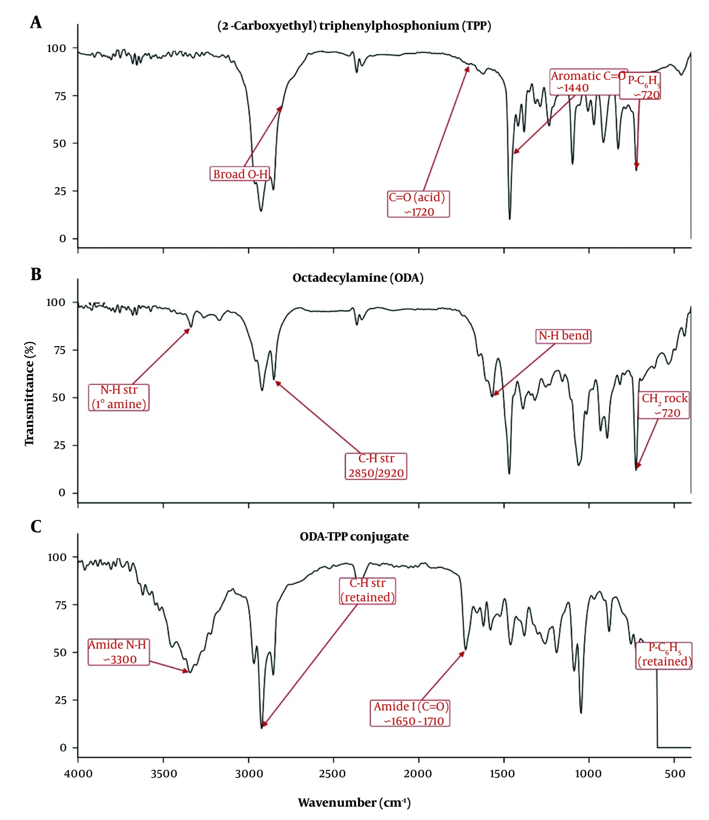
Figure 5.
Transmission electron microscopy (TEM) images of the optimized liposomal formulation. The vesicles exhibit predominantly spherical morphology with relatively smooth surfaces and slight surface undulations.
Quantitatively, fourteen individual, non-overlapping vesicles were measured (ImageJ), yielding a mean diameter of 47.22 ± 8.94 nm (range 30.3 - 67.7 nm; approximately normal, Shapiro–Wilk P = 0.57). This is markedly smaller than the DLS Z-average (129.3 nm), primarily because DLS reports an intensity-weighted hydrodynamic diameter (scaling with radius6 and dominated by the largest vesicles and aggregates), whereas TEM yields a number-weighted geometric diameter; dehydration during negative staining and loss of the hydration/counter-ion layers further contribute. Given the magnitude of this difference and the limited count (n = 14), the TEM measurements corroborate the nanoscale, spherical, non-aggregated morphology rather than providing a quantitative match to DLS.
4. Discussion
Mitochondrial drug delivery requires precisely engineered carriers capable of traversing the organelle’s double membrane. To this end, a novel octadecylamine–triphenylphosphonium (ODA-TPP) amphiphile was synthesized and anchored within the liposomal bilayer. Unlike strategies that conjugate TPP directly to the drug, which can impair potency through steric and physicochemical changes, or those relying on preformed stearyl-TPP with a narrow payload range, the present approach couples TPP to an 18-carbon amine via a mild, catalyst-free EDC/NHS reaction. The octadecyl chain provides stable, predictable bilayer anchoring while decoupling the cationic ligand from the payload, enabling drug-agnostic mitochondrial functionalization of the carrier.
Formation of the amide linkage was corroborated by two independent spectroscopic methods. In the 1H-NMR spectra, the methylene doublet adjacent to the primary amine (CH2–NH2, 2.67 ppm) disappeared upon coupling, whereas new resonances corresponding to the amide environment emerged; the aliphatic and aromatic phosphonium signals were preserved, indicating that both molecular segments remained intact. Comparable changes associated with amide-bond formation were reported by Liu et al. for phenolic–protein conjugates (36) and, regarding phosphonium retention, by Adamek et al. (37). The EDC/NHS route offers practical advantages for such amphiphiles: it forms the amide via a zero-length mechanism without additional linkers or metal catalysts, and its mild conditions limit degradation of the alkyl chain and phosphonium head group. Consistent with our results, EDC/NHS-mediated amide formation between carboxyl and amino groups is well established for bioconjugation; mechanistically, and in line with Golunova et al., EDC activates the carboxyl group as an O-acylisourea that NHS stabilizes as a more reactive ester, improving coupling efficiency and selectivity toward primary amines (38).
A 23 full factorial design identified the compositional determinants of vesicle size and EE. Cholesterol intercalates between phospholipids to increase bilayer packing, rigidity, and stability and to reduce permeability and payload leakage (39, 40); in agreement with Shaker et al., higher cholesterol content increased vesicle diameter across phospholipid levels (41). Phosphatidylcholine (PC) was the dominant variable governing EE (R2 = 80.46%, P < 0.001), consistent with its role in defining bilayer architecture and hydrophobic loading capacity (42). Increasing PC reduced particle size, in agreement with Zhao et al. (43), and thereby increased the bilayer surface area available to the lipophilic Q10, raising EE (44, 45); the drug-to-lipid ratio also influences loading and colloidal stability (46, 47). Optimisation of this kind is part of a broader effort to improve how bioactive compounds are developed and assessed, which includes clinical studies of plant-derived preparations (48) and the uptake of modern genotyping and analytical technologies (49).
Liposomes of PC and cholesterol are typically near-neutral to slightly negative; incorporation of the cationic ODA-TPP reversed the surface charge to a moderately positive value (+7.5 to +11.7 mV) (50). Cationic carriers of this type are reported to engage in electrostatically favorable interactions with the net-negative plasma membrane and, after internalization, with the strongly negative mitochondrial inner-membrane potential (−150 to −180 mV); even moderate positive potentials can suffice, such that extreme cationization is not a prerequisite and may increase toxicity (51). The moderate charge measured here therefore provides a physicochemical rationale for prospective, electrostatically driven uptake and organelle-targeting studies. Uptake and internalization were not assessed in this work and remain an inference from surface properties rather than a demonstrated result.
Colloidal stability was governed by more than surface charge alone. The measured zeta potentials (+7.5 to +11.7 mV) fall below the ± 30 - 60 mV range usually associated with purely electrostatic stabilization, indicating that non-electrostatic mechanisms predominate, including steric hydration, membrane rigidity conferred by cholesterol and saturated lipids, and Helfrich undulation repulsions supported by the small vesicle size (≈129 - 147 nm) and low-to-moderate polydispersity. Jarzynska et al. similarly found stable zwitterionic or PEG/cholesterol-rich liposomes at zeta potentials below 20 mV, where steric and hydration forces offset sub-threshold charge (52, 53). Thus, rational optimization of lipid composition and hydrodynamic parameters can secure robust stability at moderate charge while limiting charge-associated cytotoxicity. Several factorial runs showed PDI in the 0.31 - 0.34 range (moderate polydispersity that can predispose to ripening/aggregation during storage), whereas the optimized formulation (PDI 0.187) was homogeneous and more colloidally stable, consistent with the significant PDI increase observed over three months and supporting selection of the low-PDI formulation.
The optimized formulation showed a sustained in vitro release profile with diffusion-controlled (Fickian) kinetics (Higuchi R2 = 0.987; Korsmeyer–Peppas n = 0.458), indicating effective retention of Q10 within the bilayer and controlled permeation, whereas the free drug followed first-order, concentration-dependent dissolution. These observations align with Li et al., who reported delayed, composition-dependent release from Q10-loaded liposomes (54).
Study limitations. Several boundaries of the present work should be emphasized to avoid over-interpretation of the data. First, structural confirmation of the ODA-TPP conjugate rests on complementary 1H-NMR and FTIR evidence together with HPLC purity, TLC, and reaction yield; 31P-NMR and high-resolution mass spectrometry were not acquired owing to instrument-availability constraints and are designated as future structural work. Second, TEM sizing is based on n = 14 individual, non-overlapping vesicles and is therefore reported as indicative morphology corroborating the nanoscale, spherical, non-aggregated character of the vesicles rather than as a quantitative match to the intensity-weighted DLS diameter. Third, stability was assessed under ambient room-temperature storage only; ICH Q1A(R2) conditions (4 °C; 25 °C/60% RH; 40 °C/75% RH) were not employed, so these data do not support formal shelf-life assignment, and full multi-condition testing remains required future work. The HPLC assay was validated for quantification but was not established as stability-indicating because robustness, solution-stability, and forced-degradation testing were not performed; accordingly, oxidative degradation of coenzyme Q10 during preparation and storage was not formally quantified, although processing precautions (temperature maintained below 40 °C, amber glass vials, and cold storage) were applied to limit it. Cryo-electron microscopy for hydrated-state morphology and dialysis-based (sample-and-separate) release testing are planned for a later phase. The amide-linked octadecylamine anchor is presented on the basis of its synthetic modularity and its hydrolytic and anchoring stability rather than as a demonstrated functional superiority over stearyl-triphenylphosphonium (STPP), since no head-to-head comparison was performed. Finally, no cellular or in vivo data are reported here: cellular uptake, mitochondrial co-localization, cytotoxicity, and functional assays constitute the dedicated second phase of the project, currently underway.
Within these bounds, the evidence supports the rational design and reproducible manufacture of an ODA-TPP cationic liposomal carrier for coenzyme Q10 whose physicochemical properties—nanoscale size, low polydispersity, high entrapment, sustained diffusion-controlled release, and a moderate positive surface charge—are consistent with the requirements of a cationic mitochondrial delivery system. Biological and therapeutic conclusions are deferred to the ongoing second phase.
4.1. Conclusions
This study developed and optimized an ODA-TPP cationic liposomal system for coenzyme Q10 by synthesizing the ODA-TPP amphiphile and anchoring it within the liposomal bilayer. The optimized vesicles were uniform and spherical with preserved bilayer architecture, a narrow size distribution, and a low polydispersity index, and their TEM and DLS morphology were mutually consistent. Rational lipid design and structural optimization compensated for the sub-threshold surface charge while preserving dispersion homogeneity, and the formulation exhibited sustained, diffusion-controlled release. The moderately positive surface charge conferred by the lipid-anchored ODA-TPP is consistent with the electrostatic behavior reported for cationic delivery carriers and provides a physicochemical rationale for future cellular-uptake and organelle-targeting studies. Confirmation of cellular uptake, mitochondrial localization, cytotoxicity, and in vivo efficacy, the focus of the ongoing second phase, is required before any biological or therapeutic conclusion can be drawn. The optimized TPP-functionalized cationic liposomal CoQ10 formulation and its proposed mitochondrial-targeting strategy are illustrated in Figure 1.