
1. Background
Alzheimer’s disease (AD) is the leading cause of dementia in older adults and accounts for most dementia cases worldwide. It is characterized by the accumulation of β-amyloid plaques, the formation of neurofibrillary tangles composed of hyperphosphorylated tau protein, and degeneration of cholinergic neurons. These pathological changes ultimately reduce acetylcholine levels and lead to progressive cognitive decline (1, 2).
Currently available acetylcholinesterase inhibitors (AChEIs), including donepezil, rivastigmine, and galantamine, provide symptomatic relief; however, their long-term use is limited by adverse effects. These limitations have prompted increasing interest in phytomedicines as potentially safer therapeutic alternatives for AD (3, 4). However, the clinical application of many herbal preparations is restricted by the poor oral bioavailability of their bioactive constituents, mainly because of low aqueous solubility and limited membrane permeability (5, 6). Nanotechnology-based drug delivery systems have emerged as a promising approach to address these limitations by improving the stability, solubility, and bioavailability of plant-derived compounds (5, 7). Among these systems, phytosomes, biocompatible phospholipid complexes containing plant extracts, protect phytoconstituents from degradation and enhance their intestinal absorption (8).
B. sacra produces a medicinally important oleo-gum-resin (frankincense) with a complex, terpenoid-rich phytochemical profile. Frankincense has been reported to exhibit a wide range of biological activities, including anti-inflammatory, antioxidant, and neuroprotective effects (9). Boswellic acids (BAs) are the principal bioactive constituents of this resin and are largely responsible for its therapeutic properties. However, they exhibit pronounced lipophilicity and poor aqueous solubility, particularly 11-keto-boswellic acid (KBA) and 3-acetyl-11-keto-boswellic acid (AKBA). These unfavorable physicochemical properties markedly limit the oral bioavailability and clinical utility of BAs, highlighting the need for formulation strategies that enhance their intestinal absorption, such as phytosomal delivery systems (10-12).
2. Objectives
Boswellic acids have demonstrated beneficial effects in experimental models by modulating multiple pathogenic mechanisms associated with AD, including tau hyperphosphorylation, β-amyloid aggregation, acetylcholinesterase activity, neuroinflammation, and oxidative stress (13). Accordingly, this study aimed to develop and optimize a phytosomal formulation of B. sacra oleo-gum-resin extract to enhance its physicochemical properties and improve its in vitro cholinesterase inhibitory activity, as an initial step toward evaluating its potential for AD therapy.
3. Methods
3.1. Chemicals and Reagents
Silica gel (230 - 400 mesh), reversed-phase C18 (RP-C18) for column chromatography, silica gel 60 F254, and precoated reversed-phase silica gel plates for thin-layer chromatography (TLC) were purchased from Merck (Darmstadt, Germany). Polysorbate 80 (Tween 80) and polyoxyethylene sorbitan monolaurate (Tween 20) were obtained from Kimyagaran Emrooz (Tehran, Iran), and cholesterol was purchased from Sigma Chemical Co. (St. Louis, MO, USA). Organic solvents were supplied by Neutron Chemical Co. (Iran). HPLC-grade acetonitrile, double-distilled deionized water, glacial acetic acid, and deuterated chloroform (CDCl3) were obtained from J.T. Baker (USA). Soy lecithin enriched in phosphatidylcholine (≈ 70% PC) was kindly provided by Fanavaran Vanda Pharmed (Tehran, Iran). Acetylcholinesterase (AChE, EC 3.1.1.7, lyophilized powder from Electrophorus electricus), butyrylcholinesterase (BChE; from equine serum), acetylthiocholine iodide, butyrylthiocholine iodide, and 5,5′-dithiobis(2-nitrobenzoic acid) (DTNB) were purchased from Sigma-Aldrich (St. Louis, MO, USA).
3.2. Plant Material
B. sacra Flueck. oleo-gum-resin was purchased from a medicinal plant market in Tehran, Iran. The sample was authenticated at the Herbarium of the Faculty of Pharmacy, Tehran University of Medical Sciences, and deposited as the voucher specimen PMP-1837 by Professor Gholamreza Amin.
3.3. Extraction
3.3.1. Preparation of the Methanolic Extract
Powdered B. sacra oleo-gum-resin (2 kg) was macerated with methanol (3 L) at room temperature for 24 h. The extraction was subsequently repeated for 48, 72, and 120 h using a total of 12 L of methanol. The combined extracts were filtered and concentrated under reduced pressure at 40 °C using a rotary evaporator to yield a yellow-brown, syrupy extract (1500 g, 75% yield).
3.3.2. Preparation of the Boswellic Acid-Enriched Fraction
The methanolic extract was fractionated according to the method described by Mahmoudi et al. (14), with modifications to the solvent type and volume. Briefly, the methanolic extract (1,500 g) was mixed with a 2% KOH solution (2 L), and the pH was adjusted to 9 - 10 under continuous stirring. The mixture was vigorously stirred for 12 h to form a stable emulsion and then extracted with ethyl acetate (5 × 500 mL). The aqueous phase was then neutralized to pH 6 with 2% HCl and re-extracted with ethyl acetate (5 × 500 mL). The combined ethyl acetate fractions were washed with distilled water, dried over anhydrous sodium sulfate (Na2SO4), and concentrated under reduced pressure to obtain a yellow powder (210 g; 14% yield).
3.4. Isolation and Purification of Compounds
The BA-enriched fraction (80 g) was subjected to silica gel column chromatography (7.5 cm × 1.2 m, 230 - 400 mesh) using a gradient of n-hexane-ethyl acetate (10:0 to 5:5, v/v) as the mobile phase. A total of 150 fractions were collected and monitored by TLC under UV light (254 and 366 nm), yielding 15 pooled subfractions (A1-A15).
Compound 1, AKBA, was obtained from subfraction A5 (3 g) after further purification on a silica gel column (80 × 3.2 cm, 230 - 400 mesh) using a gradient of n-hexane-ethyl acetate (9:1 to 7:3, v/v). This procedure afforded 230 mg of AKBA (subfractions B18-B23).
Compound 2, KBA, was isolated from subfraction A9 (4 g). This subfraction was further purified by silica gel chromatography (90 × 4.1 cm, 230 - 400 mesh) using n-hexane-ethyl acetate (10:0 to 7:3, v/v), yielding 69 subfractions (C1-C69). Subfractions C60 and C61 (270 mg) were combined and purified on a reversed-phase silica column (20 × 1 cm) using acetonitrile-water (10:1 to 9:1, v/v) as the mobile phase. This procedure yielded 30 subfractions, from which subfractions 11 and 12 afforded purified KBA (50 mg).
Nuclear magnetic resonance spectra were recorded in CDCl3 on a 500 MHz (1H) and 125 MHz (13C) spectrometer. Residual solvent signals (δH 7.26 ppm, δC 77.0 ppm) were used as internal standards. Spectral data were processed using MestreNova version 6.0.2e-5475, and chemical shifts (δ, ppm) and coupling constants (J, Hz) were reported according to standard conventions.
3.5. HPLC Assay
Compound 1 (AKBA) was used as the marker compound for formulation standardization and calibration. Standard solutions of AKBA (1.95 - 31.2 µg/mL) were analyzed by HPLC using a C18 column (150 × 4.6 mm, 5 µm; Knauer, Berlin, Germany). The mobile phase consisted of acetonitrile and water (90:10, v/v) containing 0.1% acetic acid (pH 7.0). Chromatographic analysis was performed at a flow rate of 1.0 mL/min with an injection volume of 20 µL, a column temperature of 25 °C, a detection wavelength of 250 nm, and a run time of 20 min (15, 16). All standard and sample solutions were analyzed in triplicate, and the mean peak areas were used for quantification.
3.6. Preparation of Nanophytosome
Phytosomal formulations were prepared by high-shear homogenization (HSH) followed by ultrasonication, base on a method adapted from Puglia et al. (17). Boswellia extract, soy lecithin, and cholesterol were dissolved in chloroform to form the organic phase and stirred at 100 rpm for 15 min. As an initial screening step, 10 formulations with different extract:cholesterol:lecithin ratios were prepared and evaluated based on particle size, PDI, and zeta potential (Table S1 in the Supplementary File).
The aqueous phase consisted of Tween 20 and Tween 80 dissolved in distilled water (pH 6.5 - 7.4). The organic phase was gradually added to the aqueous phase at an oil-to-aqueous phase ratio of 1:10 under high-shear homogenization (10,000 rpm, 40 °C, 15 min) to obtain a nanosuspension. The resulting pre-emulsion was then sonicated using five 60-s cycles with 15-s intervals between cycles. Chloroform was then removed under reduced pressure using rotary evaporation, and the resulting nanosuspension was stored at 4 °C until further characterization.
3.7. Characterization of B. sacra Phytosomes
3.7.1. Particle Size Analysis
Particle size, PDI, and zeta potential were measured by DLS using a Zetasizer ZS (Malvern Instruments, UK) at 25 °C with a detection angle of 90° and a refractive index of 1.590. Samples were appropriately diluted with distilled water before analysis, and all measurements were performed in triplicate.
3.7.2. Entrapment Efficiency and Drug Loading
Entrapment efficiency (EE%) and drug loading (DL%) were determined indirectly by quantifying the amount of unencapsulated drug, as previously described (18). The phytosomal suspension was centrifuged at 20,000 rpm for 1 hour at 4 °C, and the concentration of free drug in the supernatant was quantified by HPLC. EE% and DL% were assessed only for the optimized formulation, which was selected based on particle size, PDI, and zeta potential.
3.7.3. Morphology Characterization of Phytosome Vesicles
3.7.3.1. Transmission Electron Microscopy
The morphology and size of the phytosome were examined by TEM (Zeiss EM). A 20 µL aliquot of the phytosomal suspension was placed on a 300-mesh carbon-coated copper grid, negatively stained with 2% uranyl acetate, air-dried, and examined at 100 kV.
3.7.3.2. Scanning Electron Microscopy
The surface morphology of the phytosomal formulation was examined using SEM (MIRA3, TESCAN) equipped with an InBeam detector. The diluted sample was dried on a glass slide, sputter-coated with gold, and examined at an accelerating voltage of 7 kV, a working distance of 5.26 mm, and a magnification of 175,000.
3.7.4. Differential Scanning Calorimetry
The thermal behavior of Boswellia extract, the phytosomal formulation, soy lecithin, and cholesterol was analyzed using a DSC 823 (Mettler Toledo, Switzerland). Approximately 8 mg of each sample was sealed in an aluminum pan and heated from 25 to 300 °C at a rate of 10 °C/min under a nitrogen atmosphere, with an empty aluminum pan serving as the reference. Representative thermograms were recorded for each sample.
3.7.5. Fourier Transform Infrared Spectroscopy
FTIR spectra were recorded on an Equinox 55 (Bruker, USA) using the potassium bromide (KBr) pellet method. Boswellia extract, soy lecithin, phytosomes, and cholesterol were finely ground with KBr, compressed into pellets under a pressure of 15 tons for 10 min, and scanned over the range of 400 - 4000 cm-1 at a resolution of 2 cm-1. Representative spectra were recorded for each sample.
3.8. Stability Study
The stability of the optimized phytosomal formulation was evaluated over 6 months by storing the same characterized batch in amber glass vials at 4 ± 3 °C. Particle size, PDI, and zeta potential were measured at predetermined time points (time zero, weeks 1 - 3, and months 1, 3, and 6). In addition, organoleptic properties, including appearance, color, and odor, were visually assessed throughout the storage period (Table S3 in the Supplementary File).
3.9. In Vitro Drug Release Study
The in vitro drug release profile was evaluated using the dialysis membrane method with minor modifications based on the procedure described by Badria et al. (19). The phytosomal formulation was transferred into pre-soaked dialysis bags (molecular weight cutoff, 14 kDa) and immersed in 990 mL of phosphate-buffered saline (PBS, pH 7.4) containing 1% sodium lauryl sulfate (SLS) to maintain sink conditions. The release medium was maintained at 37 ± 0.5 °C under continuous stirring at 50 rpm. Aliquots (1 mL) were withdrawn at 1-hour intervals for 12 h and immediately replaced with an equal volume of fresh medium. The collected samples were analyzed by HPLC using a 20 µL injection volume.
3.10. In Vitro Acetylcholinesterase and Butyrylcholinesterase Inhibitory Activity Assay
AChE and BChE inhibitory activities were evaluated using Ellman’s method, as adapted from Rayatzadeh et al. (20). The detailed experimental protocol is provided in Material S1 in the Supplementary File.
3.11. Statistical Analysis
All data are presented as the mean ± standard deviation (SD) of at least 3 independent experiments (n = 3). Differences in AChE and BChE inhibitory activities among the methanolic extract, boswellic acid-enriched fraction, AKBA, optimized phytosome formulation, and blank formulation were analyzed using one-way analysis of variance (ANOVA) followed by the Tukey post hoc test for multiple comparisons. A P value < 0.05 was considered statistically significant. Statistical analyses were performed using GraphPad Prism version 9.0 (GraphPad Software, San Diego, CA, USA).
4. Results
4.1. Spectroscopic Characterization of the Isolated Compounds
Chromatographic separation using silica gel and revesed phase C18 silica chromatography afforded two pure compounds: compound 1, AKBA, and compound 2, KBA (Figure 1). Purification of the boswellic acid-enriched fraction (80 g) yielded 230 mg (0.29%, w/w) of AKBA and 50 mg (0.06%, w/w) of KBA.
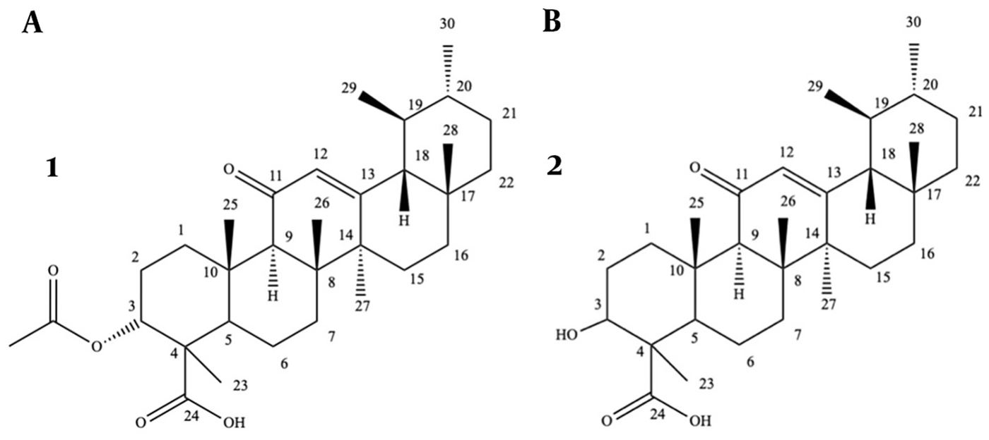
Figure 1.
The chemical structures of isolated compounds: A, 3-acetyl-11-ketoboswellic acid; and B, 11-keto-boswellic acid, isolated from the methanolic extract of B. sacra
Compound 1, AKBA: C32H48O5; white crystalline solid; Rf = 0.16 (n-hexane-ethyl acetate, 7:3 v/v) (19, 21). The 1H and 13C NMR spectroscopic data are presented in Table S2 in the Supplementary File.
Compound 2, KBA: C30H46O4; white amorphous solid; Rf = 0.10 (n-hexane-ethyl acetate, 7:3 v/v) (22). The 1H and 13C NMR spectroscopic data are presented in Table S2 in the Supplementary File.
4.2. HPLC Calibration Curve for 3-Acetyl-11-Keto-Boswellic Acid
An HPLC calibration curve was established for AKBA to quantify the extract and phytosomes (Figures S1a and S1b in the Supplementary File). It showed excellent linearity (y = 21.128x - 8.17, R2 = 0.9977), and the limit of detection (LOD) and limit of quantification (LOQ) were 0.15 and 0.46 µg/mL, respectively, based on signal-to-noise ratios of 3 and 10.
4.3. Physicochemical Characterization of the Optimized Phytosomal Formulation
As shown in Table 1, the physicochemical properties of the phytosomal formulations varied with the Boswellia extract:cholesterol:lecithin ratio. Among these, F1, F2, and F3 showed the most favorable characteristics. F3, which had the smallest particle size, the lowest PDI, and the most negative zeta potential, was selected as the optimized formulation for further evaluation (Figure 2A and B).
Table 1.
Physicochemical Characteristics of Boswellia Phytosomal Formulations a
| Formula | Boswellia Extract:Cholesterol:Lecithin | Mean particle size (nm) | PDI | Zeta Potential |
|---|---|---|---|---|
| F1 | 1:1:1 | 193 ± 10.7 | 0.33 ± 0.09 | -13.05 ± 0.05 |
| F2 | 1:1:2 | 213.67 ± 13 | 0.29 ± 0.02 | -24.33 ± 1.88 |
| F3 | 1:1.5:2 | 112 ± 12.28 | 0.23 ± 0.10 | -53 ± 5.87 |
a Values are expressed as mean ± SD; n = 3.
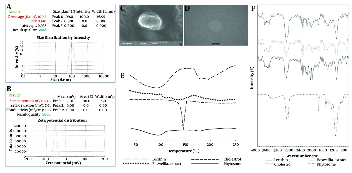
Figure 2.
A, Particle size; B, zeta potential distribution; C, SEM; and D, TEM of Boswellia phytosome; E, DSC thermograms of lecithin, Boswellia methanolic extract, cholesterol, and phytosome; and F, FTIR spectra of lecithin, Boswellia methanolic extract, cholesterol, and phytosome
4.3.1. Entrapment Efficiency and Drug Loading
The optimized Boswellia phytosomal formulation (F3) exhibited an encapsulation efficiency of 75.3 ± 1.47% and a drug loading capacity of 16.8 ± 0.53% (mean ± SD, n = 3).
4.3.2. Morphology
4.3.3. Differential Scanning Calorimetry Study
DSC analysis was performed to investigate the thermal behavior of the Boswellia extract, cholesterol, soy lecithin, and the optimized formulation (Figure 2E). The extract showed a broad endothermic peak (90 - 160 °C), while lecithin showed transitions at 92.37 °C and 192.85 °C. The phytosome thermogram showed changes, including a small peak near 180 °C and reduced extract peak intensity.
4.3.4. Fourier Transform Infrared Spectroscopy
FTIR spectroscopy confirmed complex formation between lecithin and the Boswellia extract in nanophytosomes (Figure 2F). The extract exhibited broad -OH bands (3563 - 3608 cm-1), C-H stretches at 2850 and 2922 cm-1, sp2 C-H above 3000 cm-1, and a carbonyl (C=O) band at 1740 cm-1. Lecithin displayed similar -OH bands (3566 - 3394 cm-1), fatty acid C-H stretches at 2852 and 2923 cm-1, and a carbonyl (C=O) band at 1735 cm-1. In nanophytosomes, hydroxyl bands disappeared, C-H stretches remained, sharp peaks (2000 - 2500 cm-1) vanished, and the C=O band shifted to 1732 cm-1.
4.4. Stability Study
Stability was evaluated based on particle size, PDI, and zeta potential using the same batch of optimized phytosomes (F3) that was initially characterized (Table 1). Measurements were performed at predetermined intervals over 6 months of storage at 4 ± 3 °C. The initial particle size of the freshly prepared batch was 112 ± 12.28 nm (mean ± SD, n = 3). At the first stability time point (week 1), the particle size was 100.5 nm, which fell within the standard deviation range of the initial measurement (99.72 - 124.28 nm), confirming batch consistency. Over the subsequent storage period, particle size gradually increased from 100.5 nm to 202 nm at month 6, while PDI rose from 0.14 to 0.43 and zeta potential decreased from -53.8 mV to -23 mV. No precipitation, phase separation, or visible changes in appearance were observed over 6 months of storage (Table S3 in the Supplementary File).
4.5. In Vitro Drug Release
The optimized phytosomal formulation exhibited a gradual in vitro release profile under the experimental conditions (Figure 3). Approximately 40% of AKBA was released in 3 hours, increasing to approximately 70% after 6 hours and reaching 92% after 12 hours.
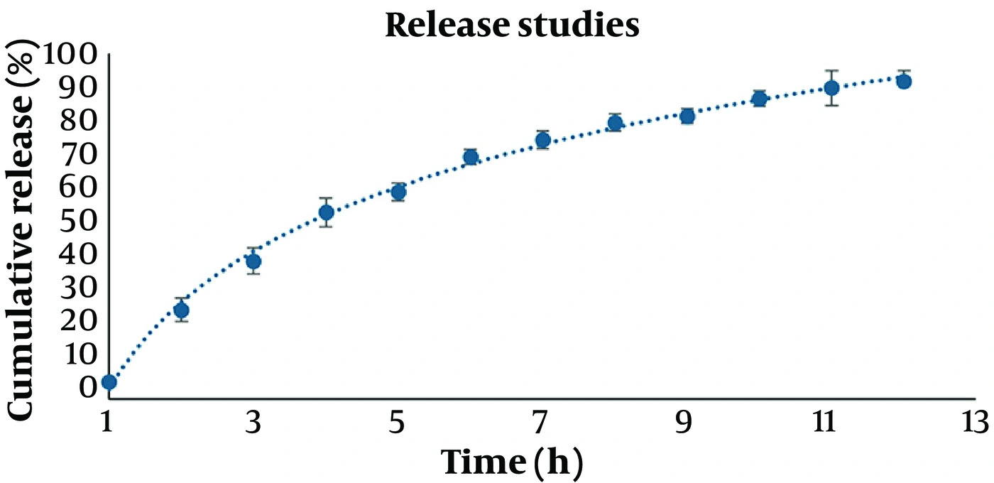
Figure 3.
in vitro release profile of optimized Boswellia phytosome in phosphate-buffered saline, pH 7.4, at 37 ± 0.5 °C (mean ± SD, n = 3)
4.6. In Vitro Acetylcholinesterase and Butyrylcholinesterase Inhibitory Activity Assay
The cholinesterase inhibitory activity of the methanolic extract, boswellic acid-enriched fraction, purified AKBA, optimized phytosome formulation, and blank formulation was assessed using Ellman’s assay, with donepezil as the reference (Table 2).
Table 2.
Comparative IC50 Values (µg/mL) of B. sacra Samples and Phytosomal Formulation Against AChE and BChE
| Samples | AChE | BChE | ||
|---|---|---|---|---|
| Inhibition (%) a | IC50 (µg/mL) | Inhibition (%) a | IC50 (µg/mL) | |
| Methanolic extract | 33.03 ± 9.91 | > 300 | 43.03 ± 8.86 | > 300 |
| Enriched boswellic acid fraction | 33.14 ± 9.2 | > 300 | 46.37 ± 7.92 | > 300 |
| 3-Acetyl-11-keto-boswellic acid | 33.94 ± 7.80 | > 300 | 48.70 ± 9.67 | > 300 |
| Optimized phytosome | 61.00 ± 10.7 b | 177.9 ± 26.8 | 42.40 ± 8.14 | > 300 |
| Blank formulation | 13.49 ± 2.9 | > 300 | 9.91 ± 1.99 | > 300 |
| Donepezil | - | 0.003 ± 0.033 | - | 3.886 ± 0.562 |
a The inhibition percentage was calculated at 300 µg/mL, mean ± SD (n = 3).
b Inhibition percentage was calculated at a concentration of 300 µg/mL. Different superscript letters within the same column indicate statistically significant differences (one-way ANOVA with Tukey post hoc test, P < 0.05). For AChE inhibition, the optimized phytosome (61.0%) was significantly higher than the methanolic extract (P = 0.028), the enriched fraction (P = 0.031), and AKBA (P = 0.041); all non-formulated samples were significantly higher than the blank formulation (P < 0.01). For BChE inhibition, no significant differences were observed among the methanolic extract, enriched fraction, AKBA, and phytosome (P > 0.05), while the blank formulation was significantly lower than all other groups (P < 0.01).
All non-formulated samples, including the methanolic extract, boswellic acid-enriched fraction, and purified AKBA, exhibited moderate BChE inhibition (~ 43 - 49%) but weaker AChE inhibition (~ 33 - 34%) at the maximum tested concentration of 300 µg/mL. In contrast, the optimized phytosome demonstrated substantially higher AChE inhibition (61.0 ± 10.7%). One-way ANOVA with Tukey post hoc test revealed that this increase was statistically significant compared with the methanolic extract (P = 0.028, 95% CI, 3.8 - 52.1), the enriched boswellic acid fraction (P = 0.031, 95% CI, 3.1 - 52.6), and AKBA (P = 0.041, 95% CI, 1.9 - 52.2). All non-formulated samples showed significantly higher AChE inhibition than the blank formulation (P < 0.01). For BChE inhibition, no statistically significant differences were observed among the extract, fraction, AKBA, and phytosome (P > 0.05), indicating that the enhancement was selective for AChE. The phytosome also achieved a measurable IC50 of 177.9 ± 26.8 µg/mL, whereas the non-formulated samples did not reach 50% inhibition within the tested concentration range (IC50 > 300 µg/mL; Figure S2 in the Supplementary File).
5. Discussion
B. sacra has emerged as a promising source of neuroactive phytochemicals; however, the poor aqueous solubility and limited oral bioavailability of BAs remain major obstacles to their therapeutic application in AD. In this study, a boswellic acid-enriched fraction was prepared, from which the 2 major BAs, AKBA and KBA, were isolated. Because AKBA was obtained in a higher yield and has been extensively reported as one of the most pharmacologically active boswellic acids, it was selected as the quantitative marker for the standardization of both the extract and the nanophytosomal formulation (23, 24).
The physicochemical behavior of the nanophytosomal formulations clearly depended on their composition. Overall, all preparations formed nanoparticles; however, the optimized formulation (F3) produced smaller, more uniformly distributed vesicles with a more negative surface charge while maintaining good drug entrapment, as summarized in Table 1. Together, these parameters are key indicators of structural integrity and colloidal stability in nanovesicular systems (25, 26). These findings suggest that achieving an appropriate balance among the extract, phospholipid, and cholesterol components is critical for obtaining a stable phytosomal formulation with favorable physicochemical properties. This observation is consistent with previous studies demonstrating that phospholipid composition is a major determinant of the stability and performance of phytosomal delivery systems (27, 28). The optimized Boswellia nanophytosomes exhibited an encapsulation efficiency of 75.3 ± 1.47%, which is comparable to that of previously reported B. serrata phytosomal formulations, while showing a smaller vesicle size and a more negative surface charge. FTIR analysis confirmed complex formation between lecithin and Boswellia extract in the nanophytosomes. The disappearance of the free hydroxyl stretching bands of the extract and lecithin, together with the unchanged C-H stretching bands of the fatty acid chains, indicated that the interaction mainly involved polar groups rather than hydrophobic chains (29, 30). In addition, the disappearance of sharp peaks in the 2000 - 2500 cm-1 region and the shift and sharpening of the C=O stretching band to 1732 cm-1 suggested hydrogen bonding between Boswellia constituents and the polar head groups of lecithin, supporting the formation of an organized nanophytosomal structure (31, 32).
The DSC findings further supported the presence of molecular interactions within the phytosomal matrix. The Boswellia extract showed a broad endothermic transition, consistent with its complex phytochemical composition (33). In contrast, lecithin exhibited two distinct thermal transitions (34). In the nanophytosome thermogram, the second lecithin transition shifted from about 192 °C to 180 °C, with reduced intensity and peak broadening, indicating structural modification of lecithin and loss of crystallinity due to interactions with the extract. Moreover, the decreased intensity of the extract-related thermal event suggested molecular dispersion of Boswellia constituents within the nanophytosomal matrix and reduced crystallinity (29, 30).
Beyond structural characterization, the physicochemical stability of the optimized nanophytosomes was monitored over a 6-month storage period. During this time, particle size gradually increased but remained within the nanoscale range. Concurrently, the increase in PDI indicated a broader particle size distribution, whereas the reduction in zeta potential reflected diminished electrostatic stabilization. Overall, these changes indicate gradual colloidal evolution during storage rather than complete physicochemical stability. Nevertheless, no visible signs of macroscopic instability, such as aggregation, precipitation, or phase separation, were observed throughout the study period. In addition to its favorable physicochemical characteristics, the optimized phytosomal formulation demonstrated gradual release, with approximately 92% cumulative release after 12 hours. A possible explanation for this behavior is the presence of the lipid-surfactant matrix, which, in previous studies, has been associated with improved apparent solubility and altered release behavior of poorly water-soluble constituents (35-38). Functionally, this release pattern may have contributed to the enhanced cholinesterase inhibitory activity observed in the present study by maintaining greater availability of the encapsulated bioactive compounds during the assay. Notably, the phytosomal formulation produced an approximately 2-fold increase in AChE inhibition (61.0 ± 10.7%) compared with the methanolic extract (≈ 33%), whereas no similar improvement was observed for BChE. The selective enhancement of AChE inhibition suggests that phytosomal encapsulation improved the functional interaction of Boswellia constituents with AChE. Although the underlying mechanism was not investigated in the present study, previous studies have suggested that phytosomal formulations enhance the apparent solubility, molecular dispersion, and accessibility of poorly soluble phytochemicals, thereby improving their biological activity (39, 40). Accordingly, the enhanced AChE inhibition observed here may be attributable to improved dispersion and availability of boswellic acids within the assay system rather than to an intrinsic increase in their inhibitory potency.
In contrast, the phytosomal formulation exhibited BChE inhibition (approximately 42%) comparable to that of the methanolic extract and purified AKBA, with no statistically significant differences among the tested samples. The active-site gorge of BChE is larger and more flexible than that of AChE and can accommodate bulky triterpenoids, such as boswellic acids, more readily (41, 42). Consequently, encapsulation within the phospholipid matrix may not confer the same functional advantage for BChE inhibition as observed for AChE. In addition, partial entrapment of the bioactive constituents within the phytosomal vesicles may transiently reduce the concentration of freely available compounds capable of directly interacting with the enzyme under the assay conditions (43).
5.1. Conclusions
This study successfully developed and characterized a nanophytosomal formulation of B. sacra extract with favorable physicochemical properties, including nanoscale size, uniform distribution, negative surface charge, and gratual release behavior. The formulation demonstrated a selective and significant enhancement of in vitro acetylcholinesterase inhibition (61.0 ± 10.7%) compared with the methanolic extract (≈ 33%), with a measurable IC50 of 177.9 ± 26.8 µg/mL, a value that could not be attained with the non-formulated samples. These results indicate that phytosomal incorporation improves the enzyme-inhibitory accessibility of Boswellia constituents in a cell-free system. Nevertheless, it is important to emphasize that these findings are strictly confined to in vitro enzyme inhibition assays. No evidence regarding blood-brain barrier penetration, neuronal uptake, cytoprotective activity, or in vivo efficacy was provided in this study. Therefore, the therapeutic relevance of this formulation for AD remains speculative at this stage. The present work serves as a foundational step, and future investigations employing appropriate cellular models (e.g., neuroblastoma cell lines), permeability assays, and animal models of cognitive impairment are necessary to validate the translational potential of this phytosomal delivery approach.
5.2. Study Limitations
The biological evaluation was restricted to in vitro cholinesterase inhibition in a cell-free system; therefore, no conclusions can be drawn regarding blood-brain barrier permeability, neuronal uptake, intracellular activity, neuroprotective effects, or in vivo therapeutic efficacy. Additionally, entrapment efficiency and AKBA content were not re-evaluated after long-term storage, precluding assessment of their stability over time. Furthermore, recovery and adsorption studies were not performed to validate the entrapment efficiency assay. Therefore, the possibility of underestimation or overestimation of EE due to matrix interference or drug adsorption to the nanocarrier surface cannot be fully excluded. A comparative in vitro release study using the non-formulated extract as a control was not conducted, and release kinetic modeling was not performed using mathematical models. Future studies should incorporate cell-based permeability assays, neuronal uptake and viability assessments, and pharmacokinetic/pharmacodynamic evaluations in appropriate animal models to establish the translational relevance of this phytosomal formulation.