
1. Background
The genus Artemisia comprises approximately 500 species that predominantly grow in the Northern Hemisphere (1). In Iran, 34 species of this genus have been identified. Artemisia turcomanica Gand., a late-flowering perennial, is one of these species. The main components of the volatile oils of A. turcomanica are oxygenated monoterpenoids (2-4). Sesquiterpene lactones have been isolated from the hydromethanolic extract of A. turcomanica (5). The antibacterial effects of its volatile oil have been reported (2, 3), as has its cytotoxic activity (4).
The PE extract of A. turcomanica inhibited heme biocrystallization in a β-hematin formation assay.
Oxidative stress is a trigger associated with neurodegeneration and with the major forms of cell death, including apoptosis, necroptosis, and parthanatos, observed in neurodegenerative diseases (6). The cytoprotective properties of different Artemisia species have been reported (7), but limited research has demonstrated the neuroprotective potential of A. turcomanica extracts.
2. Objectives
The current study investigated different fractions of the PE extract of A. turcomanica to evaluate their effects on oxidative stress and apoptosis. PC12 cells were used as an established model for in vitro neuroprotective studies. The PE extract had previously demonstrated activity in a cytoprotective assay.
3. Methods
This study was approved under ethical approval code IR.KUMS.REC.1397.554.
3.1. Plant Materials
The aerial parts of A. turcomanica Gand. were collected from Shahrabad, Maneh and Samalqan County, North Khorasan Province, Iran.
3.2. Preparation of the Extract and Fractions
The dried aerial parts of A. turcomanica (75 g) were milled and extracted with petroleum ether (40:60) at room temperature. The concentrated petroleum ether extract (2.0 g) was defatted (5) and then fractionated by NP-VLC on silica gel using a step gradient of n-hexane:ethyl acetate (94:6 to 0:100), followed by acetone and methanol. This procedure yielded 13 fractions (F1-F13).
3.3. Thin-Layer Chromatography Analysis of the Selected Fractions
The selected fractions (F6 and F10) were analyzed using TLC plates, appropriate solvent systems, and different spray reagents. The developed chromatograms were also examined under long- and short-wave UV light (8).
3.4. Determination of the Cytotoxic Effects of Different Fractions of the PE Extract and H2O2 by MTT Assay
The MTT method (9) was used to determine the cytotoxicity of H2O2 and the plant fractions against PC12 cells.
3.5. Determination of the Protective Effects of Fractions Against H2O2-Induced Cytotoxicity
Based on the cytotoxicity parameters of the fractions and their protective effects against H2O2-induced cytotoxicity, the two fractions with the lowest toxicity and greatest protective effects were selected. PC12 cells were pretreated with nontoxic concentrations (2.5 and 5.0 μg/mL) of the fractions for 24 hours and then treated with H2O2 (5.0 mg/mL) for another 24 hours. The MTT method (9) was used to determine the protective effects of the fractions against H2O2-induced cytotoxicity.
3.6. Evaluation of Intracellular Reactive Oxygen Species
Intracellular ROS levels were evaluated using the DCF-DA indicator (9). PC12 cells were pretreated with a nontoxic concentration of the selected fraction (2.5 μg/mL) for 24 hours and subsequently treated with H2O2 (5.0 mg/mL) for another 24 hours. Two additional study groups were included: 1) PC12 cells treated with H2O2 (5.0 mg/mL) and 2) the control group.
3.7. Measurement of Mitochondrial Membrane Potential
The cells were incubated for 24 hours, after which the selected fractions (2.5 μg/mL) were added to the wells. After 24 hours, the cells were treated with the IC50 of H2O2 and incubated for 4 hours. Finally, MMP was measured using Rhodamine 123.
3.8. Caspase-3 Activity Assay
PC12 cells were incubated for 24 hours. The selected fractions (2.5 μg/mL) were then added and incubated for 24 hours. Finally, H2O2 (5.0 mg/mL) was added to the wells and incubated for 4 hours. Caspase-3 activity was measured using the Sigma colorimetric caspase kit.
3.9. Statistical Analysis
All experiments in the present study were conducted in triplicate, and values are presented as the mean ± SEM. Individual groups were compared using the Tukey post hoc test or the Student t-test. A value of P < 0.05 was considered statistically significant.
4. Results
4.1. Thin-Layer Chromatography Analysis of the Selected Fractions
F6 and F10 showed fluorescence-quenching zones under UV light at 254 nm. Under long-wave UV light, F6 exhibited intense blue fluorescence, and F10 exhibited red and blue fluorescence; these signals remained stable after spraying with ethanolic potassium hydroxide reagent. The TLC plates sprayed with vanillin-sulfuric acid and Liebermann-Burchard reagents were heated for 10 minutes at 100°C. The plates were then inspected under long-wave UV and visible light, and spots with visible colors and characteristic fluorescence were observed in F6 and F10. After spraying with Dragendorff reagent, no light orange-brown areas appeared on the TLC plates of F6 and F10.
4.2. Cytotoxicity of H2O2 and Fractions on PC12 Cells
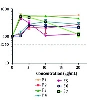
The MTT results showed that H2O2 significantly inhibited PC12 cell growth (P < 0.05). The IC50 value of H2O2 was 5.0 mg/mL. None of the PE extract fractions at concentrations up to 5.0 μg/mL induced significant toxicity in PC12 cells.
4.3. Protective Effects of the Fractions Against H2O2-Induced Cytotoxicity
F6 at a concentration of 2.5 μg/mL had a significant protective effect against oxidative stress induced by H2O2 (P < 0.01). F10 also protected PC12 cells at the same concentration (P < 0.05).
4.4. Effects of the Selected Fractions on H2O2-Induced Intracellular Reactive Oxygen Species Generation
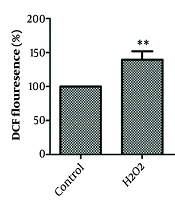
As shown in Figure 1, F6 and F10 at a concentration of 2.5 μg/mL significantly inhibited ROS production (P < 0.05).
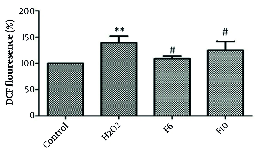
Figure 1.
Effect of pre-exposure to selected fractions of the PE extract of A. turcomanica on inhibition of ROS generation in PC12 cells. Data are presented as the mean ± SEM, n = 3. * *P < 0.01 indicates significant differences compared with the control group. # P < 0.05 indicates significant differences compared with the H2O2 group.
4.5. Effects of the Selected Fractions on Mitochondrial Membrane Potential
As shown in Figure 2, F6 and F10 (2.5 μg/mL) significantly inhibited the reduction in MMP (P < 0.05).
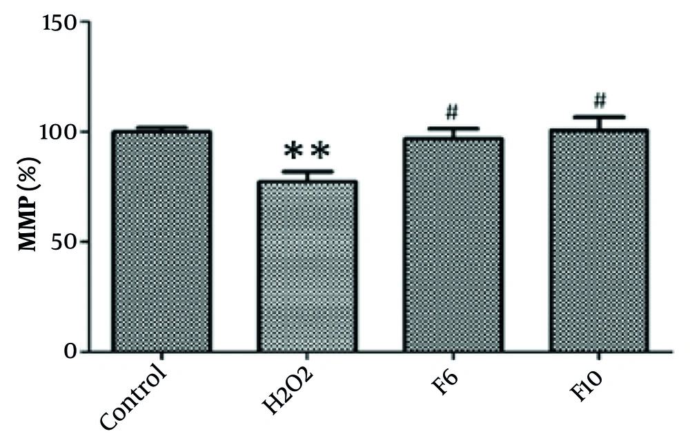
Figure 2.
Effect of pre-exposure to selected fractions of the PE extract of A. turcomanica on inhibition of MMP reduction. Data are presented as the mean ± SEM, n = 3. ** P < 0.01 indicates significant differences compared with the control group. # P < 0.05 indicates significant differences compared with the H2O2 group.
4.6. Effects of the Selected Fractions on Caspase-3 Activity
As shown in Figure 3, pretreatment of cells with F6 (P < 0.01) and F10 (P < 001) at a concentration of 2.5 μg/mL significantly decreased caspase-3 activity.
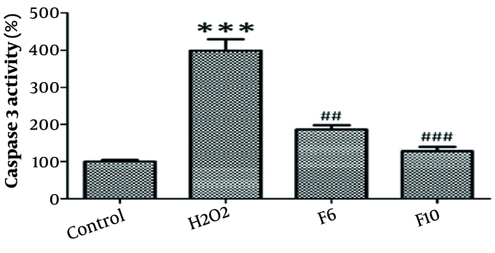
Figure 3.
Effect of pre-exposure to selected fractions of the PE extract of A. turcomanica on reduction of caspase-3 activity. *** P < 0.01 indicates significant differences compared with the control group. ## P < 0.01, ### < P < 0.001 indicate significant differences compared with the H2O2 group.
5. Discussion
Cytoprotective activities have been reported for various species across different genera (10, 11), including the genus Artemisia (7, 12).
In a recent study, the neuroprotective effects of date palm (Phoenix dactylifera L.) pollen (DPP) and fluvoxamine maleate (Flv) against H2O2-induced oxidative damage in PC12 cells were compared. The combined suppression of oxidative stress by DPP and Flv via the Nrf2 and sigma-1 signaling pathways suggested that these pretreatment regimens could be an effective modality for preventing neurodegenerative damage in an animal neurotoxicity model (11).
In the present work, the protective activity of various fractions of the PE extract of A. turcomanica in PC12 cells was investigated. A previous study examined the cytoprotective activity of different extracts of A. turcomanica in this cell line. The PE extract showed the most significant potential for reducing free radicals (123.3% reduction in ROS levels) and inhibiting caspase-3 activity (74.15% reduction in activity). The results suggested that the PE extract protected PC12 cells against apoptosis, probably via extrinsic pathways. The reduction in ROS levels, preservation of MMP, and decreased caspase-3 activity observed in the present study suggest that the PE extract of A. turcomanica may exert neuroprotective effects primarily through modulation of the intrinsic mitochondrial apoptotic pathway. Petroleum ether fractions are typically enriched in lipophilic phytochemicals, including terpenoids, sesquiterpene lactones, flavonoid aglycones, and phytosterols, which are known to possess strong antioxidant and membrane-stabilizing properties (13). These compounds may directly scavenge ROS or enhance endogenous antioxidant mechanisms, such as the Nrf2/ARE pathway, leading to the upregulation of cytoprotective enzymes, including HO-1, SOD, and catalase (14).
By reducing oxidative stress, these metabolites may help maintain mitochondrial integrity and prevent the opening of the mitochondrial permeability transition pore (mPTP), thereby preserving MMP. Stabilization of MMP is critical for maintaining ATP production and preventing the release of cytochrome c into the cytosol. Suppression of cytochrome c release consequently inhibits downstream activation of caspase-9 and executioner caspase-3, ultimately preventing apoptosis (15). No previous phytochemical study has investigated the PE extract of A. turcomanica. However, acetylenes, coumarins, flavonoids, acetophenones, and terpenoids are known as the main secondary metabolites present in the genus Artemisia (1).
Thin-layer chromatography analysis of the more potent fractions of the PE extract suggested that they are rich in terpenoids and steroids. There was no evidence of a significant presence of coumarins or alkaloids in the selected PE extract fractions. These results were consistent with previous data indicating the presence of terpenoids and widespread sterols in A. turcomanica (5). Steroids and terpenoids are plant secondary metabolites that have shown promising activity against neurodegenerative disorders (16, 17). Some isolated terpenoids from the genus Artemisia have shown neuroprotective effects in an in vivo model of Alzheimer disease or a promotive effect on NGF-induced neurite outgrowth in PC12 cells (18, 19). Recently, a known acetophenone in this genus (20) showed regulatory effects on the inflammatory microenvironment after spinal cord injury. This effect was exerted through inhibition of the NF-κB signaling pathway (21). A comprehensive phytochemical study of the PE extract of A. turcomanica should be undertaken to isolate and identify the bioactive phytochemicals.
5.1. Conclusions
The selected fractions of the PE extract of A. turcomanica had significant in vitro protective effects against oxidative stress. They inhibited ROS generation, MMP reduction, and caspase-3 activity in PC12 cells. Bio-guided isolation of phytoconstituents in the PE extract of A. turcomanica and investigation of their mechanisms of action can be considered the next steps in this research. In addition, well-designed in vivo experiments are essential to determine the efficacy, safety, and bioavailability of these fractions in a whole-organism context, which is necessary before any meaningful extrapolation to human applications can be made.