Cover image of the article: Sjogren Larsson Syndrome in Twins: Case Report

Image Credit:J Skin Stem Cell

Sjogren Larsson Syndrome in Twins: Case Report

Authors

Gabriela Roncada HaddadGabriela Roncada Haddad ORCID1,*, Mariana Righetto de Re LaiMariana Righetto de Re Lai ORCID1, Mariana Lopes Amaral BarbosaMariana Lopes Amaral Barbosa ORCID2, Andre Luis Moura BalbinoAndre Luis Moura Balbino ORCID2
1Department of Infectious Diseases, Dermatology, Imaging Diagnosis and Radioterapy, São Paulo State University, Botucatu, SP, Brazil
2São Paulo State University, Botucatu, SP, Brazil
*Corresponding Author: Department of Infectious Diseases, Dermatology, Imaging Diagnosis and Radioterapy, São Paulo State University, Botucatu, SP, Brazil. Email: gabriela.haddad@yahoo.com

Journal of Skin and Stem Cell:Vol. 10, issue 2; e135792
Published online:May 13, 2023
Article type:Case Report
Received:Feb 19, 2023
Accepted:Apr 30, 2023
How to Cite:Haddad GR, de Re Lai MR, Barbosa MLA, Balbino ALM. Sjogren Larsson Syndrome in Twins: Case Report. J Skin Stem Cell. 2023;10(2):e135792. doi: https://doi.org/10.5812/jssc-135792

Abstract

Introduction:

Sjogren-Larsson syndrome is a rare autosomal neurocutaneous disease, dermatologically manifested by congenital ichthyosis, associated with para/tetraplegia and developmental delay.

Case Presentation:

We present the case of two triplet patients, a girl, and a boy, 3 months old, admitted to the intensive care unit (ICU) due to respiratory distress, swallowing, and spasticity. The dermatological evaluation was requested due to the presence of generalized cutaneous xerosis, predominantly facial, in the girl and mild xerosis on the soles and face in the boy. The set of clinical signs and symptoms, together with the histopathological examination that revealed findings of ichthyosis, corroborated the diagnosis of this syndrome.

Conclusions:

The importance of describing this case is mainly due to the rarity of the presentation, especially in the case of triplets in which two of the brothers have the syndrome and one does not, as well as an alert to physicians for the correct diagnosis and multidisciplinary and dermatological follow-up.

Highlights

1. Introduction

Sjogren-Larsson syndrome is a rare autosomal recessive neurocutaneous disorder characterized by an inborn lipid metabolism error resulting from a mutation in the ALDH3A2 gene. This gene encodes the enzyme fatty aldehyde dehydrogenase (FALDH), and its mutation results in a decrease in the amount of FALDH present in the body, leading to a deficit in the oxidation of the long chain of fatty acids, generating a deposit of lipid metabolites in the tissues (1, 2). Deposits in the skin and central nervous system (CNS) are primarily manifested through the classic triad of congenital ichthyosis, paraplegia or quadriplegia, and mental retardation (1-3). Ichthyosis results from the disorganization and disruption of the transepidermal water barrier by the agglutination of lipid metabolites in this region, while CNS manifestations originate from disorders of myelination and demyelination of nerve fibers due to the deficiency of oxidized fatty acids (2, 4, 5). Ocular and visual changes are also frequent and result from deposits of lipofuscin pigment in the retinal epithelium (2, 6). Most reported cases have serious repercussions (1).

2. Case Presentation

We report the cases of two patients, one female and one male, aged three months, triplets from consanguineous parents (third cousins), premature at 35 weeks, presenting similar clinical conditions at birth, while the third sister did not present with cutaneous or systemic manifestations. They exhibited erythematous and xerotic skin at birth, evolving shortly afterward with apnea episodes, cyanosis, dysphagia, vomiting, and malnutrition. One patient, the girl, experienced seizure episodes. The other patient, the boy, had spasticity.
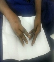
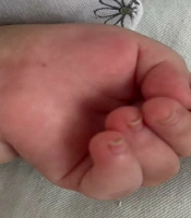
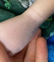
Dermatological examination revealed diffuse xerosis of the skin in both patients (Figure 1), with fine scaling and predominance on the face; an erythematous macula extending from the glabella to the right upper eyelid; a small scaly erythematous plaque in the right cervical region in the girl (Figure 2); and discolored skin, diffuse cutaneous xerosis, with fine scaling, predominantly on the face, distal third of the lower limbs and feet in the boy (Figure 3). They presented with paronychia, nail hyperkeratosis, detachment, and dystrophy of the nail plate in the nails of the hands, with no changes in the toenails.
Diffuse cutaneous xerosis. Emphasis on the abdominal region
Figure 1.
Diffuse cutaneous xerosis. Emphasis on the abdominal region
Xeroderma on the face and scaly erythematosis lesions in the glabellar region. Erythematous scaly plaque in the neck region
Figure 2.
Xeroderma on the face and scaly erythematosis lesions in the glabellar region. Erythematous scaly plaque in the neck region
Xeroderma on the face. Fine scaling on the dorsum of the left foot
Figure 3.
Xeroderma on the face. Fine scaling on the dorsum of the left foot
Together with other specialties, the diagnostic hypotheses of inborn error of metabolism, Otahara epileptic syndrome, congenital ichthyosiform erythroderma, and Sjögren-Larsson syndrome were proposed. Sjögren-Larsson syndrome was considered because of the systemic picture presented by the patients, the parents' consanguinity, and skin biopsies performed in the initial evaluation, revealing hyperkeratosis, accentuation of the granular layer and atrophy of the epidermis, with preservation of the dermis and subcutaneous tissue, compatible with ichthyosis.
Nail alterations in this syndrome have been poorly described in the literature. The acute changes observed were possibly due to inflammatory processes and metabolic stress during hospitalization.
In the reported cases, intensive skin hydration was used, which resulted in an improvement in the dermatological condition, despite serious systemic repercussions.

3. Discussion

Diffuse erythema at birth characterizes the onset of the dermatological manifestations of this syndrome, which essentially evolves with lamellar ichthyosis due to a defect in keratinization associated with pruritus. Classically, the folds, periumbilical, and neck regions are affected, while the face is generally spared, which differs from the cases presented here. Palmoplantar keratoderma can also be seen (1, 2, 7). Neurological involvement is demonstrated by progressive delay in motor development, pyramidal syndrome, and spasticity. It is possible that spasticity, in more advanced stages, affects the upper limbs and perioral region, causing speech and swallowing difficulties. Seizures, as presented by the patient in this case, are common and present in up to 40% of the cases. Premature birth has been commonly reported by researchers, as also observed in our case (7-9). The diagnosis is obtained through the presence of the classic triad and changes in the eye fundus, with crystalline maculopathy observed in up to 100% of the cases (2). Confirmation was made by enzymatic analysis, with the demonstration of FALDH deficiency in fibroblast or leukocyte cultures, which was not available in our service. It should be noted that only half of the patients with neurological symptoms similar to this syndrome, together with skin changes, have a fatty aldehyde dehydrogenase deficiency. Evaluating the urinary excretion of leukotriene B4 and its metabolites and genetic studies of the ALDH3A2 gene can help diagnose; however, it is not essential for confirmation. These tests should be performed whenever possible for diagnostic confirmation and genetic counseling (1-4, 7). Furthermore, it is necessary to make a differential diagnosis from other forms of ichthyosis, such as X-linked recessive, vulgaris, lamellar, bullous, and non-bullous congenital ichthyosiform erythroderma. The treatment is multidisciplinary, and dermatological treatment involves the topical use of emollients, keratolytics, and, in some cases, the use of systemic retinoids (1-4, 7).
We present this case due to its importance, and this description is extremely valid mainly because of the rarity of the presentation, especially because these are triplet patients in which two of the twins have the syndrome and one of the twins does not. In addition, it serves as an alert to physicians for correct investigation, correct diagnosis, and multidisciplinary and dermatological follow-up.
Footnotes

  • Authors' Contribution: GRH conceived and designed the evaluation, evaluated the clinical data, revised the manuscript, and drafted the manuscript. MRRL participated in designing the evaluation, evaluated the clinical data, revised the manuscript, and helped to draft the manuscript. MLB participated in designing the evaluation, evaluated the clinical data, revised the manuscript, and helped to draft the manuscript. ALB participated in designing the evaluation, evaluated the clinical data, revised the manuscript, and helped to draft the manuscript. All authors read and approved the final manuscript.

  • Conflict of Interests: The authors declare no conflict of interests.

  • Data Reproducibility: The dataset presented in the study is available upon request from the corresponding author during submission or after publication. The data are not publicly available due to the fact that they belong to the patient's electronic medical record, which can be accessed on the computers of the hospital where the patients were hospitalized.

  • Funding/Support: This study did not have financial support from any public or private agency. The patients were hospitalized in public service at the Hospital das Clinicas of the Faculty of Medicine of Unesp, Botucatu.

  • Informed Consent: The authors have obtained the written informed consent of the patients or subjects mentioned in the article. The authors declare that no patient data appear in this article.

References

  • 1.
    Arêdes MR, Mann EHA, de Oliveira TMFC, Lima GT, Silva MN, Simões CD, et al. [Sjögren-larsson syndrome: genetic factors, symptoms, and treatment]. Braz J Health Rev. 2021;4(2):7159-66. Portugues. https://doi.org/10.34119/bjhrv4n2-262.
  • 2.
    Benez MD, Fontenelle E, Tozzi BB, Presotto C. Do you know this syndrome? Sjogren-Larsson syndrome. An Bras Dermatol. 2010;85(6):951-3. [PubMed ID: 21308331]. https://doi.org/10.1590/s0365-05962010000600033.
  • 3.
    Assunção JBG, Resende ML, Viegas AC, Furtado T. [Sjogren Larsson Syndrome]. An Bras Dermatol. 1973;48:21. Portugues.
  • 4.
    Lynch D, Waldman AT. Sjogren-Larsson syndrome review. 2008. Available from: www.uptodate.com.
  • 5.
    Rizzo WB, Craft DA, Somer T, Carney G, Trafrova J, Simon M. Abnormal fatty alcohol metabolism in cultured keratinocytes from patients with Sjogren-Larsson syndrome. J Lipid Res. 2008;49(2):410-9. [PubMed ID: 17971613]. [PubMed Central ID: PMC3057183]. https://doi.org/10.1194/jlr.M700469-JLR200.
  • 6.
    Isaac DL, Queiroz GH, Feres CC, Avila M. [Macular crystalline dystrophy in Sjogren-Larsson syndrome: case report]. Arq Bras Oftalmol. 2009;72(2):239-42. Portuguese. [PubMed ID: 19466337]. https://doi.org/10.1590/s0004-27492009000200021.
  • 7.
    Roy U, Das U, Pandit A, Debnath A. Sjogren-Larsson syndrome: a rare disease of the skin and central nervous system. BMJ Case Rep. 2016;2016:10 1136/bcr-2016-215110. [PubMed ID: 27095813]. [PubMed Central ID: PMC4840703]. https://doi.org/10.1136/bcr-2016-215110.
  • 8.
    Willemsen MA, IJlst L, Steijlen PM, Rotteveel JJ, de Jong JG, van Domburg PH, et al. Clinical, biochemical and molecular genetic characteristics of 19 patients with the Sjogren-Larsson syndrome. Brain. 2001;124(Pt 7):1426-37. [PubMed ID: 11408337]. https://doi.org/10.1093/brain/124.7.1426.
  • 9.
    Rizzo WB. Sjogren-Larsson syndrome: explaining the skin-brain connection. Neurology. 1999;52(7):1307-8. [PubMed ID: 10227610]. https://doi.org/10.1212/wnl.52.7.1307.

Copyright

Copyright © 2023, Journal of Skin and Stem Cell. This is an open-access article distributed under the terms of the Creative Commons Attribution-NonCommercial 4.0 International License (http://creativecommons.org/licenses/by-nc/4.0/) which permits copy and redistribute the material just in noncommercial usages, provided the original work is properly cited.

Similar Articles

5
Jun
2022
Anesthetic Management of a Four-Year-Old Child Affected by Marinesco-Sjogren Syndrome Scheduled for Cataract Surgery: A Case Report

Anesthetic Management of a Four-Year-Old Child Affected by Marinesco-Sjogren Syndrome Scheduled for Cataract Surgery: A Case Report

Dr Azar Ejmalian,
Elham Memary,
Nader Nassiri

Ejmalian DA, Memary E, Nassiri N. Anesthetic Management of a Four-Year-Old Child Affected by Marinesco-Sjogren Syndrome Scheduled for Cataract Surgery: A Case Report. J Cell Mol Anesth. 2023;8(1):e151427. doi: https://doi.org/10.22037/jcma.v7i4.38067

16
Mar
2023
General Anesthesia in Schwartz-Jampel Syndrome: A Case Report

General Anesthesia in Schwartz-Jampel Syndrome: A Case Report

Pooya Derakhshan,
Azadeh Habibi,
Saied Amniati

Derakhshan P, Habibi A, Amniati S. General Anesthesia in Schwartz-Jampel Syndrome: A Case Report. Anesth Pain Med. 2023;13(3):e129305. doi: https://doi.org/10.5812/aapm-129305

28
Jun
2021
Normo-Calcemic Tetany in a Patient with Hypokalemic Periodic Paralysis of Sjogren’s Syndrome: Neuromuscular Excitability Secondary to the ‘Forgotten Cation’

Normo-Calcemic Tetany in a Patient with Hypokalemic Periodic Paralysis of Sjogren’s Syndrome: Neuromuscular Excitability Secondary to the ‘Forgotten Cation’

Urmila Anandh,
Pradeep Mishra,
Hilal Malla

Anandh U, Mishra P, Malla H. Normo-Calcemic Tetany in a Patient with Hypokalemic Periodic Paralysis of Sjogren’s Syndrome: Neuromuscular Excitability Secondary to the ‘Forgotten Cation’. Arch Neurosci. 2021;8(2):e105542. doi: https://doi.org/10.5812/ans.105542

12
Nov
2024
Schaaf-Yang Syndrome Presenting with Prolonged Hyperthermia in a Child: A Case Report

Schaaf-Yang Syndrome Presenting with Prolonged Hyperthermia in a Child: A Case Report

Pooya Poormehr,
Arya Behzadi,
Nazanin Farahbakhsh,
Amir Reza Bahadori

Poormehr P, Behzadi A, Farahbakhsh N, Bahadori AR. Schaaf-Yang Syndrome Presenting with Prolonged Hyperthermia in a Child: A Case Report. Arch Pediatr Infect Dis. 2025;13(2):e155654. doi: https://doi.org/10.5812/apid-155654

22
Oct
2022
Anesthetic Management of a 9-Years-Old Child Affected by Al-Raqad Syndrome Scheduled for Cataract Surgery: A Case Report

Anesthetic Management of a 9-Years-Old Child Affected by Al-Raqad Syndrome Scheduled for Cataract Surgery: A Case Report

Azar Ejmalian,
Nader Nassiri,
Shahram Sayadi,
Dariush Abtahi,
Elham Memary

Ejmalian A, Nassiri N, Sayadi S, Abtahi D, Memary E. Anesthetic Management of a 9-Years-Old Child Affected by Al-Raqad Syndrome Scheduled for Cataract Surgery: A Case Report. J Cell Mol Anesth. 2023;8(2):e149687. doi: https://doi.org/10.22037/jcma.v8i2.39304

More by these authors

Gabriela Roncada HaddadPubMedScholar
Mariana Righetto de Re LaiPubMedScholar
Mariana Lopes Amaral BarbosaPubMedScholar
Andre Luis Moura BalbinoPubMedScholar
Share
Cited by
Metrics
Indexed in