
1. Background
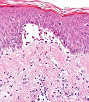
Pemphigus refers to a group of chronic, potentially life-threatening autoimmune blistering disorders characterized by intraepidermal blisters and erosions of the skin and mucosae. Disease activity is driven by circulating IgG autoantibodies against desmogleins, cadherin-family proteins that anchor keratinocytes to one another within desmosomes (1, 2). Antibodies against desmoglein 3 predominate when mucosal surfaces bear the brunt of disease, whereas combined desmoglein 1 and 3 reactivity underlies disease that also involves the skin. Binding of these antibodies to their targets leads to loss of keratinocyte-to-keratinocyte adhesion (acantholysis), resulting in flaccid, easily ruptured blisters and painful erosions (1).
Pemphigus vulgaris (PV) accounts for the majority of cases, whereas pemphigus foliaceus (PF), in which more superficially directed anti-desmoglein-1 antibodies cause crusted, scaly plaques without mucosal involvement, is less common. Systemic corticosteroids have historically been the mainstay of treatment, but the cumulative toxicity of prolonged high-dose steroid exposure is itself a major source of morbidity in this disease. Steroid-sparing immunosuppressants such as azathioprine and mycophenolate mofetil are therefore commonly added to reduce this burden (3).
Rituximab is a chimeric monoclonal antibody against CD20, a surface marker on B lymphocytes; by depleting this pool, it eliminates the source of pathogenic autoantibody production and interrupts the autoimmune process driving pemphigus (4, 5). The pivotal reports by Ahmed et al. and Joly et al. first established rituximab as an effective option for refractory pemphigus and as a potent steroid-sparing agent (4, 5). This evidence base was subsequently strengthened by the Ritux 3 trial, a prospective, multicentre, open-label randomised controlled trial conducted across French dermatology departments, which showed that first-line rituximab administered with a short course of prednisone outperformed prednisone alone; this led to the adoption of rituximab as a first-line option for moderate-to-severe pemphigus in several international guidelines (6). A subsequent long-term follow-up of the Ritux 3 cohort, at a median of 7.3 years, confirmed durable corticosteroid-free remission with first-line rituximab (7). Despite this robust evidence base, real-world data from Indian tertiary-care settings—where patients often present later in the disease course, dosing protocols vary by cost and availability, and rituximab is more often used as a second-line rather than a first-line agent—remain limited (8).
2. Objectives
The present study was undertaken to evaluate the clinical efficacy of rituximab in a consecutive series of patients with pemphigus treated at our institution, including both newly diagnosed cases and those with longer-standing, relapsed, or refractory disease, and to characterise its adverse-effect profile, remission and relapse patterns, and outcomes across different dosing protocols and disease phenotypes (PV and PF) encountered in real-world practice.
3. Methods
3.1. Study Design and Setting
This single-centre, retrospective, observational study was conducted in the Department of Dermatology, Venereology and Leprosy, B. J. Government Medical College and Sassoon General Hospital, Pune, a tertiary-care teaching hospital in western India. Case records of all patients with pemphigus who received rituximab infusions from September 2022 to November 2024 (approximately 26 months) were retrieved from the medical records section and reviewed. The study was conducted after approval from the Institutional Ethics Committee.
3.2. Study Population and Diagnostic Criteria
Patients were eligible for inclusion if they had pemphigus confirmed clinically, together with skin biopsy and Tzanck smear per institutional protocol; had received rituximab; and had adequately documented case records. Adequate documentation was defined as complete baseline demographic, diagnostic, dosing, and follow-up outcome data sufficient to classify the patient’s treatment response. Patients with incomplete medical records for any of these parameters were excluded. Direct immunofluorescence and quantitative anti-desmoglein 1/3 ELISA were not performed as routine diagnostic requirements at our centre during the study period; diagnosis therefore relied on clinicopathological correlation rather than a full immunological work-up, and this is discussed further as a limitation. Among the 30 patients analysed, no patient with active systemic infection, malignancy, or pregnancy at the time of rituximab initiation was identified; these conditions were therefore not separately tabulated as exclusions. A total of 30 patients were included in the final analysis, comprising a mixed population of newly diagnosed, relapsed, and refractory patients.
3.3. Baseline Evaluation and Treatment Protocol
Baseline investigations before rituximab administration included a complete haemogram, renal and liver function tests, viral markers, Mantoux test, chest radiograph, ultrasonography of the abdomen and pelvis, and electrocardiography. After written informed consent and premedication with intravenous hydrocortisone 100 mg, intravenous pheniramine maleate 22.5 mg, and oral paracetamol 500 mg, rituximab was infused under strict monitoring over 5 - 6 hours. The infusion rate was initiated at 30 mL/h and escalated by 30 mL/h every 30 minutes to a maximum of 250 mL/h. Rituximab was administered according to the rheumatoid arthritis (RA) protocol (1 g given 2 weeks apart), the lymphoma protocol (375 mg/m2 weekly for 4 weeks), or via the intralesional route; one patient received intralesional rituximab. The choice of protocol was not dictated by a fixed institutional algorithm but reflected physician judgement, disease severity, and cost or affordability, representing a pragmatic, real-world pattern of protocol selection and a source of heterogeneity in this cohort. Concurrent immunosuppressive or adjuvant medications were continued as clinically indicated.
3.4. Outcome Definitions
Clinical endpoints followed the consensus statement of the International Pemphigus Committee (3). Disease control, the early endpoint, was defined as the point at which new lesions stopped forming and existing lesions began to heal. Complete remission (CR) was defined as the absence of new or established lesions for at least 2 months and was subclassified as CR off therapy, defined as no systemic therapy of any kind, or CR on minimal therapy. CR on minimal therapy was defined as maintenance on 10 mg/day or less of prednisolone or the equivalent and/or a low-dose adjuvant agent, specifically no more than half the dose that would define treatment failure, sustained for at least 2 months. Partial remission was defined as transient lesions healing within 1 week, either off therapy or while on minimal therapy as defined above. Relapse, synonymous with flare, was defined as the appearance of 3 or more new lesions in a month that failed to heal spontaneously within 1 week, or extension of an existing lesion, in a patient who had already achieved disease control. Failure of therapy denoted persistence of active disease despite full-dose systemic treatment; no patient in this cohort met this definition.
3.5. Follow-Up Assessment
Patients were reviewed in the dermatology outpatient clinic at routine intervals after rituximab infusion, with disease status, new lesion counts, and adverse effects recorded at each visit and relapse ascertained clinically against the definition above. Patients unable to attend in person were followed up telephonically for interim symptoms, where documented in the case records. Because the cohort was enrolled on a rolling basis across the study period, the duration of observable follow-up differed between the earliest- and latest-enrolled patients, and an exact median follow-up duration for the full cohort could not be centrally tabulated from the retrospective records. The time-to-remission and time-to-relapse data reported in the Results reflect the subset of patients in whom these intervals were unambiguously documented. This heterogeneity in follow-up duration is acknowledged as a limitation.
3.6. Data Collection and Statistical Analysis
Data on patient demographics, diagnosis, duration of symptoms before rituximab, dosing protocol, concurrent therapy, comorbidities, current disease status, and adverse effects were extracted using a structured proforma. Continuous variables are expressed as mean ± standard deviation (SD) and range; categorical variables are expressed as frequencies and percentages, with 95% confidence intervals (Wilson score method) for the principal outcome proportions. Given the retrospective, descriptive design and the small size of the PF subgroup (n = 6), prespecified formal inferential testing was not undertaken; however, a post hoc 2-sided Fisher exact test was performed for the primary PV-versus-PF relapse and remission comparison, as the raw counts permitted this without overinterpreting the wider dataset. SPSS version 22 was used.
3.7. Ethical Considerations
The study was conducted after approval from the Institutional Ethics Committee, B. J. Government Medical College and Sassoon General Hospital, Pune. Informed consent was obtained from all participants.
4. Results
4.1. Patient Demographics and Disease Characteristics
This retrospective institutional analysis included 30 patients with pemphigus who received rituximab therapy. The cohort comprised 24 patients with PV (80.0%) and 6 with PF (20.0%). The mean age at rituximab initiation was 44.8 ± 12.6 years (range, 18 - 65 years). There were 18 females (60.0%) and 12 males (40.0%); the female-to-male ratio was 1.5:1.
Regarding disease extent, 17 patients (56.7%) had combined cutaneous and mucosal (C+M) involvement, 12 patients (40.0%) had exclusively cutaneous disease, and 1 patient (3.3%) had isolated mucosal involvement. Most patients (n = 20, 66.7%) had no significant comorbidities. Diabetes mellitus was the most frequently recorded comorbidity (n = 7, 23.3%), followed by concomitant hypertension and diabetes (n = 2, 6.7%), hepatitis B surface antigen (HBsAg) positivity (n = 1, 3.3%), and a history of cerebrovascular accident (n = 1, 3.3%). Comorbidity categories were not mutually exclusive. No patient had a recorded history of active infection, malignancy, or pregnancy at treatment initiation.
All patients had previously received systemic corticosteroids (prednisolone), consistent with rituximab being used as a second-line, steroid-sparing agent in this cohort rather than as first-line monotherapy. Azathioprine was co-administered in 25 patients (83.3%). Additional adjuvant therapies included cyclophosphamide (n = 4), dapsone (n = 3), intralesional steroids (n = 5), and entecavir (n = 1) in the HBsAg-positive patient. The duration of pemphigus symptoms before rituximab initiation ranged from 1 month to 4 years; 33.3% of patients had a symptom duration of 3 months or less, whereas 10.0% had symptoms persisting for more than 12 months before rituximab was commenced, reflecting a cohort spanning both relatively early and long-standing disease. Table 1 summarizes the patient demographics and disease characteristics (Figures 1-5).
Table 1.
Patient Demographics and Disease Characteristics (N = 30) a
| Variables | Values |
|---|---|
| Age (y) | 44.8 ± 12.6 (18 - 65) |
| Gender | |
| Female | 18 (60.0) |
| Male | 12 (40.0) |
| Diagnosis | |
| PV | 24 (80.0) |
| Standard PV | 18 (75.0 of PV) |
| PVeg | 2 (8.3 of PV) |
| PF | 6 (20.0) |
| Disease extent | |
| Cutaneous + mucosal (C+M) | 17 (56.7) |
| Cutaneous only (C) | 12 (40.0) |
| Mucosal only (M) | 1 (3.3) |
| Comorbidities | |
| None | 20 (66.7) |
| DM | 7 (23.3) |
| Hypertension + DM | 2 (6.7) |
| HBsAg positive | 1 (3.3) |
| History of stroke | 1 (3.3) |
| Rituximab protocol | |
| RA dosing: 1000 mg × 2 infusions | 24 (80.0) |
| Lymphoma dosing: 375 mg/m2 × 4 | 5 (16.7) |
| Intralesional (10 mg/cm2) | 1 (3.3) |
| Symptom duration before rituximab (mo) | |
| ≤ 3 | 10 (33.3) |
| 4 - 6 | 12 (40.0) |
| 7 - 12 | 5 (16.7) |
| > 12 | 3 (10.0) |
| Booster doses | |
| Received booster 1 | 6 (20.0) |
| Received booster 2 | 2 (6.7) |
| Concurrent / prior therapy | |
| Prednisolone (systemic steroid) in varying doses | 30 (100.0) |
| AZA 100 mg/d | 25 (83.3) |
| CP 100 mg/d | 4 (13.3) |
| Dapsone 100 mg/d | 3 (10.0) |
| ILS | 5 (16.7) |
| Entecavir (HBsAg prophylaxis) | 1 (3.3) |
a Values are expressed as mean ± SD (range) or No. (%) unless otherwise indicated. Comorbidity categories are not mutually exclusive; therefore, category counts may sum to more than the total cohort size. Abbreviations: PV, pemphigus vulgaris; PF, pemphigus foliaceus; PVeg, pemphigus vegetans; RA, rheumatoid arthritis; C+M, cutaneous and mucosal; DM, diabetes mellitus; HBsAg, hepatitis B surface antigen; AZA, azathioprine; CP, cyclophosphamide; ILS, intralesional steroids.
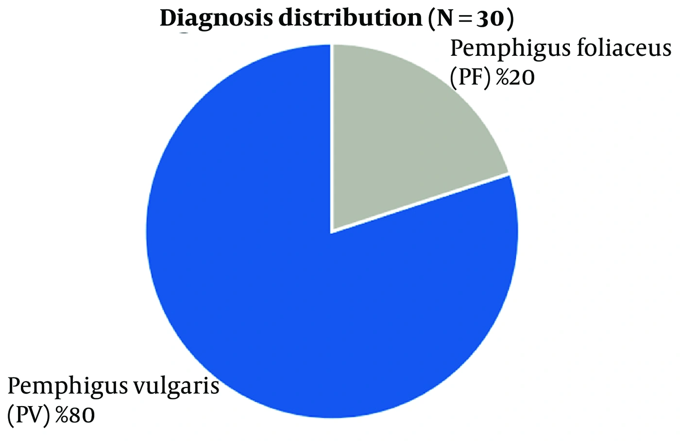
Figure 1.
Distribution of pemphigus diagnoses: PV (n = 24, 80%) versus PF (n = 6, 20%).
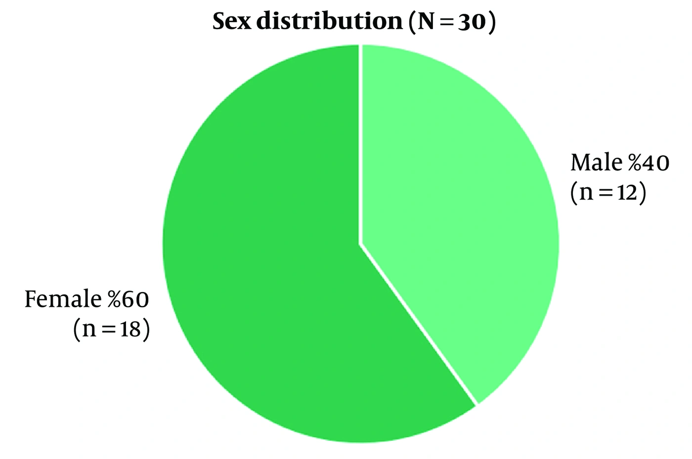
Figure 2.
Sex distribution among the 30 patients: female (n = 18, 60%) and male (n = 12, 40%).
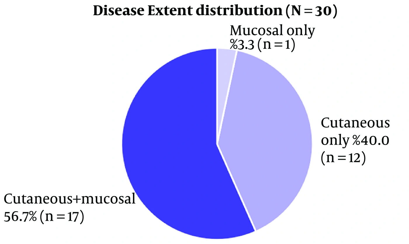
Figure 3.
Distribution of disease extent: C+M (n = 17, 56.7%), cutaneous only (n = 12, 40.0%), and mucosal only (n = 1, 3.3%).
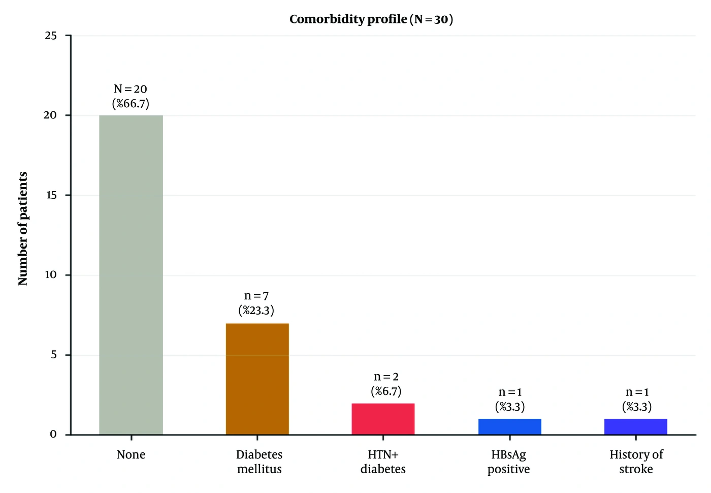
Figure 4.
Comorbidity distribution among the 30 patients; diabetes mellitus was the most prevalent recorded comorbidity (23.3%).
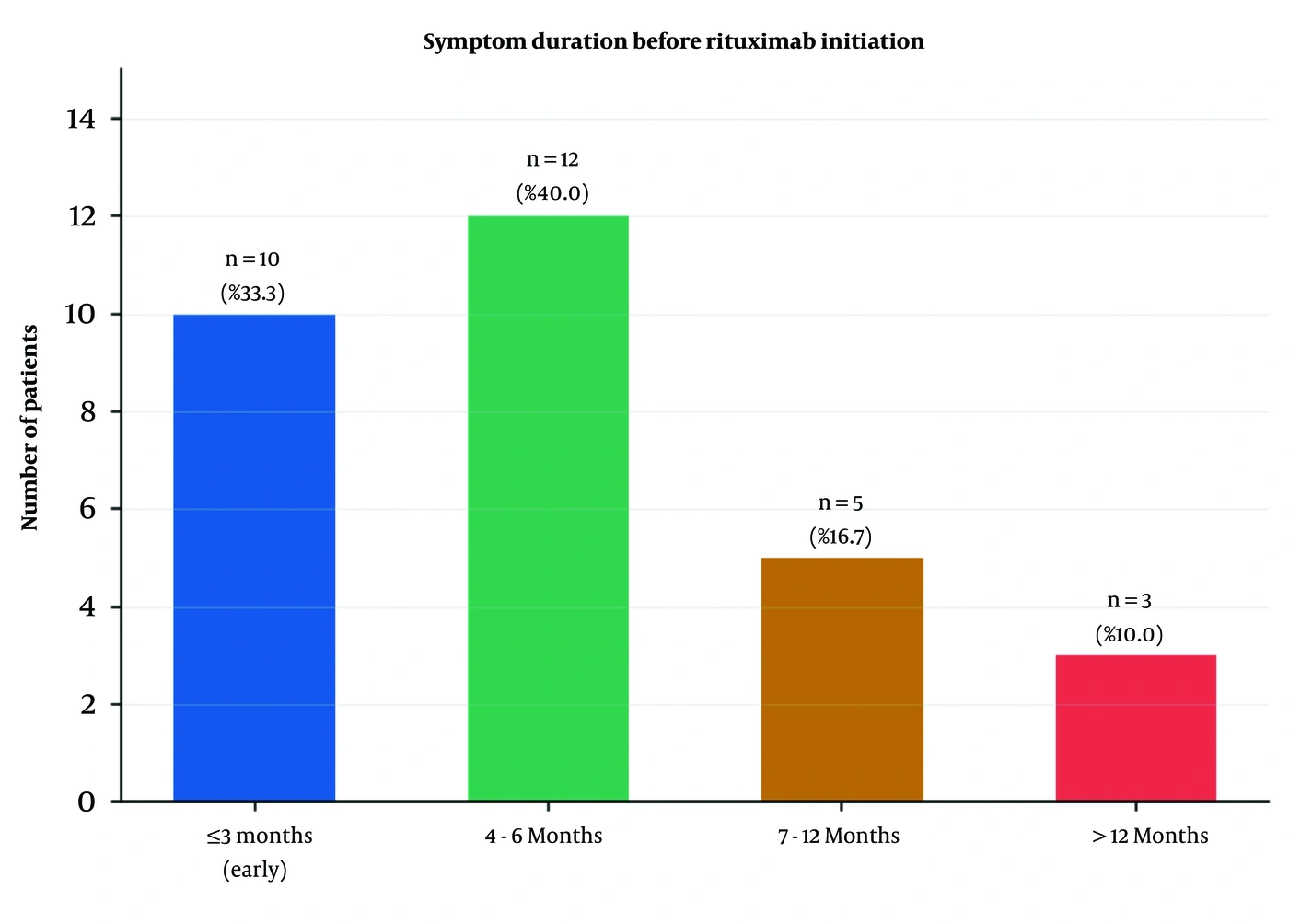
Figure 5.
Distribution of symptom duration before rituximab initiation; 40% of patients had 4 - 6 months of symptoms before treatment commencement.
4.2. Final Complete-Remission and Relapse Status
At the final documented assessment, 26 of 30 patients (86.7%, 95% CI 70.3 - 94.7%) were in complete remission. Of these, 12 patients (40.0%) were in CR on minimal maintenance therapy, whereas 14 patients (46.7%, 95% CI 30.2 - 63.9%) were in CR off all therapy, indicating sustained disease-free status at the final assessment. Four patients (13.3%, 95% CI 5.3 - 29.7%) were in relapse after previously achieving complete remission. No patient was classified as a non-responder; all patients demonstrated some degree of clinical response to rituximab. These final complete-remission rates compare favourably with published data reporting overall CR rates ranging from 37% to 77% across various rituximab dosing regimens (9, 10) (Figure 6).
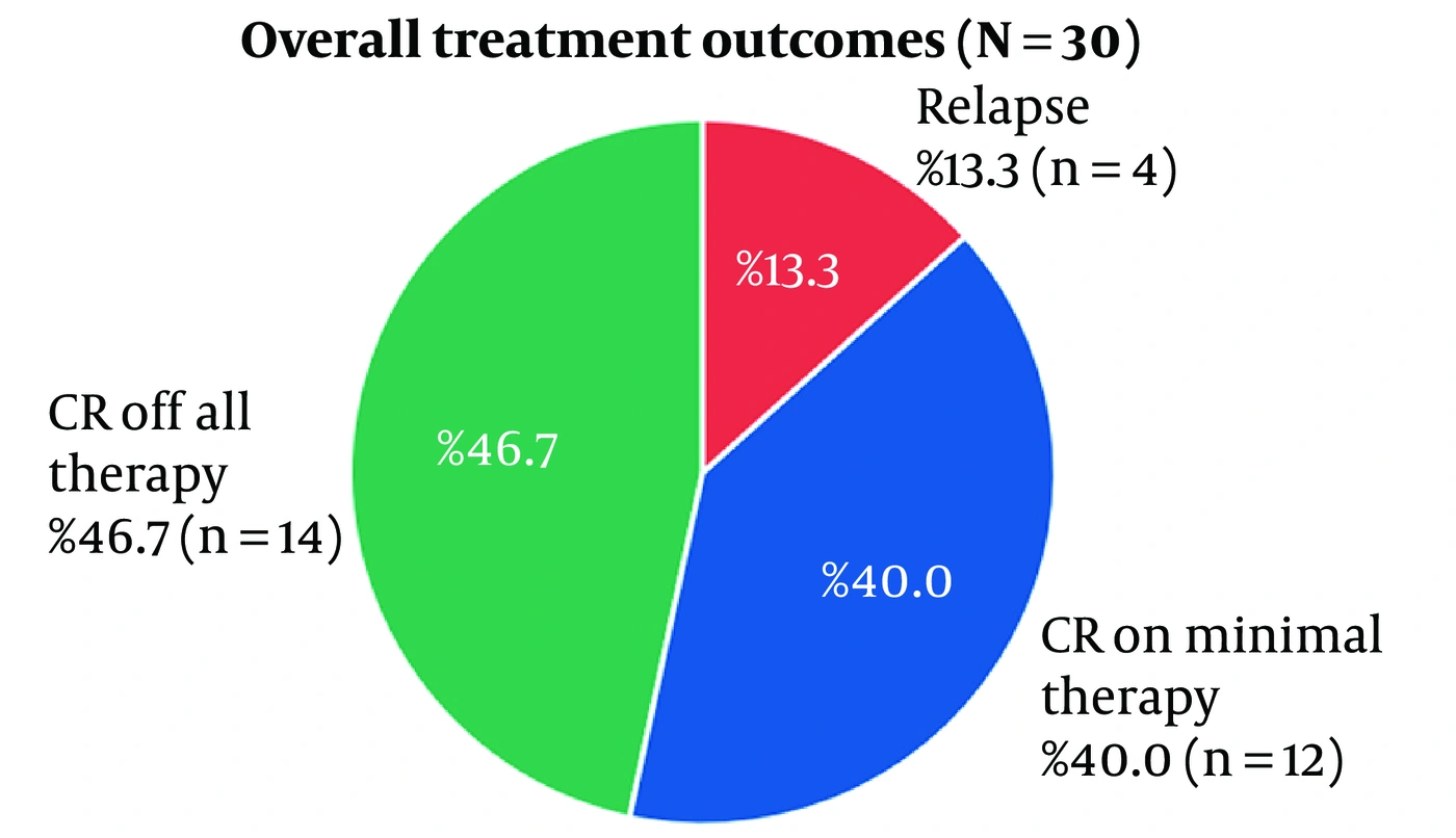
Figure 6.
Final documented treatment status: 86.7% of patients were in complete remission (CR on + CR off), and 13.3% were in relapse after prior complete remission.
4.3. Time to Complete Remission
Among the 13 patients with documented time-to-CR data, remission was achieved within 6 months in 5 patients (38.5%), between 7 and 12 months in 4 patients (30.8%), and between 13 and 24 months in the remaining 4 patients (30.8%). The remaining patients who were in CR at the final assessment could not be assigned an exact time to remission because the case records did not consistently capture the precise date of complete remission onset relative to rituximab initiation. This is a recognised limitation of retrospective chart review and is discussed further under Limitations. Within the subset with documented data, this distribution reflects the known variable latency of rituximab’s B-cell-depleting effect, which typically reaches its nadir 6 - 8 weeks after infusion, with clinical remission often lagging behind the immunological response by several additional months (Figure 7).
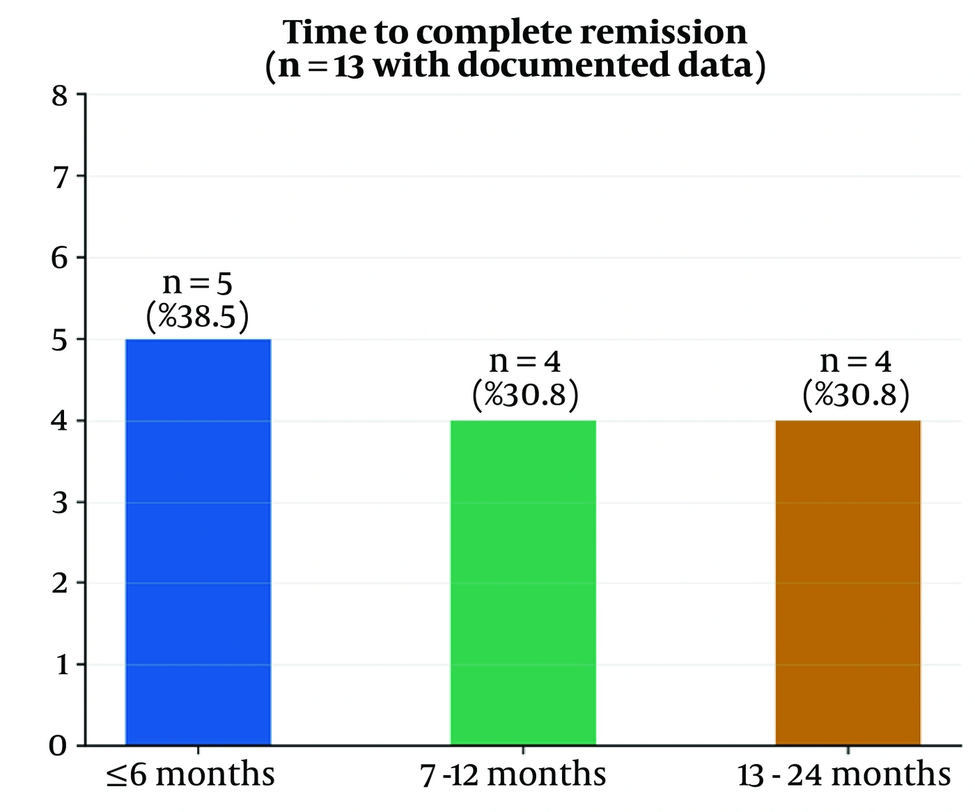
Figure 7.
Distribution of time to complete remission among the 13 patients with documented data.
4.4. Relapse
Four patients (13.3%) had a documented relapse at the final assessment after prior complete remission. Two patients, 1 with PV on the RA protocol (8 months' symptom duration before rituximab) and 1 with PF on the RA protocol (2 months' duration), relapsed at approximately 1 year; a third patient with PF, hypertension, and diabetes relapsed at 14 months; and a fourth patient with PF who was HBsAg-positive and received the lymphoma protocol relapsed at 9 months. Notably, 3 of the 4 relapsing patients had PF, consistent with published reports of higher relapse rates in PF than in PV. The relapse rate among PF patients in our cohort was 50.0% (3/6, 95% CI 18.8 - 81.2%) versus 4.2% (1/24, 95% CI 0.7 - 20.2%) among PV patients; this difference was statistically significant on a post hoc 2-sided Fisher exact test (P = 0.018), notwithstanding the very small PF subgroup, which limits the precision of this estimate (Figure 8).
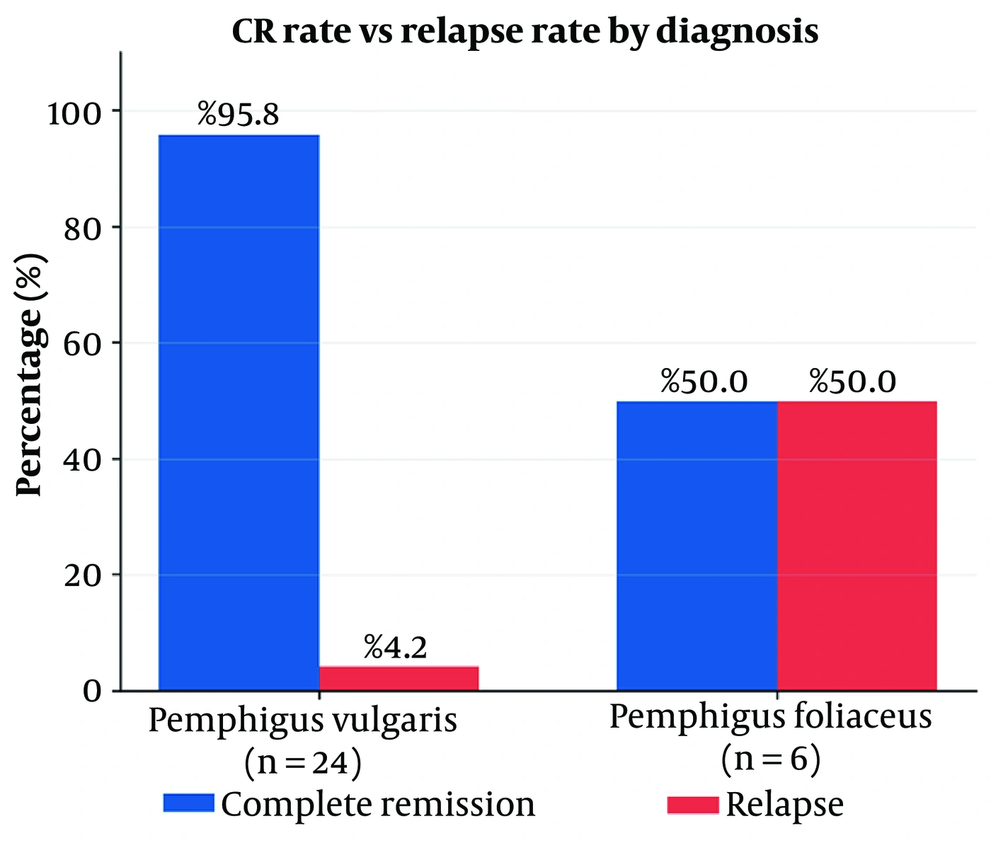
Figure 8.
Comparison of final complete-remission and relapse rates between PV and PF; PF demonstrated a markedly higher relapse rate (50% vs 4.2%; Fisher exact P = 0.018).
4.5. Outcomes by Protocol and Disease Extent
Patients receiving the RA dosing protocol (1000 mg × 2 infusions) were in CR in 87.5% (21/24) of cases at the final assessment. Those receiving the lymphoma protocol (375 mg/m2 × 4 weekly infusions) were in CR in 80.0% (4/5) of cases, with 1 patient in relapse; this difference was not statistically significant (Fisher exact P = 0.55), consistent with the small lymphoma-protocol subgroup. The single patient treated with intralesional rituximab, who had pemphigus vegetans and diabetes, achieved CR off therapy at 9 months. By disease extent, the final CR rate was 94.1% in C+M disease, 75.0% in cutaneous-only disease, and 100% in the single patient with mucosal-only disease (Figures 9 and 10).
Table 2 summarizes the clinical outcomes following rituximab therapy.
Table 2.
Clinical Outcomes Following Rituximab Therapy (N = 30) a
| Outcome variable | Values |
|---|---|
| Final treatment status | |
| Complete remission (CR) | 26 (86.7); (70.3 - 94.7) |
| CR on minimal therapy | 12 (40.0) |
| CR off all therapy | 14 (46.7); (30.2 - 63.9) |
| Relapse after prior CR | 4 (13.3); (5.3 - 29.7) |
| Non-responders / lost to follow-up | 0 (0.0) |
| Time to complete remission (n = 13); (mo) | |
| ≤ 6 | 5 (38.5) |
| 7 - 12 | 4 (30.8) |
| 13 - 24 | 4 (30.8) |
| Relapse details (n = 4) | |
| Time to relapse: 9 (mo) | 1 (25.0) |
| Time to relapse: approximately 12 (mo) | 2 (50.0) |
| Time to relapse: 14 (mo) | 1 (25.0) |
| Adverse effects | |
| No adverse effects | 29 (96.7) |
| Raised LFT (×2 ULN) | 1 (3.3) |
| Severe infections | 0 (0.0) |
| Hypogammaglobulinaemia | 0 (0.0) |
| Progressive multifocal leucoencephalopathy | 0 (0.0) |
| Outcomes by diagnosis | |
| PV - complete remission | 23/24 (95.8); (79.8 - 99.3) |
| PV - relapse | 1/24 (4.2); (0.7 - 20.2) |
| PF - complete remission | 3/6 (50.0); (18.8 - 81.2) |
| PF - relapse | 3/6 (50.0); (18.8 - 81.2) |
| PV vs PF relapse, Fisher exact | P = 0.018 |
| Outcomes by protocol | |
| RA protocol (1 g × 2) - CR rate | 21/24 (87.5) |
| Lymphoma protocol (375 mg/m2 × 4) - CR rate | 4/5 (80.0) |
| RA vs lymphoma CR, Fisher exact | P = 0.55 |
| Outcomes by disease extent | |
| C+M - complete remission | 16/17 (94.1) |
| Cutaneous only - complete remission | 9/12 (75.0) |
| Mucosal only - complete remission | 1/1 (100.0) |
a Values are expressed as No. (%) or 95% (CI) unless otherwise indicated. Abbreviations: CR, complete remission; PV, pemphigus vulgaris; PF, pemphigus foliaceus; RA, rheumatoid arthritis; LFT, liver function test; ULN, upper limit of normal; C+M, cutaneous and mucosal; CI, confidence interval.
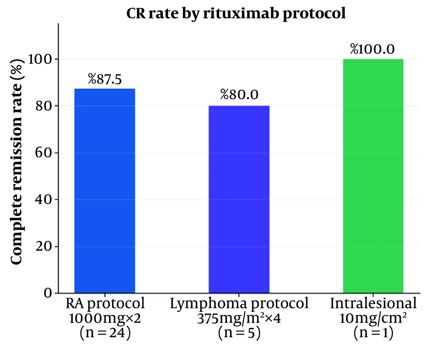
Figure 9.
Final complete-remission rates stratified by rituximab dosing protocol.
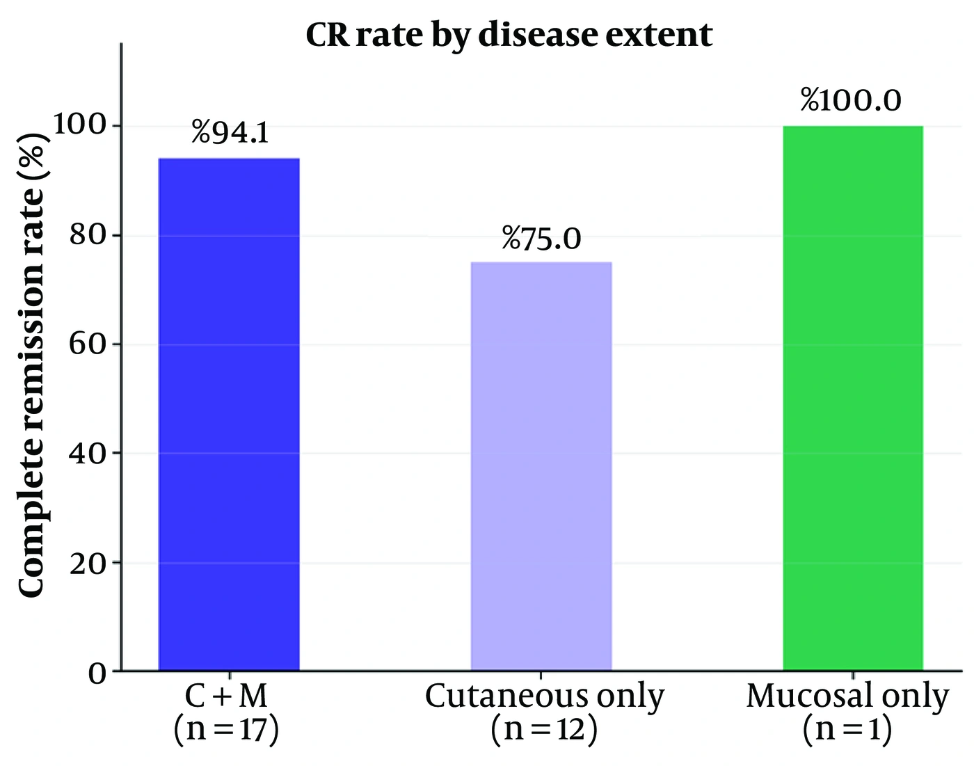
Figure 10.
Final complete-remission rates stratified by disease extent; among groups containing more than 1 patient, C+M involvement had the highest CR rate (94.1%).
4.6. Safety and Adverse Effects
Rituximab was well tolerated across the cohort. Twenty-nine of 30 patients (96.7%) reported no adverse effects attributable to rituximab. One patient with PF who received the RA protocol developed a transient elevation of liver function tests to twice the upper limit of normal, which resolved spontaneously without dose modification or treatment discontinuation. No severe adverse events were recorded, including serious infections such as sepsis, pneumonia, or opportunistic infections; hypogammaglobulinaemia or neutropenia; progressive multifocal leucoencephalopathy; or infusion reactions requiring cessation of therapy. This safety profile is consistent with published prospective studies of rituximab in pemphigus, which similarly report low rates of serious adverse events at standard dosing (4, 5). The absence of severe infection in our cohort may partly reflect careful patient selection and prophylactic antimicrobial use, as well as the generally lower cumulative corticosteroid burden associated with earlier rituximab introduction.
5. Discussion
In this retrospective single-centre analysis of 30 patients, 86.7% were in complete remission at the final documented assessment (95% CI 70.3 - 94.7%), with almost half (46.7%) in remission off all systemic therapy and no patients failing to respond. These outcomes are at the upper end of the published range. To contextualise our findings, we compared our principal outcomes, including final complete remission, remission off therapy, relapse, protocol-specific response, time to remission, and safety, with those of major published series and trials, including several recent studies; these comparisons are summarised in Table 3.
Table 3.
Comparison of the Present Study with Major Published Rituximab-in-Pemphigus Series and Trials a
| Studies | n | Protocol | Complete remission (95% CI where available) | Relapse | Reference |
|---|---|---|---|---|---|
| Present study | 30 | RA / lymphoma / IL | 86.7% (26/30); 95% CI 70.3 - 94.7% | 13.3% (4/30); 95% CI 5.3 - 29.7% | — |
| Ahmed et al., 2006 (4) | 11 | Lymphoma + IVIG | 82% (9/11) | Low; remission 22 - 37 mo | 4 |
| Joly et al., 2007 (5) | 21 | RA-type, single cycle | 86% (18/21) | Not primary endpoint | 5 |
| Kim et al., 2011 (10) | 27 | Lymphoma (2 vs ≥ 3 inf.) | — | 67% (2 inf.) vs 0% (≥ 3 inf.) | 10 |
| Lunardon et al., 2012 (11) | 31 | RA / lymphoma | 58% (18/31) | Median remission 19 mo | 11 |
| Wang et al., 2015 (meta-analysis) (12) | 578 | Various | 76% (pooled) | 40% (pooled) | 12 |
| Joly et al., 2017 (Ritux 3) (6) | 46 | RA + boosters | 89% off therapy at 24 mo | Very low with boosters | 6 |
| Tedbirt et al., 2024 (Ritux 3, 7.3-year follow-up) (7) | 83 | RA + boosters, first-line | Sustained CR off steroids, long-term | 2-fold lower vs prednisone alone | 7 |
| Tedbirt et al., 2025 (PF vs PV) (13) | — | Various, long-term | PV > PF for CR off therapy | Higher/earlier relapse in PF | 13 |
| Wang Y et al., 2025 (induction dose) (14) | 53 | High / low / ultralow dose | Dose-dependent; cost-influenced choice | Under evaluation by dose | 14 |
a Complete-remission definitions, follow-up durations, and, for some recent studies, exact point estimates differ between studies; figures are therefore indicative rather than strictly equivalent, and 95% CIs are reported only where they could be calculated from published raw counts. Abbreviations: RA, rheumatoid arthritis protocol (1 g × 2 infusions); IL, intralesional; IVIG, intravenous immunoglobulin; inf., infusions; mo, months; CR, complete remission; CI, confidence interval.
5.1. Overall Complete-Remission Rate
Our final complete-remission rate of 86.7% (26/30) was close to the 86% (18/21) reported by Joly et al. in their landmark single-cycle rituximab series and to the 82% (9/11) durable-remission rate achieved by Ahmed et al. using rituximab combined with intravenous immunoglobulin in refractory disease (4, 5). It was also comparable to the 89% (41/46) complete-remission-off-therapy rate reported at 24 months in the rituximab arm of the Ritux 3 trial and exceeded both the pooled complete-remission rate of 76% derived from 578 patients across 30 studies in the meta-analysis by Wang et al. and the 58% (18/31) reported by Lunardon et al. (6, 11, 12). The higher rate in our cohort relative to some older adjuvant series may reflect a real-world case mix that included both newly diagnosed and recalcitrant patients, together with the generally favourable natural history of PV, which predominated in our sample.
5.2. Complete Remission Off Therapy
Complete remission off all systemic therapy was present in 46.7% (14/30) of our patients at the final assessment, closely matching the 42% (13/31) reported by Lunardon et al. and the approximately 39% - 45% therapy-free remission rates described across other prospective cohorts (11). It was lower than the 89% off-therapy figure from the Ritux 3 trial; this gap is readily explained by that trial’s protocolised maintenance booster infusions at 12 and 18 months and its first-line, newly diagnosed population, whereas most patients in our cohort received the 2-infusion RA schedule without routine scheduled boosters (6). A subsequent long-term follow-up of the Ritux 3 cohort reinforced this point, showing that first-line rituximab with structured maintenance sustained corticosteroid-free remission over a median of 7.3 years, an outcome our largely booster-naive, second-line cohort was not positioned to replicate (7).
5.3. Relapse and Disease Subtype
The final relapse rate in our cohort was 13.3% (4/30), lower than the pooled 40% relapse rate reported by Wang et al. and lower than the 22% - 81% range described across various long-term series (12). The most striking finding was the markedly disproportionate relapse burden in PF: the relapse rate was 50.0% (3/6) in PF versus 4.2% (1/24) in PV, a difference that reached statistical significance on Fisher exact testing (P = 0.018) despite the very small PF subgroup. This pattern is corroborated by a recent French cohort study specifically comparing long-term rituximab outcomes in PF and PV, which similarly found that patients with PV were more likely than those with PF to achieve complete remission off therapy (13). This finding is biologically plausible given the more superficial, anti-desmoglein-1-driven pathology of PF and is further supported by the observation by Kim et al. that 3 or more rituximab infusions produced a markedly lower relapse rate than 2 infusions (0% vs 67%), directly relevant here because most of our patients, including 3 of the 4 who relapsed, received only 2 infusions (10). Collectively, these data support closer long-term surveillance and a lower threshold for booster dosing, particularly in patients with PF; however, our very small PF subgroup means this estimate should be regarded as hypothesis-generating rather than definitive.
5.4. Dosing Protocol and Time to Remission
In our study, the RA protocol (1 g × 2), used in 80% of patients, yielded a final complete-remission rate of 87.5% (21/24), not significantly different from the 80% (4/5) achieved with the lymphoma protocol (Fisher exact P = 0.55), although the analysis was underpowered because of the small lymphoma-protocol subgroup. This aligns with the meta-analysis by Wang et al., which likewise found no significant difference in complete-remission or relapse rates between the RA and lymphoma dosing regimens; thus, protocol choice can reasonably be guided by cost and availability (12). A recent real-world Chinese cohort comparing high-, low-, and ultralow-dose rituximab induction regimens similarly found that dosing strategy in practice is often driven by cost and physician discretion, echoing the pragmatic protocol selection in our cohort (14). Regarding response latency, 38.5% of patients with documented data reached complete remission within 6 months, consistent with the mean time to remission of approximately 5.8 months reported by Wang et al.; the remainder reached remission over 7 - 24 months, reflecting the known lag in clinical benefit after B-cell depletion (12). The single patient treated with intralesional rituximab achieved remission off therapy, consistent with emerging interest in localised delivery for limited or recalcitrant disease.
5.5. Safety
The safety profile in our cohort was reassuring: 96.7% of patients experienced no adverse effects, and the sole event, a transient hepatic enzyme elevation to twice the upper limit of normal, resolved without intervention. No serious infections occurred. This compares favourably with the 3.3% rate of serious adverse events across 578 patients in the meta-analysis by Wang et al. and is consistent with the absence of significant infections or infusion-related side effects reported by Ahmed et al. and Joly et al. (4, 5, 12). The favourable profile likely reflects careful baseline screening, including viral markers and entecavir prophylaxis in the HBsAg-positive patient, and the reduced cumulative corticosteroid burden afforded by effective B-cell depletion.
5.6. Early Intervention
One-third of our patients received rituximab within 3 months of symptom onset. Earlier introduction of rituximab has been associated with a greater likelihood of therapy-free remission: Lunardon et al. found that patients reaching complete remission off therapy had a median disease duration before rituximab of 19 months, compared with 86 months in those who did not (11). This supports a gradual shift away from reserving rituximab solely for refractory disease and towards earlier use, in line with the first-line indication established by the Ritux 3 trial and its long-term follow-up (6, 7).
5.7. Selection, Information, and Missing-Data Bias
As with any retrospective chart-review study, our findings are subject to several forms of bias that warrant explicit acknowledgement. Selection bias may arise because only patients who were both offered rituximab and had sufficiently complete records were analysed; patients managed without rituximab, or whose records were incomplete, are not represented, and the true denominator of all patients with pemphigus seen at our centre during the study period could not be reliably reconstructed. Information bias is possible because outcome ascertainment relied on retrospectively recorded clinical notes rather than a prospectively applied, standardised assessment tool, which may have resulted in inconsistent documentation of milder relapses or transient lesions across records generated by different clinicians. Missing-data bias is most apparent in the time-to-remission analysis, for which only 13 patients had an unambiguous remission date recorded. If patients with undocumented dates differed systematically, for example, if slower or more gradual responses were less likely to be precisely dated, the reported time-to-remission distribution may not be fully representative of the cohort as a whole. We did not impute these missing values and instead report results for this defined subset with the corresponding caveat.
5.8. Study Limitations
This study has several limitations that should be considered when interpreting its findings. First, its retrospective, single-centre design carries an inherent risk of selection, information, and missing-data bias, as detailed above. Second, the overall sample size is modest, and the PF subgroup in particular is very small (n = 6); the statistically significant PV-versus-PF relapse difference (P = 0.018) should therefore be interpreted cautiously and treated as hypothesis-generating rather than conclusive. Third, disease severity was not formally quantified using a standardised instrument such as the Pemphigus Disease Area Index, so our characterisation of cases as broadly moderate-to-severe rests on clinical impression rather than a validated severity score; this also precluded severity-adjusted comparisons across protocols or subtypes. Fourth, diagnosis relied on clinical assessment, skin biopsy, and Tzanck smear rather than direct immunofluorescence or quantitative anti-desmoglein 1/3 ELISA, so a small risk of diagnostic misclassification and an inability to correlate serological titres with clinical outcomes cannot be excluded. Fifth, exact concurrent corticosteroid and adjuvant-agent doses were not uniformly extractable from the retrospective records, limiting our ability to adjust for differences in background immunosuppression across patients. Sixth, follow-up duration varied across the cohort as a consequence of rolling enrolment, and time-to-remission data were available for only a subset of patients. Finally, the primarily descriptive statistical approach, supplemented by a post hoc comparison for the PV-versus-PF relapse contrast, precludes formal multivariable analysis of predictors of remission or relapse. Prospective studies with uniform dosing, objective severity indices, serological monitoring, and predefined follow-up schedules are warranted to build on these real-world findings and refine patient selection and re-treatment strategies.
5.9. Conclusions
In this real-world cohort of 30 patients, rituximab was associated with a high final complete-remission rate (86.7%, 95% CI 70.3 - 94.7%) and an excellent safety profile, with no treatment failures. These findings support rituximab as an effective and well-tolerated steroid-sparing agent for both PV and PF. The significantly higher relapse rate observed in PF (50.0% vs 4.2%, P = 0.018) highlights the need for closer long-term monitoring and individualised maintenance strategies in this subgroup, notwithstanding its small size in our cohort. Earlier introduction of rituximab, coupled with appropriate baseline screening, may offer a greater prospect of sustained, therapy-free remission.