
1. Introduction
Bullous disorders constitute a heterogeneous group of dermatoses characterized by vesicles and bullae arising from diverse etiologies, including allergic, autoimmune, inflammatory, infectious, mechanical, and metabolic causes. In most cases, an accurate diagnosis can be established through a detailed history, careful clinical examination, and supportive histopathological findings.
Bullous Sweet’s syndrome is a rare morphological variant of Sweet’s syndrome in which vesicles and bullae develop over tender erythematous plaques rather than as the typical edematous papules and nodules. This variant is clinically important because it closely resembles other vesiculobullous disorders, erythema multiforme, and drug-induced reactions. It is frequently associated with underlying hematological malignancies, necessitating a comprehensive systemic evaluation.
Herein, we report a case of localized bullous Sweet’s syndrome associated with underlying metabolic syndrome.
2. Case Presentation
A 40-year-old woman presented with multiple painful, erythematous, fluid-filled lesions over both legs for 15 days, associated with swelling. She had undergone bilateral tubectomy after completing her family and had been receiving treatment for hypothyroidism for 4 years. She denied fever, joint pain, abdominal complaints, and recent infection. There were no oral or genital lesions, and she reported no drug intake before the onset of the lesions. The patient denied weight loss, fatigue, night sweats, anorexia, and any history of underlying cancer.
On examination, multiple erythematous, blanchable, tender papules and plaques of varying sizes, with overlying clear, tense vesicles and bullae, were observed on both lower legs. A few had ruptured, forming erosions with hemorrhagic and necrotic crusts. She also had round-to-oval, punched-out ulcers measuring 2 - 3 cm, with erythematous edges and yellowish slough on the anterolateral aspects of both lower legs, accompanied by nonpitting edema (Figure 1). Nikolsky and bulla-spread signs were negative. The remainder of the cutaneous examination was unremarkable. Her body mass index was 36.8 kg/m2 (obese), with a waist circumference of 110 cm and no lymphadenopathy. Sensory and motor examinations were unremarkable, with no peripheral nerve thickening. The clinical differential diagnoses included bullous erythema multiforme, erythema nodosum leprosum, nodular vasculitis, pretibial bullous pemphigoid, Sweet’s syndrome, and pyoderma gangrenosum.
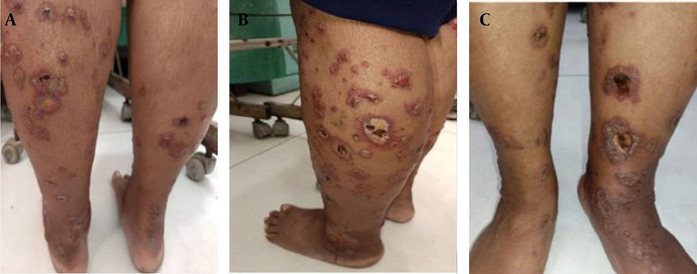
Figure 1.
Clinical presentation of localized bullous Sweet’s syndrome: Multiple erythematous, tender papules and plaques of varying sizes with overlying clear, tense vesicles and bullae, some with central hemorrhagic and necrotic crusts, over both lower legs. A few round-to-oval, punched-out ulcers measuring 2 - 3 cm, with erythematous edges and yellowish slough, were present over the anterolateral aspects of both lower legs.
Laboratory tests revealed a low hemoglobin level of 10 g/dL. The absolute neutrophil count (ANC) was elevated at 16,000 cells/µL, with no atypical white blood cells on peripheral blood smear. The erythrocyte sedimentation rate (ESR) was elevated at 25 mm/h by the Westergren method, whereas the C-reactive protein level was normal. The Mantoux test and slit-skin smear were negative. Fasting blood glucose (343 mg/dL) and HbA1c (13.1%) levels were markedly elevated. Further evaluation showed a deranged fasting lipid profile, with triglycerides of 170 mg/dL. Thyroid function tests were within the normal range. Routine urine microscopy showed +1 sugar and 6 - 7 pus cells. Other blood and radiological investigations, including ultrasonography, abdominal computed tomography, and chest radiography, were normal. The urine pregnancy test was negative.
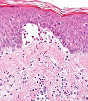
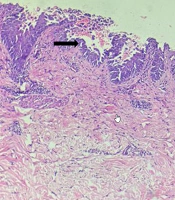
Gram staining of bulla fluid showed gram-positive cocci in pairs, and microbial culture demonstrated methicillin-sensitive Staphylococcus aureus sensitive to beta-lactams. Predominant polymorphonuclear leukocytes were observed on the Tzanck smear. Direct immunofluorescence (DIF) studies were not performed because of financial constraints. Histopathological examination revealed a subepidermal cleft containing multiple neutrophils, a few eosinophils, and erythrocytes, together with a dense perivascular infiltrate of neutrophils and eosinophils and extravasation of erythrocytes in the dermis (Figure 2). These findings were consistent with bullous Sweet’s syndrome, with an incidental diagnosis of metabolic syndrome.
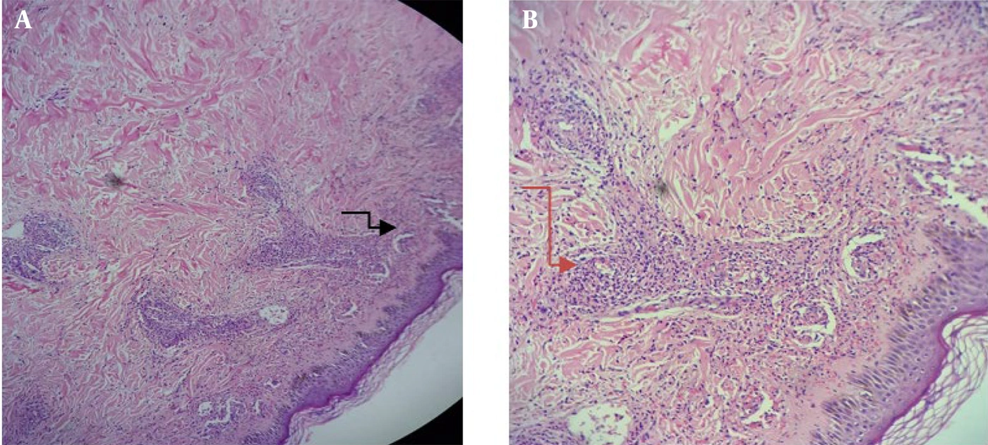
Figure 2.
A, Photomicrograph (hematoxylin and eosin, 10×); B, Photomicrograph (hematoxylin and eosin, 40×). Histopathological findings in Sweet’s syndrome: A subepidermal cleft containing multiple neutrophils, a few eosinophils (black arrowhead), and erythrocytes, together with a dense perivascular infiltrate of neutrophils and eosinophils and extravasation of erythrocytes in the dermis (red arrowhead), without evidence of true vasculitis.
The patient was advised to implement lifestyle and dietary changes. She was started on oral prednisolone at a weight-based dose of 0.5 mg/kg/day and systemic antibiotics according to microbial culture and sensitivity results, with strict glucose monitoring. Thyroxine 50 µg once daily for hypothyroidism and hematinics for low hemoglobin were administered.
Although the lesions began to regress within 2 weeks of treatment, persistently elevated blood glucose levels prompted early tapering of prednisolone and the addition of steroid-sparing anti-inflammatory therapies. With hemoglobin levels consistently maintained between 11 - 12 g/dL, dapsone 100 mg once daily, colchicine 0.5 mg twice daily, and doxycycline 100 mg twice daily were administered for 5 weeks. After complete resolution of the vesiculobullous lesions, colchicine was discontinued; however, deep punched-out ulcers persisted, necessitating pentoxifylline 400 mg three times daily and thalidomide 100 mg twice daily, after explicit counseling and written informed consent, to facilitate healing. Regular cleaning and dressing were performed using zinc oxide compounded in phenytoin paste and a topical antibiotic. The patient’s hemoglobin levels and liver function tests were monitored throughout treatment.
The patient achieved complete resolution of all lesions over 1.5 months, with atrophic scars and post-inflammatory dyspigmentation (Figures 3 and 4). She is currently off all medication and is under regular follow-up, with no recurrence or adverse effects during a 6-month follow-up period. In addition, diabetes mellitus and the deranged lipid profile were managed according to the physician’s recommendations, using insulin injections, oral hypoglycemic agents, and statins. The clinical course is summarized in Table 1.
Table 1.
Timeline of Clinical Events
| Timeline | Clinical events |
|---|---|
| On presentation | Multiple erythematous, blanchable, tender papules and plaques with overlying clear, tense vesicles and bullae on both lower legs, along with erosions with hemorrhagic and necrotic crusts. Multiple deep, round-to-oval, punched-out ulcers (2 - 3 cm) with erythematous edges and yellowish slough over the anterolateral aspects of both lower legs. Investigations included ANC, metabolic profile, Tzanck smear, and histopathology. |
| Final diagnosis | Localized bullous Sweet’s syndrome with underlying metabolic syndrome. |
| Treatment initiated | Oral prednisolone 40 mg once daily, tapered over 3 weeks, with regular glucose monitoring. |
| 2 weeks after initiation of treatment | Blood glucose levels remained deranged; dapsone 100 mg once daily, colchicine 0.5 mg twice daily, and doxycycline 100 mg twice daily were started. |
| 3 weeks after initiation of treatment | Complete resolution of vesiculobullous lesions was noted; therefore, colchicine was stopped, but deep punched-out ulcers persisted. Pentoxifylline 400 mg three times daily and thalidomide 100 mg twice daily were administered to facilitate ulcer healing. |
| 1.5 months after initiation of treatment | Complete resolution with atrophic scars and post-inflammatory dyspigmentation, with no adverse effects observed. |
| 3 months after initiation of treatment | All medications were gradually discontinued. |
| Follow-up at 6 months from treatment initiation | No new lesions or treatment-related adverse effects were documented. |
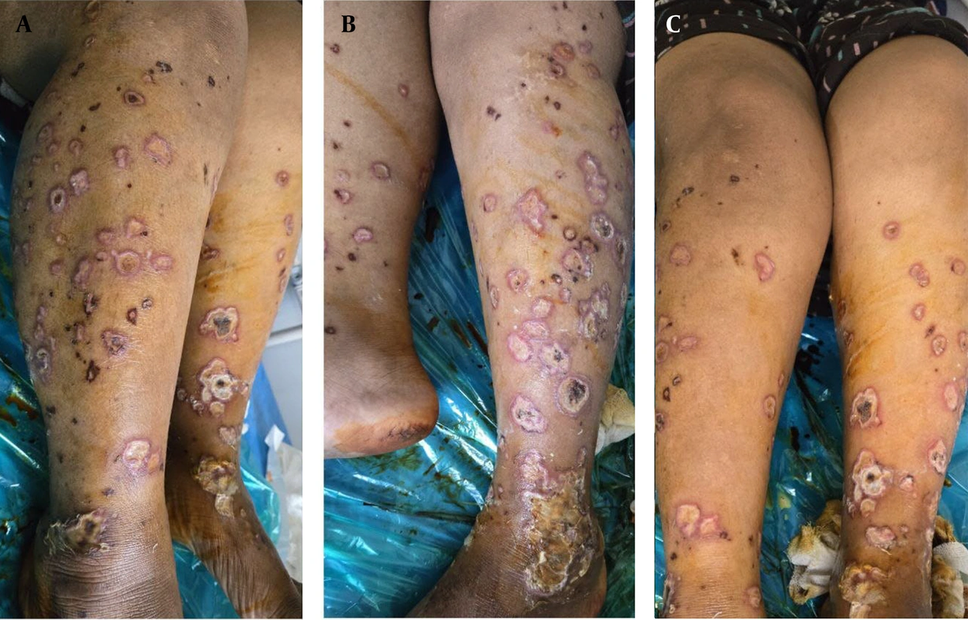
Figure 3.
One-month follow-up clinical outcome: Multiple erythematous plaques of varying sizes, some with central necrotic crusts, over both lower legs. A few round-to-oval, punched-out ulcers measuring 2 - 3 cm, with erythematous edges and yellowish slough, were present over the anterolateral aspects of both lower legs.
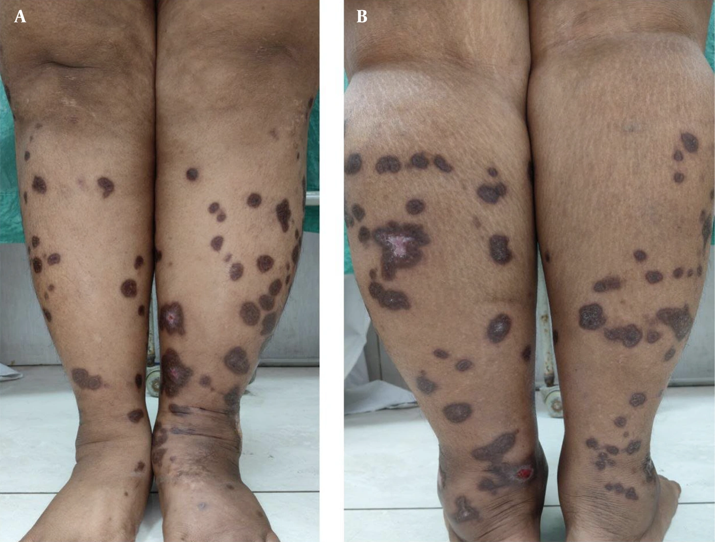
Figure 4.
Three-month follow-up clinical outcome: Multiple hyperpigmented patches, a few with central hypopigmented areas of atrophic scarring, over both lower legs.
3. Discussion
Neutrophilic dermatoses comprise a heterogeneous group of inflammatory skin disorders that are histologically characterized by sterile neutrophilic infiltration. Variations in the distribution of this infiltrate, together with clinical morphology and disease course, help differentiate the entities within this group. The spectrum of neutrophilic dermatoses includes Sweet’s syndrome, pyoderma gangrenosum, Behçet syndrome, and neutrophilic eccrine hidradenitis (1).
Sweet’s syndrome, also referred to as acute febrile neutrophilic dermatosis, is a prototype condition characterized by the sudden appearance of tender erythematous plaques, often accompanied by fever, arthralgia, and systemic features. First described in 1964, it shows a female predominance, with a ratio of approximately 4:1, and is most frequently observed in individuals aged 30 - 50 years (2). Based on etiology, it has been categorized into idiopathic, inflammatory, malignancy-associated, and pregnancy-related forms, with drug-induced cases reported less commonly (3).
Although the classic presentation includes erythematous plaques or nodules, several atypical morphological variants have been documented, including bullous, subcutaneous, pustular, erythema multiforme-like, and cellulitis-like forms. The bullous variant, in particular, is often associated with malignancy and may occur as a paraneoplastic manifestation in patients with known or occult neoplasms (4). Previous reports have described classic Sweet’s syndrome in association with diabetes mellitus (5); however, it has not been described as a direct diagnostic sign of diabetes. In contrast, this case highlights a bullous variant occurring in the setting of previously undiagnosed metabolic syndrome, with lesions confined to the lower legs, an unusual localization compared with the typically widespread distribution.
The novelty of this case lies in the atypical bullous morphology and distribution restricted to the lower legs, together with underlying metabolic syndrome. However, a bullous variant of Sweet’s syndrome localized to the palms and soles has previously been documented in association with a hematological malignancy (6).
Histopathological examination in bullous Sweet’s syndrome typically demonstrates a dense neutrophilic infiltrate within the dermis, accompanied by papillary dermal edema, leukocytoclasia, and subepidermal bulla formation, without evidence of true vasculitis (7), consistent with the findings in this patient.
The clinical differential diagnoses of bullous Sweet’s syndrome include bullous erythema multiforme, vasculitis, bullous erythema nodosum leprosum, autoimmune vesiculobullous disorders such as pretibial bullous pemphigoid, bullous pyoderma gangrenosum, irritant contact dermatitis, and bullous fixed drug eruption. Bullous erythema multiforme was ruled out because of the absence of classic target lesions and mucosal involvement, and the lack of basal cell vacuolation on histopathology. Histopathological examination did not reveal fibrinoid necrosis of vessel walls or significant vascular damage; therefore, cutaneous vasculitis was considered unlikely. There were no granulomas, lymphocytes, foamy histiocytes, acid-fast bacilli, or dermal perineural infiltrates to support erythema nodosum leprosum. Autoimmune vesiculobullous disorders, particularly bullous pemphigoid, were considered unlikely because of the absence of significant pruritus and the presence of dense dermal and perivascular neutrophilic infiltrates, together with neutrophilia on the peripheral blood smear. Bullous pyoderma gangrenosum was considered less likely because of the absence of typical undermined ulcers with violaceous borders. Bullous fixed drug eruption was excluded because of the absence of a clear drug trigger and site-specific recurrence. Histopathological examination did not reveal interface changes or necrotic keratinocytes. Although DIF could have further strengthened the diagnosis, it could not be performed. Nevertheless, the findings supported the diagnosis of bullous Sweet’s syndrome.
Su and Liu proposed diagnostic criteria consisting of 2 major and 4 minor components (8). In the present case, the patient fulfilled both major criteria: painful erythematous plaques with vesiculation and histopathological evidence of a dense dermal neutrophilic infiltrate without leukocytoclastic vasculitis. In addition, 3 of 4 minor criteria were satisfied: leukocytosis exceeding 8000 cells/µL with neutrophilia greater than 70%, elevated erythrocyte sedimentation rate, and a favorable response to systemic corticosteroids, supporting the diagnosis of Sweet’s syndrome.
The diagnosis of metabolic syndrome was established based on the National Cholesterol Education Program Adult Treatment Panel III (NCEP ATP III) guidelines (9). The patient met 3 of the 5 required parameters, including increased waist circumference (110 cm), elevated triglyceride levels (170 mg/dL), and elevated fasting blood glucose (> 100 mg/dL).
Systemic corticosteroids remain the cornerstone of therapy for Sweet’s syndrome, with prednisolone administered at a dose of 0.5 - 1.5 mg/kg/day, followed by gradual tapering over 2 - 4 weeks. The usefulness of steroid-sparing anti-inflammatory drugs in complicated cases is well recognized (2). This case, with uncontrolled diabetes and dyslipidemia, highlights a scenario in which corticosteroids, although effective, could not be continued long term because of their potential adverse effects, particularly metabolic derangements. In such circumstances, alternative anti-inflammatory agents play a crucial role in maintaining disease control and achieving a favorable clinical outcome. In cases of localized disease, topical or intralesional corticosteroids may be considered (10). Recent case reports have described alternative therapeutic agents, including thalidomide, pentoxifylline, dapsone, potassium iodide, colchicine, and minocycline. Immunosuppressive agents such as colchicine, dapsone, and methotrexate may also be used as steroid-sparing therapies, particularly in patients who do not tolerate corticosteroids well or in whom corticosteroid therapy alone is insufficient (2).
The usual duration of treatment ranges from 2 to 6 weeks, beginning with higher doses of systemic corticosteroids followed by gradual tapering to minimize the risk of recurrence. In certain cases, extended maintenance therapy with low-dose corticosteroids or steroid-sparing agents, such as colchicine, dapsone, or thalidomide, may be required for an additional 4 - 8 weeks to sustain remission, as in this patient (2).
3.1. Patient Perspective
The patient expressed concern regarding the diagnosis. However, following treatment, she experienced significant improvement in her symptoms and was satisfied with the overall outcome. She was also relieved to learn that her skin disease had led to the detection and subsequent management of previously undiagnosed metabolic derangements.