Cover image of the article: The Abnormal and Rapid Movement of Sewing Needle From Chest Toward Heart

Image Credit:Medical - Surgical Nursing Journal

The Abnormal and Rapid Movement of Sewing Needle From Chest Toward Heart

Authors

Changiz Azadi Ahmad Abadi1,*, Hamed Kiani2
1The Cardiac Surgery Department of Ali Ibn Abi Taleb Hospital, Faculty of Surgical, Zahedan University Of Medical Sciences, Zahedan, Iran
2Surgical Technologist of Ali Ibn Abi Taleb Hospital, Zahedan, Iran
*Corresponding Author: The Cardiac Surgery Department of Ali Ibn Abi Taleb Hospital, Faculty of Surgical, Zahedan University Of Medical Sciences, Zahedan, Iran. Tel: +98-9122448521, Email: dr.changiz_azadi@yahoo.com

Medical-Surgical Nursing Journal:Vol. 9, issue 2; e107850
Published online:Oct 04, 2020
Article type:Case Report
Received:Jul 25, 2020
Accepted:Sep 22, 2020
How to Cite:Azadi Ahmad Abadi C, Kiani H. The Abnormal and Rapid Movement of Sewing Needle From Chest Toward Heart. Med Surg Nurs J. 2020;9(2):e107850. doi: https://doi.org/10.5812/msnj.107850

Abstract

We herein introduced a 3 years old girl, who is a rare case of penetrating intracardiac damage severely induced by the abnormal movement of a sewing needle form the right lung toward the heart and entering in the right ventricle, which created Right Pneumothorax and right side bleeding. In clinical examinations, all vital symptoms were normal, and there was no sign of Cardiac tamponade, except reduced sound in the right lung base. Also, the external object was retained in the midclavicular line between the third rib, which was significant. In the Simple posterior-anterior and lateral graphics of chest radio opacity linear image on the lower part of the heart, the shadow was also significant. This is a rare case of abnormal needle movement from the right thorax to the right ventricular apex, which caused pulmonary problems. Following necessary diagnostic tests, a surgery was performed to remove the needle from the patient’s chest without open-heart surgery and CPB. Finally, 3 days after the operation, the patient was discharged from the cardiac surgery service in good general condition.

Highlights

1. Introduction

Blunt or sharp trauma may cause damages to or may penetrate different parts of the heart (1). Right ventricle trauma is the most common damage, which may be associated with other cardiac cavities (2). Many cases of penetrating trauma have been recorded, such as glass, sharp metals, bullet, and needle, while mainly led to significant damages to the anterior place of the right ventricle (3). Although penetrating trauma may be asymptomatic, but its most common symptoms include dyspnea and chest pain. Since needles are sharp, if left untreated, they can easily migrate to adjacent tissues, leading to hemothorax, cardiac tamponade, and pneumothorax. Even in the absence of migration, it may cause the formation of mural thrombi, which leads to repeated embolization. Rarely, valvular regurgitation may be observed, especially when the needle is embedded in the ventricular septum. Besides, in longstanding cases, it may cause complications such as infective endocarditis. Therefore, removing foreign bodies is essential. However, in most cases, open-heart surgery with CPB is required for removing foreign bodies (4). From 1967 to 2013, there were only 40 reported cases of sewing needle penetration in the heart (4). In the present study, the target case is a rare abnormal needle movement from the right thorax to the right ventricular apex, who referred with pulmonary manifestations of the lung. Following necessary diagnostic tests, a surgery was performed to remove the needle from the patient’s chest, without open-heart surgery and CPB.

2. Case Presentation

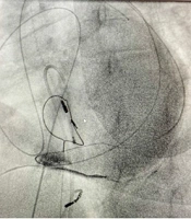
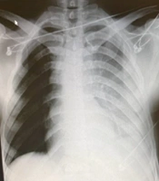
The patient is a 3 years old girl who was complaining of right chest pain and mild shortness of breath, while fell on the house carpet two days ago. The pain was in the front of the right thorax, which spread to the right shoulder of the patient, and the breath shortness of the patient became more severe as acting. The patient had no history of heart surgery or any specific disease. In clinical examinations, all vital symptoms were normal, and there was no sign of cardiac tamponade, except reduced sound in the right lung base. Besides, the external object was retained in the midclavicular line between the third rib, which was significant. In the Simple posterior-anterior and lateral graphics of chest radio opacity linear image on the lower part of the heart, the shadow was also significant. Echocardiography was performed to confirm the location of the foreign body and an ocular linear object resembling a large needle was observed in the right ventricular cavity (Figure 1).
Existence of a foreign object in a spiral CT scan
Figure 1.
Existence of a foreign object in a spiral CT scan
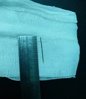
The CT scan spiral lung without contrast revealed a metal linear density image in the lower anterior part of the chest. The CT also showed that the needle pathway is in developing from the anterior part toward the posterior and tangent on the diaphragm. Therefore, first, the surgery was performed on the anterior thoracotomy fourth intercostal of the right side. The external object was not found after removing about 150 CC of blood and clots and lung exploration. Hence, the pericardium on the right longitudinally was opened, and the mediation and heart were explored. After observing the effects of the needle near the right ventricular apex and cutting on the site, the needle came out about 3.5 cm in size and after repairing the ventricle and closing the pericardium partially and placement of the chest tube in the right thorax, thoracotomy was closed. Finally, 3 days after the operation, the patient was discharged from the cardiac surgery department in good general condition (Figure 2).
Sewing needle removed from right ventricle after surgery
Figure 2.
Sewing needle removed from right ventricle after surgery

3. Discussion

Foreign bodies damage the heart and can migrate to adjacent tissues through the venous system or direct penetrating trauma, which mainly occurs in the right ventricle (5, 6). Signs and symptoms of foreign body penetration in the heart depend on the shape, size, and entering the location of the foreign object. However, it may be asymptomatic or include false aneurysm, cardiac tamponade, embolism, conduction disorders, etc. (7). Probable risks of the foreign body determine the necessity of using surgery, such as Pericardial effusion, bleeding, valve damage, embolism, etc. (8). In the current study, the patient was emergently operated due to the abnormal and rapid movement of the needle and the possibility of damaging to valves and heart cavities. Early diagnosis and intervention are always life-saving. These objects should be removed immediately by surgical procedures. Determining the exact location of the foreign body in the heart is important to ensure its complete removal. Moreover, it helps to avoid open-heart surgery and using CPB.
Footnotes

  • Authors' Contribution: HK did study concept and design and acquisition of data. CA did study supervision.

  • Conflict of Interests: The authors declare no conflict of interest.

  • Funding/Support: This study did not receive any funds.

  • Informed Consent: After expressing the possible risks and complications of the surgery to the parents of the patient, their written informed consent was obtained.

References

Copyright

Copyright © 2020, Medical - Surgical Nursing Journal. This is an open-access article distributed under the terms of the Creative Commons Attribution-NonCommercial 4.0 International License (http://creativecommons.org/licenses/by-nc/4.0/) which permits copy and redistribute the material just in noncommercial usages, provided the original work is properly cited.

Similar Articles

26
May
2026
Emergent Surgical Intervention for a Right Ventricular Stab Wound in a Young Man: A Case Report

Emergent Surgical Intervention for a Right Ventricular Stab Wound in a Young Man: A Case Report

Roozbeh Shadidi Asil,
Arshia Zardoui,
Leila Vatuizade,
Amir Zamani,
Kasra Hatampour Geraviani

Shadidi Asil R, Zardoui A, Vatuizade L, Zamani A, Hatampour Geraviani K. Emergent Surgical Intervention for a Right Ventricular Stab Wound in a Young Man: A Case Report. Int Cardiovasc Res J. 2026;20(1):e167339. doi: https://doi.org/10.5812/icrj-167339

4
Jun
2013
Penetrating Heart Injury: a case report

Penetrating Heart Injury: a case report

Ali Karbalaikhani,
Azita shishegar,
Salman Dehkhoda

Karbalaikhani A, shishegar A, Dehkhoda S. Penetrating Heart Injury: a case report. Ann Mil Health Sci Res. 2013;11(2):e66698. doi:

17
May
2022
Life Threatening Right Ventricle and Atrium Perforation and Tricuspid Valve Injury Due to Pacemaker Lead Dislodgment- A Case Report

Life Threatening Right Ventricle and Atrium Perforation and Tricuspid Valve Injury Due to Pacemaker Lead Dislodgment- A Case Report

Roxana Rastravan,
Rasoul Azarfarin,
Touraj Babaei,
Kourosh Tirgarfakheri,
Maryam Ghanbari

Rastravan R, Azarfarin R, Babaei T, Tirgarfakheri K, Ghanbari M. Life Threatening Right Ventricle and Atrium Perforation and Tricuspid Valve Injury Due to Pacemaker Lead Dislodgment- A Case Report. Multidiscip Cardio Annal. 2022;13(1):e123135. doi: https://doi.org/10.5812/mca-123135

30
Dec
2014
An Atypical Suicide Attempt: Self-Inflicted Intra-Cardiac Injury with Sewing Needle

An Atypical Suicide Attempt: Self-Inflicted Intra-Cardiac Injury with Sewing Needle

Nazila Shahmansouri,
Mahmood Shirzad,
Sam Zeraatiannejad Davani,
Fetemeh Heidari

Shahmansouri N, Shirzad M, Zeraatiannejad Davani S, Heidari F. An Atypical Suicide Attempt: Self-Inflicted Intra-Cardiac Injury with Sewing Needle. Iran J Psychiatry Behav Sci. 2014;8(4):. doi:

16
Oct
2016
Emergent CT Diagnosis of Right Ventricular Rupture Caused by Central Venous Catheter Injury

Emergent CT Diagnosis of Right Ventricular Rupture Caused by Central Venous Catheter Injury

Sang Hoon Kim,
Seung Min Yoo,
Hwa Yeon Lee,
Gun Lee,
Hyung Jong Kim,
Minjung Shim
,et al.

Kim SH, Yoo SM, Lee HY, Lee G, Kim HJ, et al. Emergent CT Diagnosis of Right Ventricular Rupture Caused by Central Venous Catheter Injury. I J Radiol. 2017;14(1):e13483. doi: https://doi.org/10.5812/iranjradiol.39484

More by these authors

Changiz Azadi Ahmad AbadiPubMedScholar
Hamed KianiPubMedScholar
Share
Cited by
Metrics