Emergent CT Diagnosis of Right Ventricular Rupture Caused by Central Venous Catheter Injury

Authors

Sang Hoon Kim1, Seung Min Yoo2,*, Hwa Yeon Lee3, Gun Lee4, Hyung Jong Kim1, Minjung Shim1, Sung-ik Kim1, In Song Song5, Hye Rin Kim2, Charles S White6
1Department of Internal Medicine, CHA University College of Medicine, Bundang, Korea
2Department of Diagnostic Radiology, CHA University College of Medicine, Bundang, Korea
3Smile Radiologic Clinic, Seoul, Korea
4Department of Cardiothoracic Surgery, CHA University College of Medicine, Bundang, Korea
5Department of Diagnostic Radiology, Chunju Jesus Hospital, Chunju, Korea
6Department of Diagnostic Radiology, University of Maryland Medical Center, Baltimore Maryland, USA
*Corresponding Author: Corresponding author: Seung Min Yoo, Department of Diagnostic Radiology, CHA University College of Medicine, Bundang, Korea. Tel: +82-317805423, Fax: +82-317804800, E-mail: Email: smyoo68@hanmail.net

Iranian Journal of Radiology:Vol. 14, issue 1; e13483
Published online:Oct 16, 2016
Article type:Case Report
Received:May 26, 2016
Accepted:Aug 12, 2016
How to Cite:Kim SH, Yoo SM, Lee HY, Lee G, Kim HJ, et al. Emergent CT Diagnosis of Right Ventricular Rupture Caused by Central Venous Catheter Injury. I J Radiol. 2017;14(1):e13483. doi: https://doi.org/10.5812/iranjradiol.39484

Abstract

Diagnosis of cardiac tamponade has been mainly based on clinical features and echocardiography. However, CT may be an initial tool for the evaluation of cardiac tamponade because cutting edge CT is increasingly being installed in the emergency room, and presentation of cardiac tamponade can be nonspecific. Importantly, CT has better spatial resolution compared with echocardiography. Thus, identification of the etiology of cardiac tamponade is possible on CT. We report a case of cardiac tamponade demonstrating direct leakage of contrast material from the right ventricle into the pericardial space caused by central venous catheter injury diagnosed on CT.

1. Introduction

Acute cardiac tamponade is a life-threatening condition caused by a variety of etiologies including blunt trauma, stab wounds, Stanford type A aortic dissection, or cardiac rupture as a complication of acute myocardial infarction or central venous catheter injury (1-6). Cardiac tamponade is accompanied by hemodynamic instability such that computed tomography (CT) is relatively contraindicated. Thus, the diagnosis of cardiac tamponade is based mainly on clinical features (i.e., the classic triad of hypotension, neck vein distension, and muffled heart sounds) and bedside echocardiography. However, at times CT may be an initial tool for evaluation of cardiac tamponade because state-of-the-art CT scanners are increasingly being installed within the emergency department (ED), and the presentation of cardiac tamponade is often nonspecific. Importantly, CT has better spatial resolution and no limitation of viewing windows compared with echocardiography. Thus, precise identification of the etiology of cardiac tamponade and localization of the site of rupture may be possible on CT, assisting the cardiac surgeon in establishing an appropriate surgical plan. Notably, direct visualization of leakage of contrast material from a cardiac chamber into the pericardial space causing cardiac tamponade on CT has not been reported in the literature (1-6). We report a case of cardiac tamponade caused by central venous catheter injury, demonstrating direct leakage of contrast material from the right ventricle into the pericardial space on CT.

2. Case Presentation

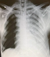
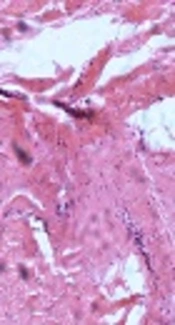
A 75-year-old woman presented to the ED with sudden dyspnea and loss of consciousness during hemodialysis. The patient had no history relevant to cardiac tamponade other than end stage renal disease due to hypertension. Because an arteriovenous fistula in her left arm did not mature fully, hemodialysis had been performed through a right subclavian vein catheter for a year. Figure 1. Blood pressure was 163/68 mmHg at the time of initiation of hemodialysis, but the blood pressure dropped suddenly to 63/45 mmHg two hours into the hemodialysis session. On chest radiography performed in the ED, the tip of 14.5 Fr central venous catheter (Hemostar® Long-term Homodialysis Catheter, BARD Access Systems, Inc., Salt Lake City, USA) projected between the right atrium and ventricle (Figure 1A). As the primary concern of the ED physician was massive pulmonary embolism, a pulmonary CT angiography was performed. The study showed severe compression of the right ventricle and atrium by a large hemopericardium and distension of the inferior vena cava, consistent with cardiac tamponade (Figure 1B and 1C). Direct leakage of contrast material (Figure 1C) from the free wall of the right ventricle into the pericardial space was demonstrated on coronal reformatted CT image. A diagnosis of right ventricular rupture caused by central venous catheter injury was made, and emergency surgery was recommended based on CT findings. Impaction of the central venous catheter tip within the right ventricular free wall was demonstrated on the intra-operative echocardiography, suggesting central venous catheter injury as the etiology of cardiac tamponade. At surgery, a 3-4 mm linear tear with active bleeding was found on the right ventricular free wall (Figure 1D), and direct closure of the defect was performed.

3. Discussion

Right ventricular rupture caused by central venous catheter injury is extremely rare (4, 5). It is known that the tip of the central venous catheter can be moved by changes in neck motion, respiration, or cardiac rhythm (4). Thus, placement of the tip of the central venous catheter in the superior vena cava is optimal to avoid serious complications. Although there are limitations due to a selected set of circumstances (i.e., infants and older catheters), one study reported that the incidence of cardiac tamponade occurring ≥ 7 days after insertion of a central venous catheter was 18% (7). On further investigation, the central venous catheter in this patient had been located in the right cardiac chamber for approximately one year, leading to an increased risk of cardiac tamponade. Typical CT findings of cardiac tamponade were demonstrated in the present case (i.e., large hemopericardium compressing the right atrium and ventricle and marked dilatation of the inferior vena cava). A previous study has shown that compression of the right ventricle is highly specific for the diagnosis of cardiac tamponade (3). However, there is no prior report demonstrating direct leakage of contrast material from a cardiac chamber to the pericardial space on CT as a finding of cardiac tamponade. Detection of leakage of contrast material into the pericardial space on CT in the present case may have been aided by two factors. First, the size of the defect in the right ventricle by central venous catheter injury may not have been large enough to cause sudden death, thus allowing sufficient time for identification on CT. The second potential reason is that the high injection rate of contrast material in the right ventricle on pulmonary CT angiography may have facilitated detection of leakage into the pericardium. It should be stressed that if only axial CT images are reviewed, the leakage of contrast materials from the right ventricle into the pericardial space may be misinterpreted as an artifact caused by cardiac motion and therefore overlooked. Thus, thorough evaluation of coronal and sagittal CT reformatted images is mandatory in the evaluation of cardiac tamponade.
In summary, the location of the tip of a central venous catheter should be carefully evaluated to avoid the risk of rupture of the right atrium or ventricle. Moreover, ED physicians and radiologists should assess for leakage of contrast material from cardiac chambers into the pericardial space on CT in patients with hemopericardium and an indwelling central venous catheter.
Acknowledgments

Footnotes

  • Authors’ Contributions:None.

  • Financial Disclosure:None.

  • Funding/Support:None.

References

  • 1.
    Kim MH, Lee DJ, Kim MC. Bilateral hydrothorax and cardiac tamponade after right subclavian vein catheterization -A case report. Korean J Anesthesiol. 2010;59 Suppl:S211-7. [PubMed ID: 21286444]. https://doi.org/10.4097/kjae.2010.59.S.S211.
  • 2.
    Nichols J, Berger N, Joseph P, Datta D. Subacute right ventricle perforation by pacemaker lead presenting with left hemothorax and shock. Case Rep Cardiol. 2015;2015:983930. [PubMed ID: 25785204]. https://doi.org/10.1155/2015/983930.
  • 3.
    Spodick DH. Acute cardiac tamponade. N Engl J Med. 2003;349(7):684-90. [PubMed ID: 12917306]. https://doi.org/10.1056/NEJMra022643.
  • 4.
    Ellis LM, Vogel SB, Copeland EM. Central venous catheter vascular erosions. Diagnosis and clinical course. Ann Surg. 1989;209(4):475-8. [PubMed ID: 2930292].
  • 5.
    Greenall MJ, Blewitt RW, McMahon MJ. Cardiac tamponade and central venous catheters. Br Med J. 1975;2(5971):595-7. [PubMed ID: 1131628].
  • 6.
    Schiavone WA. Cardiac tamponade: 12 pearls in diagnosis and management. Cleve Clin J Med. 2013;80(2):109-16. [PubMed ID: 23376916]. https://doi.org/10.3949/ccjm.80a.12052.
  • 7.
    Giacoia GP. Cardiac tamponade and hydrothorax as complications of central venous parenteral nutrition in infants. JPEN J Parenter Enteral Nutr. 1991;15(1):110-3. [PubMed ID: 1901100].

Copyright

Copyright © 2016, Tehran University of Medical Sciences and Iranian Society of Radiology. This is an open-access article distributed under the terms of the Creative Commons Attribution-NonCommercial 4.0 International License (http://creativecommons.org/licenses/by-nc/4.0/) which permits copy and redistribute the material just in noncommercial usages, provided the original work is properly cited.

Similar Articles

30
Jun
2014
Ruptured Valsalva Sinus Aneurysm to Pericardium Simulated Aortic Root Dissection

Ruptured Valsalva Sinus Aneurysm to Pericardium Simulated Aortic Root Dissection

Tahereh Davarpasand,
Ali Hosseinsabet,
Kumars Abassi,
Sorya Arzhan

Davarpasand T, Hosseinsabet A, Abassi K, Arzhan S. Ruptured Valsalva Sinus Aneurysm to Pericardium Simulated Aortic Root Dissection. Int Cardiovasc Res J. 2014;8(2):e12038. doi:

31
Oct
2017
Successful Detachable Coil Embolization of Iatrogenic Superior Vena Cava Penetration by Central Venous Catheter: A Case Report

Successful Detachable Coil Embolization of Iatrogenic Superior Vena Cava Penetration by Central Venous Catheter: A Case Report

Byung Jin Park,
Jae Kyu Kim,
Hyoung Ook Kim,
Nam Yeol Yim,
Yang Jun Kang

Jin Park B, Kyu Kim J, Ook Kim H, Yeol Yim N, Jun Kang Y. Successful Detachable Coil Embolization of Iatrogenic Superior Vena Cava Penetration by Central Venous Catheter: A Case Report. I J Radiol. 2017;14(4):e63481. doi: https://doi.org/10.5812/iranjradiol.63481

26
May
2026
Emergent Surgical Intervention for a Right Ventricular Stab Wound in a Young Man: A Case Report

Emergent Surgical Intervention for a Right Ventricular Stab Wound in a Young Man: A Case Report

Roozbeh Shadidi Asil,
Arshia Zardoui,
Leila Vatuizade,
Amir Zamani,
Kasra Hatampour Geraviani

Shadidi Asil R, Zardoui A, Vatuizade L, Zamani A, Hatampour Geraviani K. Emergent Surgical Intervention for a Right Ventricular Stab Wound in a Young Man: A Case Report. Int Cardiovasc Res J. 2026;20(1):e167339. doi: https://doi.org/10.5812/icrj-167339

30
Dec
2008
Migration of the Tip of a Central Venous Catheter

Migration of the Tip of a Central Venous Catheter  

Masoud Shariat,
M. Zahiah,
P.K. Chan

Shariat M, Zahiah M, Chan P. Migration of the Tip of a Central Venous Catheter  . I J Radiol. 2008;5(4):e79034. doi:

28
Jun
2017
Right Para Cardiac Pericardial Cyst

Right Para Cardiac Pericardial Cyst

Aghigh Haydari,
Feridoun Sabzi,
Samsam Dabiri,
Alireza Poormotaabed

Haydari A, Sabzi F, Dabiri S, Poormotaabed A. Right Para Cardiac Pericardial Cyst. Multidiscip Cardio Annal. 2018;9(2):e68382. doi: https://doi.org/10.5812/mcardia.68382

More by these authors

Sang Hoon KimPubMedScholar
Seung Min YooPubMedScholar
Hwa Yeon LeePubMedScholar
Hyung Jong KimPubMedScholar
Minjung ShimPubMedScholar
Sung-ik KimPubMedScholar
In Song SongPubMedScholar
Hye Rin KimPubMedScholar
Charles S WhitePubMedScholar
Share
Cited by
Metrics