
Dear Editor,
Hemiscorpius lepturus, locally known in parts of Iran as Gadim, can cause cutaneous necrosis and severe systemic envenomation. It has been reported from Iran, Iraq, Oman, Saudi Arabia, Yemen, Pakistan, and the United Arab Emirates. In Iran, documented records include Khuzestan, Semnan, Fars, Kurdistan, Hormozgan, Sistan and Baluchestan, Isfahan, Bushehr, Kohgiluyeh and Boyer-Ahmad, Ilam, Chaharmahal and Bakhtiari, Lorestan, Hamadan, Kermanshah, and Kerman. The species has been collected in both indoor and outdoor environments, including beneath rocks, within narrow rock crevices, in cracks and burrows in mud walls, and beneath tree bark in areas with moist soil (1). These records document the species’ distribution and do not establish its absence from other provinces.
A historical study from Khuzestan involving 26,397 scorpion-sting records found that H. lepturus accounted for approximately 10 - 15% of stings, 89% of deaths, and 92% of hospitalized cases in that study population. These region-specific historical findings should not be extrapolated to contemporary national epidemiology (2).
Clinical manifestations of H. lepturus stings can be categorized into 3 forms: cutaneous, systemic, and combined cutaneous-systemic (1, 3, 4). Cutaneous manifestations include erythema, edema, ecchymosis, blistering, cellulitis, and skin necrosis. Systemic manifestations may develop after a latent period of approximately 2 to 7 days and include constitutional symptoms, hemolysis, hemoglobinuria, thrombocytopenia, coagulopathy, disseminated intravascular coagulation (DIC), hemolytic uremic syndrome (HUS), and acute kidney injury (1, 3). In the combined form, systemic manifestations may likewise appear after a similar latent period, together with local skin injury. Thus, in a patient presenting with apparently isolated cutaneous manifestations and no evidence of systemic envenomation, constitutional symptoms such as fever, malaise, nausea, vomiting, weakness, or diaphoresis are less likely; their subsequent development should raise concern for progression to systemic envenomation (1, 3). H. lepturus stings are typically painless initially or cause only a mild, localized burning sensation. However, pain may develop later and intensify as tissue damage progresses toward necrosis (1, 3, 4).
Among Hemiscorpius species for which published human reports document cutaneous necrosis in Iran, H. lepturus and H. acanthocercus are currently implicated. Although H. lepturus remains the best-documented species, fatal human envenomation by H. acanthocercus with local necrosis and systemic toxicity has also been reported (5). H. enischnochela has been associated with severe hemolysis and hemoglobinuria in a human case report, but its clinical spectrum remains insufficiently characterized (6). Thus, clinical evidence for these species remains very limited, although their reported manifestations appear similar to those of H. lepturus (5, 6).
The Mediterranean recluse spider, Loxosceles rufescens, has been documented in Iran, including Tehran, Golestan, Razavi Khorasan, Fars, Hormozgan, Kohgiluyeh and Boyer-Ahmad, Mazandaran, Qom, and Alborz. It has been collected from houses, apartments, parks, caves, and other sheltered environments (7, 8). It has also been documented in Iraq, Pakistan, Yemen, Saudi Arabia, and the United Arab Emirates (9-12). These countries, along with Iran, are therefore areas of potential geographic overlap with H. lepturus.
Loxoscelism has cutaneous and systemic forms. L. rufescens bites may initially be painless or cause only local burning; pain may subsequently develop and increase as the lesion evolves toward necrosis, as with H. lepturus stings (10, 13). Cutaneous loxoscelism may cause erythema, edema, ecchymosis, blistering, and dermonecrosis. Constitutional symptoms such as fever, malaise, nausea, vomiting, and weakness are common in patients with cutaneous loxoscelism caused by L. rufescens (10, 13). Systemic loxoscelism may include hemolysis, rhabdomyolysis, coagulation abnormalities, and acute kidney injury, but progression from cutaneous to systemic disease appears uncommon in confirmed L. rufescens bites (13). Figures 1 and 2 show documented cases of cutaneous necrosis following an L. rufescens bite and an H. lepturus sting, respectively, illustrating that the appearance of the necrotic lesions alone may not reliably distinguish between these 2 etiologies.
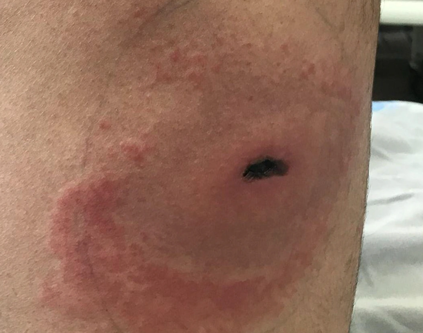
Figure 1.
Representative clinical photograph of a confirmed Loxosceles rufescens bite on the flank, captured approximately 4 - 5 days after envenomation. The patient witnessed the spider at the time of the bite. Before photography, the patient had received conservative treatment. Informed consent for publication was obtained. Photograph by the author.
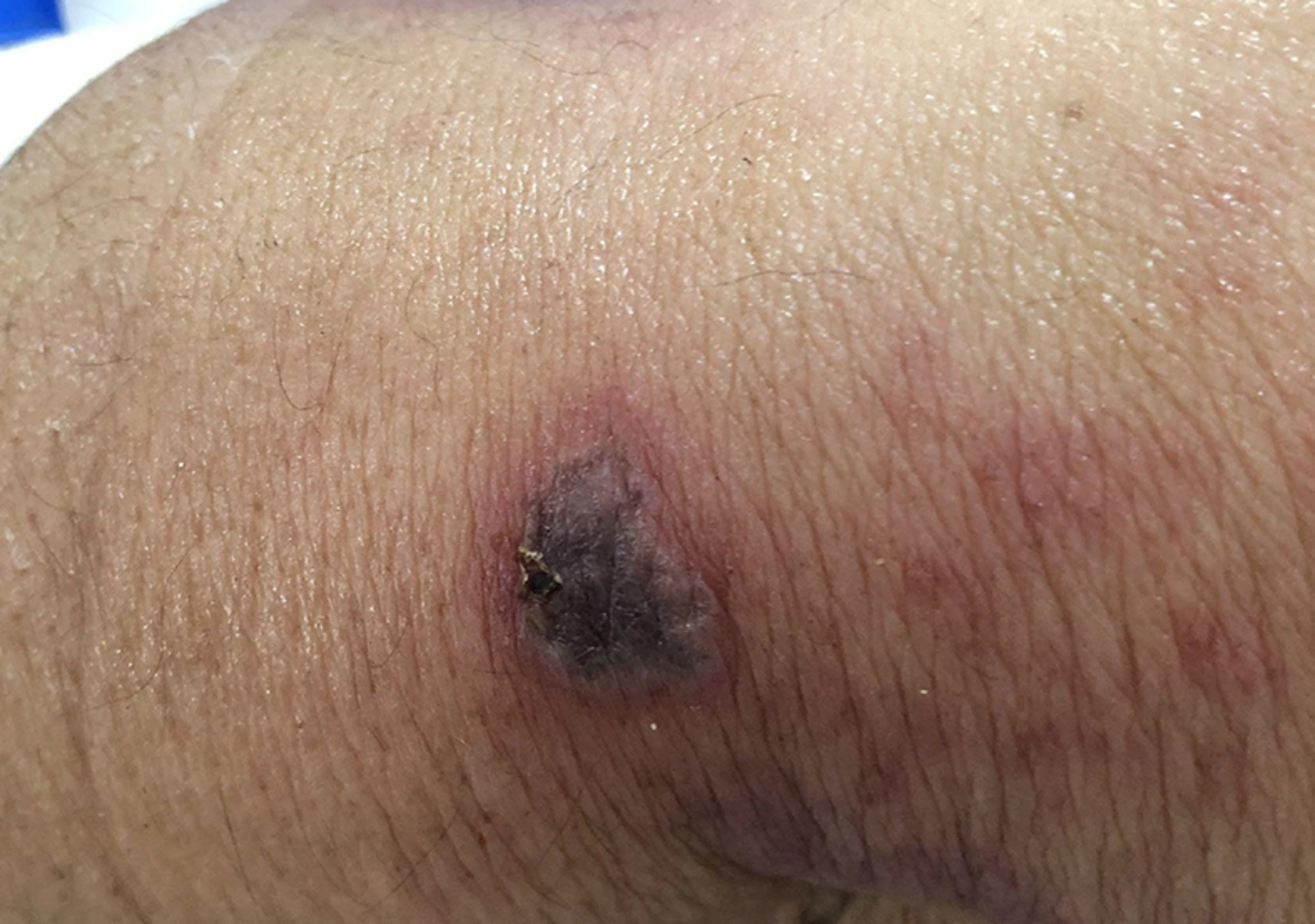
Figure 2.
Representative clinical photograph of a confirmed Hemiscorpius lepturus (Gadim) sting on the elbow, captured approximately 2 - 3 days after the sting. The patient brought a photograph of the scorpion taken with a mobile phone. No treatment was administered before photography. Informed consent for the publication of this photograph was obtained. Photograph by the author.
When H. lepturus and L. rufescens occur in the same geographic setting and the arthropod is not witnessed, constitutional symptoms accompanying a necrotic lesion may increase suspicion for loxoscelism; however, they are a nonspecific clinical clue rather than a diagnostic criterion. The sensitivity, specificity, likelihood ratios, and reliability of these symptoms across disease severity have not been adequately established. Diagnostic assessment should integrate the exposure setting, geographic location, witnessed arthropod exposure, expert identification when available, evidence of hemolysis, hemoglobinuria, anemia, thrombocytopenia, coagulopathy, and renal abnormalities (1, 9, 10, 13, 14). An unwitnessed necrotic lesion should not automatically be attributed to either arthropod. Important differential diagnoses include bacterial cellulitis or necrotizing soft-tissue infection, ecthyma gangrenosum, cutaneous anthrax, pyoderma gangrenosum, vasculitis, vascular skin necrosis, calciphylaxis, traumatic or pressure-related injuries, drug-associated skin necrosis, and other arthropod envenomations (14). Rapidly progressive necrosis, severe pain out of proportion to examination, hemodynamic instability, systemic inflammatory toxicity, extensive tissue destruction, hemolysis, coagulopathy, or acute kidney injury should prompt appropriate surgical, infectious-disease, nephrology, hematology, or medical-toxicology assessment.
For suspected skin necrosis caused by either an L. rufescens bite or an H. lepturus sting, wound cleansing, tetanus prophylaxis when indicated, analgesia, antipruritic treatment, and appropriate wound care are reasonable. Antibiotics should be reserved for documented or strongly suspected secondary bacterial infection rather than sterile venom-induced inflammation (10, 14). In suspected loxoscelism, local heat and topical corticosteroids should not be routinely recommended because their benefit is inadequately established (10, 14). Systemic corticosteroids should not be considered routine treatment for uncomplicated cutaneous loxoscelism. They may be considered selectively in cases of significant systemic hemolysis, particularly when a warm autoimmune hemolytic anemia phenotype is demonstrated, although evidence is derived mainly from case series and observational experience, particularly involving L. reclusa (15). However, L. reclusa has not been reported from Iran.
Accordingly, when a necrotic lesion is compatible with either H. lepturus or L. rufescens in an area where both species are distributed, empiric antivenom may be considered when clinical suspicion for H. lepturus is substantial, particularly before systemic manifestations or laboratory evidence of hemolysis and renal injury develop. According to Iranian expert opinion, a single-vial dose of intravenous polyvalent scorpion antivenom may be considered (16). However, the optimal dose, timing, route, and dose adjustment according to severity, age, body mass, or time since exposure have not been established by high-quality comparative clinical trials (16-18). Earlier administration has a biological and experimental rationale, but its clinical effectiveness remains incompletely established (17, 18). There is no national Iranian protocol specifically addressing an unidentified necrotic lesion potentially caused by H. lepturus or L. rufescens. Therefore, empiric treatment should be reserved for cases in which the probability and potential consequences of H. lepturus envenomation are substantial, rather than for uncertainty alone. Administration of antivenom does not preclude simultaneous clinical assessment for loxoscelism or evaluation for infectious, vascular, inflammatory, traumatic, drug-related, or other causes of necrosis. Evidence is insufficient to establish the cost-effectiveness of empiric antivenom solely because the 2 envenomations cannot initially be distinguished.
Because the available Iranian polyvalent antivenom is equine-derived, intravenous administration requires appropriate clinical observation and the immediate capability to manage hypersensitivity reactions. Routine pre-administration skin testing is not recommended because it does not reliably predict immediate hypersensitivity reactions (19, 20). Antivenom should therefore be administered intravenously under appropriate monitoring, with readiness to treat anaphylaxis (19, 20).
Patients with suspected H. lepturus or L. rufescens envenomation associated with cutaneous necrosis should remain hospitalized and under observation because systemic toxicity may be delayed. Baseline evaluation should include a complete blood count, hemoglobin, platelet count, electrolytes, blood urea nitrogen, creatinine, creatine kinase, coagulation studies, and urinalysis, with serial reassessment during hospitalization. In suspected H. lepturus stings, urinalysis once or twice daily is appropriate for monitoring evolving hemolysis, hemoglobinuria, and renal involvement (1, 3). Fever, malaise, or vomiting developing after a suspected H. lepturus sting should be regarded as concerning for systemic envenomation and should prompt escalation of monitoring and treatment. Dark urine, falling hemoglobin, thrombocytopenia, coagulopathy, rising creatinine, hemodynamic instability, or progressive tissue injury should similarly prompt specialist assessment. Because delayed systemic toxicity may occur, the author's conservative expert-practice recommendation is to continue hospitalization and observation for at least 1 week in cases with sufficient clinical suspicion of either H. lepturus or L. rufescens envenomation, with longer observation warranted when systemic abnormalities or progressive local injury persist. This period is a conservative expert-practice recommendation rather than a nationally standardized evidence-based protocol.
The literature supporting this letter was identified through PubMed/MEDLINE and supplemented by reference-list review, with an emphasis on primary clinical studies, species-specific case reports, regional reviews, and recent literature concerning loxoscelism and acute cutaneous necrosis. Epidemiological and clinical descriptions are based mainly on observational studies, case reports, and reviews; antivenom recommendations reflect regional Iranian practice supported by limited clinical and experimental evidence; hypersensitivity recommendations are supported by clinical studies; and loxoscelism treatment recommendations are based mainly on observational evidence and case series. Diagnostic discrimination, optimal antivenom dosing, cost-effectiveness, and the appropriate observation period therefore remain areas of uncertainty rather than established standards (1, 3, 9, 10, 13, 14, 15, 16, 17, 18, 19, 20).
In summary, cutaneous necrosis after an unwitnessed arthropod exposure does not reliably distinguish between an H. lepturus sting and an L. rufescens bite. Constitutional symptoms accompanying necrosis may increase suspicion for loxoscelism but are not specific and should not be used alone to distinguish the 2 envenomations. When the epidemiological and clinical probability of H. lepturus is substantial, early antivenom may be considered while evaluation for loxoscelism and other causes of necrosis proceeds concurrently. Continued hospitalization, serial clinical assessment, and laboratory monitoring are warranted because systemic toxicity may be delayed.