Hematological and morphological findings have been demonstrated in
Table 1 and
Figures 1 -
5. As shown in
Table 1, the number of leukocytes was significantly lower in group III (8.36 ± 0.23 × 10
3/µL) and group IV (7.85 ± 0.18 × 10
3/µL) in comparison with group I (8.78 ± 0.14 × 10
3/µL) and group II (8.50 ± 0.20 × 10
3/µL) (P value III:I = 0.004, and P value IV:II < 0.0001). In case of using curcumin following CFL exposure, the levels of leukocytes were reached to the normal levels both at 10 and 30 days in group V (8.85 ± 0.29 × 10
3/µL) and group VI (8.30 ± 0.16 × 10
3/µL); there were no significant differences after 10 and 30 days between control and curcumin groups (P value V:I = 0.79, and P value VI:II = 0.09).
| Group I | Group III | Group V | Group II | Group IV | Group VI | P Value III:I | P Value V:I | P Value IV:II | P Value VI:II | P Value V:III | P Value VI:IV | P Value IV:III | P Value VI:V |
|---|
| WBC × 103/µL | 8.78 ± 0.14 | 8.36 ± 0.23 | 8.85 ± 0.29 | 8.50 ± 0.20 | 7.85 ± 0.18 | 8.30 ± 0.16 | 0.004* | 0.790 | < 0.0001* | 0.090 | 0.001* | < 0.0001* | < 0.0001* | < 0.0001* |
| N × 103/µL | 1.71 ± 8.32 | 1.84 ± 30.32 | 1.73 ± 14.06 | 1.91 ± 9.83 | 2.24 ± 15.76 | 2.04 ± 55.13 | < 0.0001* | 0.307 | < 0.0001* | < 0.0001* | < 0.0001* | < 0.0001* | < 0.0001* | < 0.0001* |
| E × 103/µL | 0.09 ± 1.20 | 0.02 ± 1.31 | 0.02 ± 0.76 | 0.09 ± 1.51 | 0.04 ± 1.31 | 0.02 ± 0.76 | < 0.0001* | < 0.0001* | < 0.0001* | < 0.0001* | 0.195 | < 0.0001* | < 0.0001* | 0.482 |
| M × 103/µL | 0.09 ± 1.20 | 0.05 ± 33.70 | 0.09 ± 1.31 | 0.02 ± 0.76 | 0.04 ± 1.31 | 0.02 ± 0.76 | 0.004* | >0.99 | < 0.0001* | >0.99 | 0.004* | < 0.0001* | 0.435 | < 0.0001* |
| L × 103/µL | 6.89 ± 6.05 | 6.45 ± 62.69 | 7.01 ± 42.31 | 6.48 ± 4.54 | 5.53 ± 38.03 | 6.21 ± 55.17 | < 0.0001* | < 0.0001* | < 0.0001* | < 0.0001* | < 0.0001* | < 0.0001* | < 0.0001* | < 0.0001* |
| PLTt × 103/µL | 851 ± 3.06 | 848 ± 4.50 | 846 ± 5.60 | 846 ± 2.73 | 750 ± 6.52 | 808 ± 2.13 | 0.243 | 0.053 | < 0.0001* | < 0.0001* | 0.688 | < 0.0001* | < 0.0001* | < 0.0001* |
Abbreviations: E, eosinophil; L, lymphocyte; M, monocyte; N, neutrophil; PLT, platelet; WBC, white blood cell.
aValues are expressed as means ± SD.
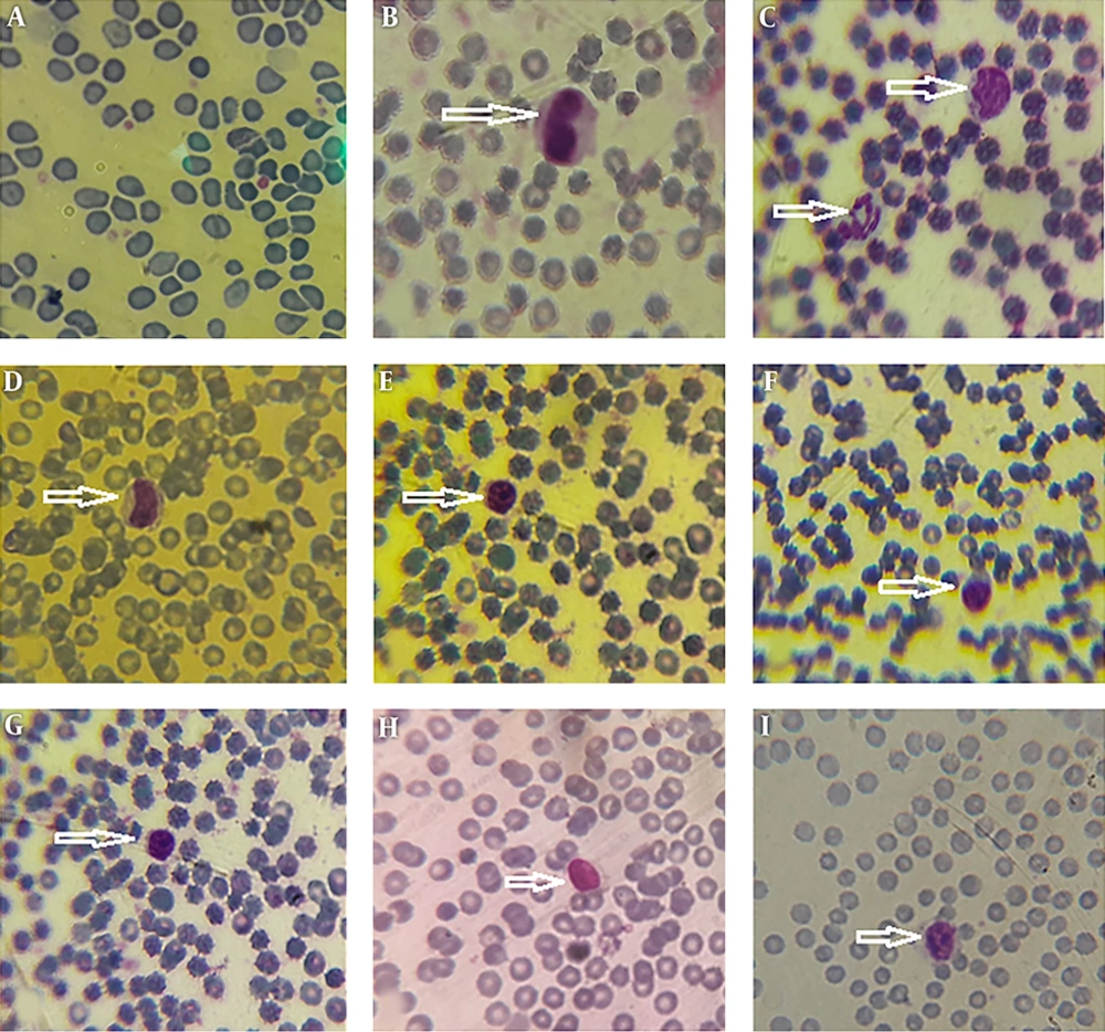
Wright’s-stained smears of leukocytes in control groups (I and II). Neutrophil (C), monocytes (B - D), and lymphocytes (E - I) are seen. There are no leukocytes in (A). Magnification × 400.
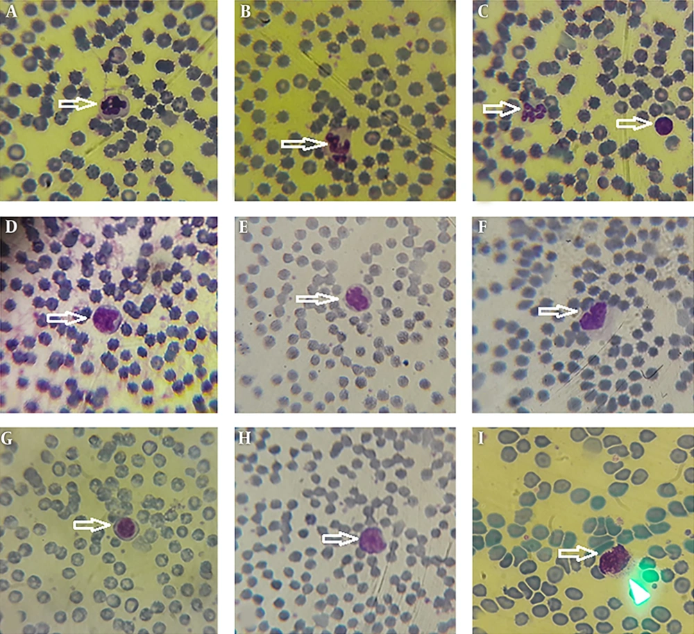
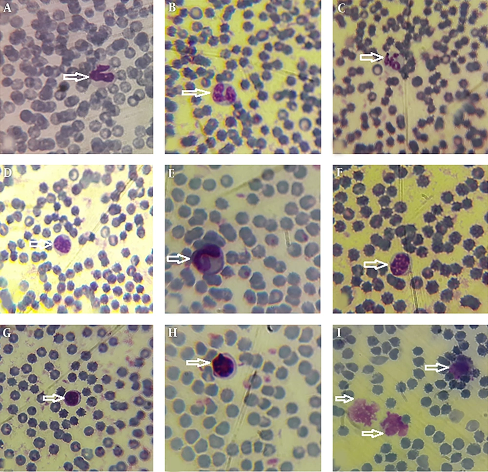
Wright’s-stained smears of leukocytes in fluorescent group 10 days after exposure to fluorescent light exposure (group III). Neutrophils (A - C), monocytes (D - F) and lymphocytes (C, G - I) are seen. Magnification × 400.
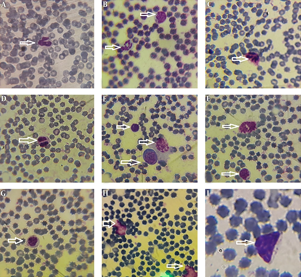
Wright’s-stained smears of leukocytes in fluorescent group 30 days after exposure to fluorescent light (group IV). Neutrophils (A - C), eosinophil (D), monocytes (B, E, and F) and lymphocytes (E - I) are seen. Magnification × 400.
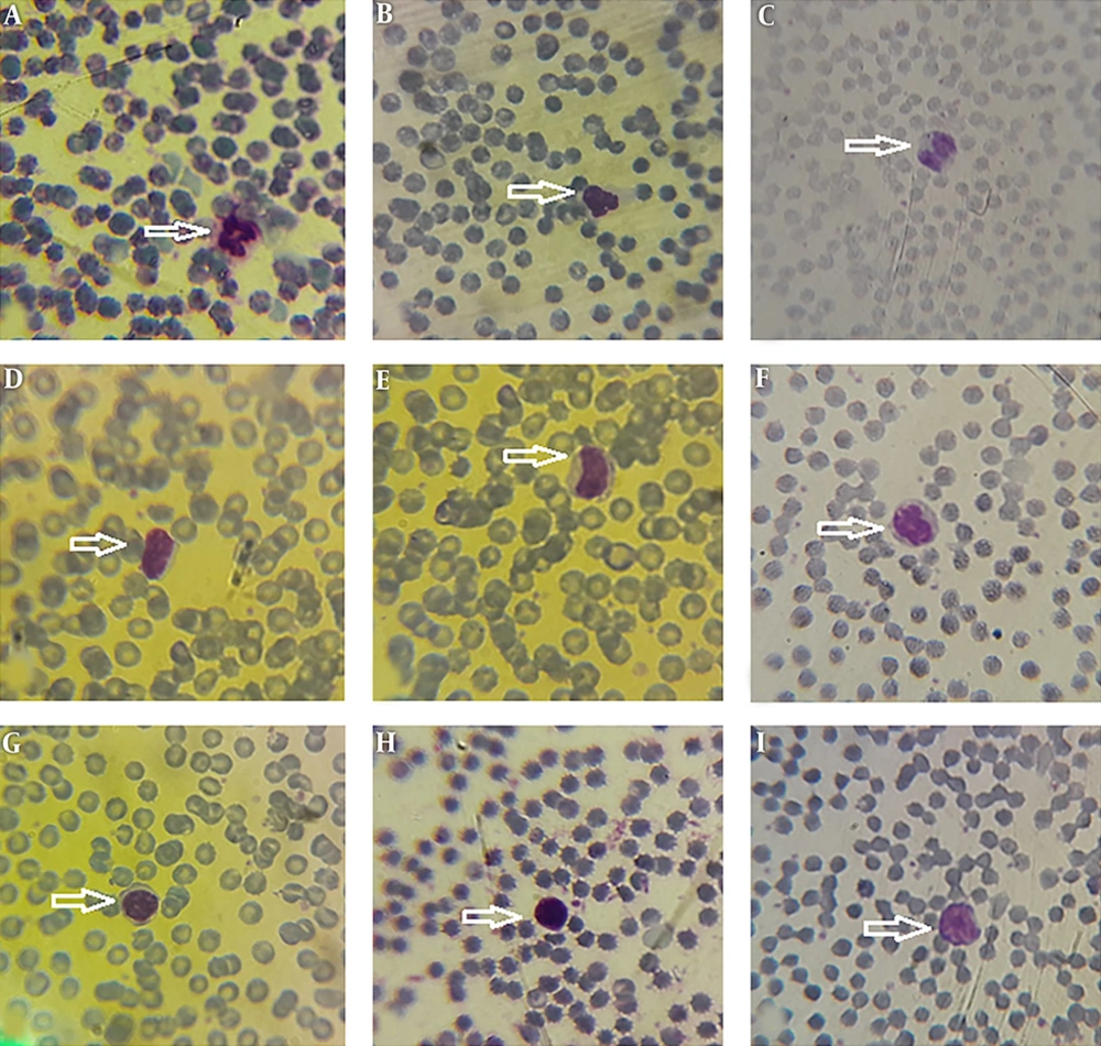
Wright’s-stained smears of leukocytes in curcumin group 10 days after both fluorescent light exposure and curcumin administration (group V). Neutrophils (A - C), monocytes (D - F) and lymphocytes (G - I) are seen. Magnification × 400.
Wright’s-stained smears of leukocytes in curcumin group 30 days after both fluorescent light exposure and curcumin administration (group VI). Neutrophils (A - C), monocytes (D - F) and lymphocytes (G - I) are seen. Magnification × 400.
The subtypes of leukocytes including neutrophil, eosinophil, monocyte, and lymphocyte were changed by exposure to CFL (
Table 1).
The number of neutrophils was significantly higher in group III (1.84 ± 30.32 × 10³/µL) and group IV (2.48 ± 15.76 × 10³/µL) compared to group I (1.71 ± 8.32 × 10³/µL) and II (1.91 ± 9.83 × 10³/µL) (P value III:I < 0.0001, and P value IV:II < 0.0001). In case of administration of curcumin following CFL exposure, the level of neutrophils was reached to a normal level in 10 days in group V (1.73 ± 14.06 × 10³/µL) (P value V:I = 0.31); however, it was not reached to a normal level in 30 days in group VI (2.04 ± 55.13 × 10³/µL) (P value VI:II < 0.0001). There were not significant differences after 10 days between the control and curcumin groups (P value V:I = 0.307). However, there was a significant difference after 30 days between the two groups (P value VI:II < 0.0001).
In addition, the number of eosinophils was significantly lower in groups III (0.02 ± 1.31 × 10³/µL), IV (0.04 ± 1.31 × 10³/µL), V (0.02 ± 0.76 × 10³/µL), and VI (0.02 ± 0.76 × 10³/µL) in comparison with groups I (0.09 ± 1.20 × 10³/µL) and II (0.09 ± 1.51 × 10³/µL) (P value III:I < 0.0001, P value IV:II < 0.0001, P value V:I < 0.0001, and P value VI:II < 0.0001).
The number of monocytes was significantly lower in group III (0.05 ± 33.70 × 10³/µL) compared to group I (0.09 ± 1.20 × 10³/µL) (P value III:I = 0.004). Whereas, the number of monocytes was significantly higher in group IV (0.04 ± 1.31 × 10³/µL) in comparison with group II (0.02 ± 0.76 × 10³/µL) (P value III:I < 0.0001). After application of curcumin following CFL exposure, the level of monocytes was reached to a normal level both after 10 and 30 days in groups V (0.09 ± 1.31 × 10³/µL) and VI (0.02 ± 0.76 × 10³/µL) (P value V:I > 0.99 and P value VI:II > 0.99). Thus, there were no significant differences after 10 and 30 days between the control and curcumin groups.
The number of lymphocytes was significantly lower in groups III (6.45 ± 62.69 × 10³/µL) and IV (5.53 ± 38.03 × 10³/µL) compared to the groups I (6.89 ± 6.05 × 10³/µL) and II (6.48 ± 4.54 × 10³/µL) (P value III:I < 0.0001 and P value IV:II < 0.0001). After usage of curcumin following CFL exposure, the level of lymphocytes was increased to a normal level after 10 days in group V (7.01 ± 42.31 × 10³/µL) (P value V:III < 0.0001). The number of lymphocytes was considerably lower in group V compared to group I, however, it is reached near the normal level after 30 days in group VI (6.21 ± 55.17 × 10³/µL) (P value VI:II < 0.0001).
The number of platelets (PLTs) was significantly lower in group IV (750 ± 6.52 × 10³/µL) and VI (808 ± 2.13 × 10³/µL) in comparison to group II (846 ± 2.73 × 10³/µL) (P value IV:II < 0.0001 and P value VI:II < 0.0001). There were no significant differences after 10 days in groups I (851 ± 3.06 × 10³/µL), III (848 ± 4.50 × 10³/µL), and V (846 ± 5.60 × 10³/µL) (P value III:I = 0.24 and P value V:I = 0.05).
Morphological data of this study in the control group without CFLs exposure showed that the size and shape of leukocytes and their subtypes were normal. Hemolysis and atopic cells were not observed (
Figure 1A-
I). The number of neutrophils (
Figure 1C), monocytes (
Figure 1B-
D), and lymphocytes (
Figure 1E-
I) were observed. After exposure to CFLs, atopic cells, bond cells, and hemolysis of leukocytes were observed after 10 and 30 days (
Figures 2 and
3). After exposure to CFLs, atopic cells in lymphocyte was observed after 10 days (
Figure 2I). After 30 days, the bond cells (
Figure 3A), lysis of monocytes (
Figure 3E) and lymphocytes (
Figure 3H), abnormal monocytes (
Figure 3F) and atopic lymphocytes (
Figure 3G and
I) were seen, but during treatment with curcumin, there were no changes in leukocytes after 10 and 30 days (
Figures 4 and
5). We only observed lysis of lymphocytes after 30 days (
Figure 5I).