Full Text
Full text is available in PDF
An Innovative Journal in the Field of Radiology
Authors
Deep seated cavernous angioma (CA) is a very rare entity, those occurring in the hypothalamus are still less common. We present a case of a 52-year-old man who presented with behavior and memory disturbance attacks. He had a CA of the hypothalamus as revealed by magnetic resonance imaging (MRI). We will discuss the role and importance of imaging in such scenario and also the differential diagnoses of this rare entity.
© 2012, Author(s). This open-access article is available under the Creative Commons Attribution 4.0 (CC BY 4.0) International License (https://creativecommons.org/licenses/by/4.0/), which allows for unrestricted use, distribution, and reproduction in any medium, provided that the original work is properly cited.
Sayadnasiri M, Fadai F. Multiple Cavernous Angiomas Associated With Psychotic Symptoms: A Case Report. Zahedan J Res Med Sci. 2016;18(10):e3479. doi: https://doi.org/10.17795/zjrms-3479
Jung WS, Yoo CY, Park Y, Ihn YK. Hemangioma of the Maxillary Sinus Presenting as a Mass: CT and MR Features. I J Radiol. 2015;12(2):e6923. doi: https://doi.org/10.5812/iranjradiol.6923
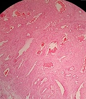
Moradi B, Izadi-Mood N, Chavoshi M, Shirali E, Yarandi F, et al. Magnetic Resonance Imaging Features of Diffuse Cavernous Hemangioma of the Uterus Misdiagnosed as a Low Grade Sarcoma: A Case Report. I J Radiol. 2020;17(3):e99416. doi: https://doi.org/10.5812/iranjradiol.99416
Rezaei O, Sharifi G, Ebrahimzadeh K, Hallajnejad M. Symptomatic Occlusion of Internal Carotid Artery Caused by Pituitary Macro Adenoma Among the Patients Without Apoplectic Signs; Case Report and Review of the Literature. Int J Cancer Manag. 2017;10(3):e5301. doi: https://doi.org/10.5812/ijcp.5301
Agrawal A, Kakani A, Vagha S, Meshram N, Paliwal A. Neurofibroma with Bone Destruction Masquerading Extracranial Meningioma. Int J Cancer Manag. 2010;3(1):e80659. doi:
Last Update: 1 month ago
Last Update: 1 month ago
Last Update: 1 month ago
Last Update: 4 days ago