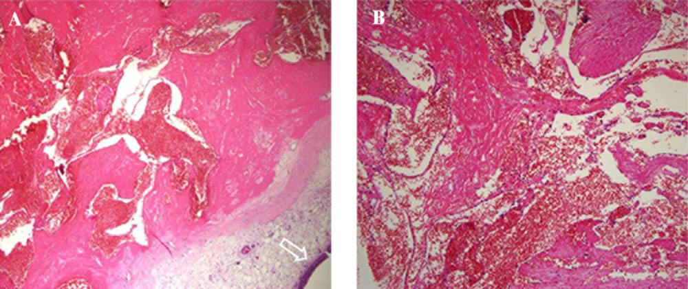
Hemangiomas of the head and neck are benign vascular lesions, and although they are common in these locations, they occur infrequently in the paranasal sinuses. Hemangiomas are divided into capillary and cavernous types, depending on the dominant vessel size seen on microscopic examination. Capillary hemangioma is the more common type and is composed of capillary-sized vessels lined with flattened epithelium. Cavernous hemangiomas are composed of large, endothelium-lined vascular spaces (
5). The common symptoms of the sinonasal hemangiomas include nasal obstruction, epistaxis, and, occasionally, a visible nasal mass (
6). Although there have been multiple clinical studies of nasal hemangioma (
2), only less than 20 cases of maxillary hemangioma have been reported (
1,
3).
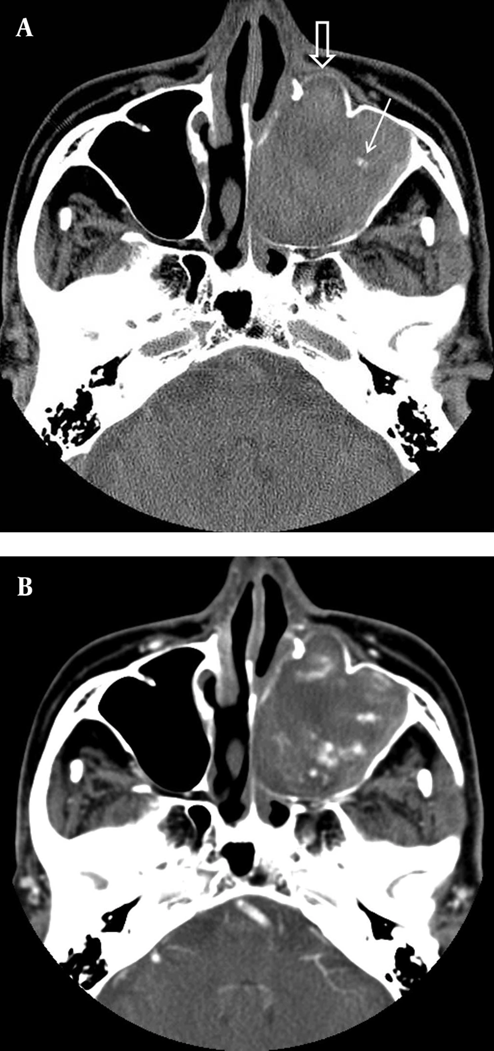
The radiographic appearance of hemangiomas has been reported in the literature. Previously reported CT features of maxillary sinus hemangioma can be summarized as depicting a highly vascularized, soft-tissue mass. Although several enhancing portions have been noted on CT scans, even larger areas did not enhance because of necrosis and hemorrhage within the tumor (
3,
4). Furthermore, hemangiomas have been reported to cause changes in adjacent bone. According to Dillon et al. capillary hemangiomas of the nasal vault had benign appearing bone changes, consisting of remodeling or expansion (
7). Kim et al. reported two cases of cavernous hemangioma that caused erosion of the wall of the maxillary sinus, nasal turbinate, and orbit (
8). According to Weiss et al. amorphous or curvilinear calcification is a non-specific finding, whereas phlebolith formation is a more specific finding of cavernous hemangioma (
5). In our case, CT also showed a soft tissue mass, which caused expansile bone remodeling of the maxillary sinus, with a small calcification within the lesion, similar to previously reported CT findings. We, therefore, assume that this CT finding offers a diagnostic clue regarding the presence of a hemangioma. According to Itoh et al. (
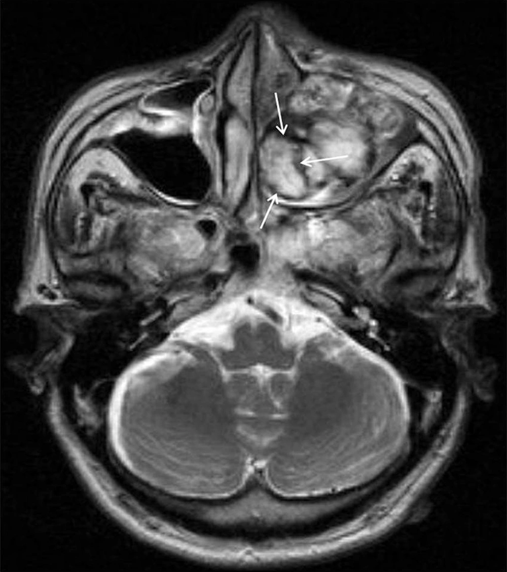
9), MR images of cavernous hemangioma reflect the signal of unclotted blood, and they have prolonged T1 and T2 relaxation times, which result in low signal intensity on T1-weighted images and high signal intensity on T2-weighted images. However, these lesions do not show any of the signal voids associated with the hypervascularity, often seen in other types of vascular malformation. Dillon et al. reported that, on T2-weighted sequences, capillary hemangioma of the nasal vault shows a hypointense rim surrounding a mass of mixed signal intensities. Pathology examination revealed that these signals were apparently correlated with areas of clotted blood (
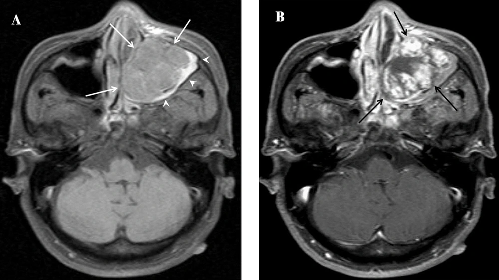
7). Our patient’s MR scan also showed a peripheral, hypointense rim, surrounding a central mass, on the T2-weighted MR sequence. Although an exact MR-pathology correlation could not be obtained, because the lesion was removed in pieces, we believe that the heterogeneous signal intensity seen on the MR images reflects the various components, such as hemorrhage, fibrosis, and vascular proliferation.
Organized hematoma (OH) is the most difficult lesion to differentiate, both clinically and radiologically, from sinonasal cavernous hemangioma. Although Yagisawa et al. suggested that hemangioma and sinonasal OH are the same pathologic entity, the fact that the vascular lumina of cavernous hemangioma are usually larger than those of OH on histologic examination still raises the question of the probability of the different nature of the two lesions (
10). In addition to OH, mucocele, fungus ball, inflammatory polyp, cholesterol granuloma, inverted papilloma, and carcinoma can present as an expansile mass (
11). Contrast material administration is useful because mucocele, fungus ball, polyp, and cholesterol granuloma are not usually enhanced. Carcinoma has a pattern of nodular enhancement (
12).
The surgical resection is the main treatment for maxillary sinus hemangioma (
4). According to tumor extent different surgical resection methods can be used for maxillary sinus hemangioma, such as lateral rhinotomy, the Caldwell-Luc operation or endoscopic sinus surgery (
4). Preoperative transarterial embolization can decrease the tumor size and reduce the risk of hemorrhage during surgery (
6).
In conclusion, sinonasal cavernous hemangioma may be mistaken for a locally aggressive neoplasm. The CT and MR images have demonstrated an expansile, well-enhanced heterogeneous mass, with bone remodeling and erosive change. Internal calcification may also be visible. Although it is a very unusual tumor, based on the imaging findings, cavernous sinus hemangioma should be considered in the differential diagnosis of a paranasal sinus tumor.