Full Text
Full text is available in PDF
An Innovative Journal in the Field of Radiology
Authors
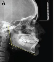
The gonial angle is one of the most important measurements required for orthodontic treatment and orthognathic surgery. It is difficult to determine the accurate measurement of each gonial angle on cephalometric radiographs because of superimposition of the left and right angles.
The aim of the present study was to determine the right and left gonial angles on panoramic radiographs and to compare them with an evaluated cephalometric sample.
A total of 80 panoramic and 80 cephalometric radiographs were obtained from 6 to 12-year-old children and the gonial angle was determined by the tangent of the inferior border of the mandible and the most distal aspect of the ascending ramus and the condyleon both panoramic and cephalometric radiographs. We used Pearsons correlation coefficient and paired t-test for comparison.
The mean gonial angle was 127.07 6.10 and 127.5 6.67 degrees on panoramic and cephalometric radiographs, respectively. There was no statistically significant difference between the measured gonial angles on panoramic and cephalometric radiographs and also no difference between the right and left (both Ps = 0.18)
The value of the gonial angle measured on panoramic radiography was the same as that measured on the routinely used cephalometric radiography.
© 2012, Author(s). This open-access article is available under the Creative Commons Attribution 4.0 (CC BY 4.0) International License (https://creativecommons.org/licenses/by/4.0/), which allows for unrestricted use, distribution, and reproduction in any medium, provided that the original work is properly cited.
Hoseini Zarch SH, Bagherpour A, Javadian Langaroodi A, Ahmadian Yazdi A, Safaei A. Evaluation of the Accuracy of Panoramic Radiography in Linear Measurements of the Jaws. I J Radiol. 2011;8(2):e93931. doi:
Vasheghani Farahani M, Mashhadi Ismaeeli R, Mirzaii-Dizgah I, Isazadeh M. Gender Determination Using Mandibular Angle Measurement in Iranian Adult Graphics in a Dental Center. Ann Mil Health Sci Res. 2021;19(3):e114608. doi: https://doi.org/10.5812/amh.114608
Nouri M, Saffarpour M, Azimi S. Longitudinal cephalometric study of mandibular growth in Qazvin children with normal occlusion. J Inflamm Dis. 2024;10(2):e155209. doi:
Toode Zaeim M, Yassaei S, Aghili H, Tefakh S. Comparison of some cephalometric indices in skeletal class III with skeletal class I patients. J Inflamm Dis. 2024;15(3):e155623. doi:
Nouri M, Rezaei B, Azimi S. Evaluating the effect of reference line on interpretation of cephalometric analysis. J Inflamm Dis. 2024;11(4):e155355. doi:
Last Update: 2 weeks ago
Last Update: 2 weeks ago
Last Update: 2 weeks ago
Last Update: 4 days ago
Last Update: 1 day ago
Last Update: 2 days ago