Cover image of the article: Real-Time Ultrasound Guided the New Standard Technique for Percutaneous Dilatational Tracheostomy (PDT)

Image Credit:Anesth Pain Med

Real-Time Ultrasound Guided the New Standard Technique for Percutaneous Dilatational Tracheostomy (PDT)

Authors

Mohsen Ziyaeifard1, Ziya Totonchi1,*
1Rajaie Cardiovascular, Medical and Research Center, Iran University of Medical Sciences , Tehran, Iran
*Corresponding Author: Corresponding author: Ziya Totonchi, Rajaie Cardiovascular, Medical and Research Center, Iran University of Medical Sciences , Tehran, Iran. Tel: +98-2123922153, Fax: +98-22663293, E-mail: Email: ziya189@yahoo.com

Anesthesiology and Pain Medicine:Vol. 5, issue 3; e24653
Published online:Jun 22, 2015
Article type:Editorial
Received:Oct 18, 2014
Accepted:Nov 22, 2014
How to Cite:Ziyaeifard M, Totonchi Z. Real-Time Ultrasound Guided the New Standard Technique for Percutaneous Dilatational Tracheostomy (PDT). Anesth Pain Med. 2015;5(3):e24653. doi: https://doi.org/10.5812/aapm.24653v2

Several studies demonstrated that percutaneous dilatational tracheostomy (PDT) is a safe, easy, low-cost and minimally-invasive bedside method with numerous known advantages more than surgical tracheostomy such as fewer adjacent tissue injury, lesser bleeding risk, wound infectivity (morbidity) and mortality (1, 2). PDT technique is usually done with the help of fiberscope guide for more security and accurateness of the practice.
Blind PDT technique has several disadvantages including failure to recognize accurate point of opening and position of tracheostomy pipe, chance of damage to nearby structures and false passageway. PDT with bronchoscopic guide can lessen these hesitations (3). However, it has its own difficulties such as disorder of ventilation - particularly in patients with elevated of Positive End-Expiratory Pressure (PEEP) - and considerable hypercarbia through risk of increased intracranial pressure. Therefore, patients with spinal cord or head damage may weakly tolerate this method (2, 3).
Recently, portable ultrasonography is used in these cases for pre-intubation evaluation of upper airway, post-intubation confirmation of endo-tracheal tube position, and extubation tracheal tube result (4). However, there is limited information and observational study data that supports the efficacy and safety of ultrasonography guided of PDT (5, 6).
Ultrasonography has numerous advantages. It is individually portable, reliable, safe, pain-free, low-cost and simply available (7). Portable and real-time ultrasound has further advantage for facilitating the clinicians toward identifying the needle passageway by presentation dislocation of tissues in front of the needle (6, 8) (Figure 1). Sharp groove in anterior tracheal wall with incursion of needle and following flattening might be more visible by this procedure (5).
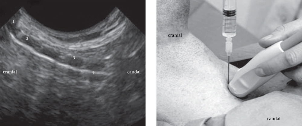
This figure shows Thyroid cartilage (1), Cricoid cartilage (2), second tracheal cartilage ring (3), and the echogenic line is consistent with an air-tissue interface at the anterior tracheal wall. The cartilages are echolucent. Location of needle insertion is between first and second cartilage ring.
Figure 1.
This figure shows Thyroid cartilage (1), Cricoid cartilage (2), second tracheal cartilage ring (3), and the echogenic line is consistent with an air-tissue interface at the anterior tracheal wall. The cartilages are echolucent. Location of needle insertion is between first and second cartilage ring.
Real- time ultrasound guided PDT preserves as a safe and cost-effective bedside method for superior use in critical ill patients, particularly in patients with morbid obesity and spinal damage (6, 7).
Michigan researchers have shown that this technique can reduce the rate of major complications. Some studies showed that the complications are 10 times less in patients who received ultrasonography guided PDT comparing with patients that had conventional procedure approach (Box 1) (9, 10).
Box 1.
Complications Associated to Conventional Procedure or Non-use of Ultrasound in PDT
Hemorrhage
Failure of airway in procedure
Early dislodgement
Infectivity
Pneumomediastinum
Pneumothorax
Failure to complete procedure
Switch to surgical tracheostomy /require for change of tracheostomy
Posterior wall damage
Tracheomalacia, tracheal stenosis
Tracheo-cutaneous, tracheo-innominate, and tracheo-esophageal fistula
Nasir Bhatti, MD (director of adult Tracheostomy and Airway Service in the Hospital affiliated to Johns Hopkins University, Baltimore, USA) supported the use of ultrasound guided PDT by non-surgeons; including pulmonologists and intensivists performing arts of PDTs (10).
Ultrasound guided PDT facilitate clinicians to preserve adjacent structures r; demonstrate the guide wire location and the correct level of cannula. It also enables clinicians to assess the space between the anterior tracheal wall and the skin, to evaluate extends length of tube, and suitably put the endo-tracheal tube in the correct position. However, randomized clinical trials are required to evaluate ultrasound guided PDT as if can be a standard technique, similar to the previous method which is used in central venous catheter placement (10-12).


Acknowledgments

References

  • 1.
    Delaney A, Bagshaw SM, Nalos M. Percutaneous dilatational tracheostomy versus surgical tracheostomy in critically ill patients: a systematic review and meta-analysis. Crit Care. 2006;10(2):R55. [PubMed ID: 16606435]. https://doi.org/10.1186/cc4887.
  • 2.
    Youssef TF, Ahmed MR, Saber A. Percutaneous dilatational versus conventional surgical tracheostomy in intensive care patients. N Am J Med Sci. 2011;3(11):508-12. [PubMed ID: 22361497]. https://doi.org/10.4297/najms.2011.3508.
  • 3.
    Rajajee V, Fletcher JJ, Rochlen LR, Jacobs TL. Real-time ultrasound-guided percutaneous dilatational tracheostomy: a feasibility study. Crit Care. 2011;15(1):R67. [PubMed ID: 21342494]. https://doi.org/10.1186/cc10047.
  • 4.
    Rodriguez SJ, Esteves LE. Real-time ultrasound-guided percutaneous dilatational tracheostomy. Crit Care. 2011;15(5):443. [PubMed ID: 21996442]. https://doi.org/10.1186/cc10344.
  • 5.
    Chacko J, Nikahat J, Gagan B, Umesh K, Ramanathan M. Real-time ultrasound-guided percutaneous dilatational tracheostomy. Intensive Care Med. 2012;38(5):920-1. [PubMed ID: 22349428]. https://doi.org/10.1007/s00134-012-2514-3.
  • 6.
    Joelsons D, Ho YL, Park M. Ultrasound-guided percutaneous tracheostomy: a feasible alternative for tetanus patients. Crit Care. 2014;18(3):441. [PubMed ID: 25043740]. https://doi.org/10.1186/cc13946.
  • 7.
    Mehta Y. Percutaneous dilatational tracheostomy: Guided well with real-time ultrasound. Indian J Crit Care Med. 2013;17(6):335-6. [PubMed ID: 24501483]. https://doi.org/10.4103/0972-5229.123434.
  • 8.
    Kleine-Brueggeney M, Greif R, Ross S, Eichenberger U, Moriggl B, Arnold A, et al. Ultrasound-guided percutaneous tracheal puncture: a computer-tomographic controlled study in cadavers. Br J Anaesth. 2011;106(5):738-42. [PubMed ID: 21414983]. https://doi.org/10.1093/bja/aer026.
  • 9.
    Mitra S, Kapoor D, Srivastava M, Sandhu H. Real-time ultrasound guided percutaneous dilatational tracheostomy in critically ill patients: A step towards safety!. Indian J Crit Care Med. 2013;17(6):367-9. [PubMed ID: 24501489]. https://doi.org/10.4103/0972-5229.123449.
  • 10.
    Kate OR. Ultrasound: The New Standard of Care for Percutaneous Dilatational Tracheostomy? J Clin Anesthesiol. 2014;40(3).
  • 11.
    Dinh VA, Farshidpanah S, Lu S, Stokes P, Chrissian A, Shah H, et al. Real-time sonographically guided percutaneous dilatational tracheostomy using a long-axis approach compared to the landmark technique. J Ultrasound Med. 2014;33(8):1407-15. [PubMed ID: 25063406]. https://doi.org/10.7863/ultra.33.8.1407.
  • 12.
    Ziyaeifard M, Azarfarin R. Ultrasound is a new and reliable technique for central venous cannulation. Res Cardiovasc Med. 2014;3(3). eee17328. [PubMed ID: 25478542]. https://doi.org/10.5812/cardiovascmed.17328.

Copyright

Copyright © 2015, Iranian Society of Regional Anesthesia and Pain Medicine (ISRAPM). This is an open-access article distributed under the terms of the Creative Commons Attribution-NonCommercial 4.0 International License (http://creativecommons.org/licenses/by-nc/4.0/) which permits copy and redistribute the material just in noncommercial usages, provided the original work is properly cited.

Similar Articles

1
Sep
2018
Comparison of the frequency of short-term complications of percutaneous dilatation tracheostomy and versus surgical tracheostomy: A clinical trial

Comparison of the frequency of short-term complications of percutaneous dilatation tracheostomy and versus surgical tracheostomy: A clinical trial

Gholamreza Mohajeri,
Vahid Goharian,
Mohsen Kolahdouzan

Mohajeri G, Goharian V, Kolahdouzan M. Comparison of the frequency of short-term complications of percutaneous dilatation tracheostomy and versus surgical tracheostomy: A clinical trial. koomesh. 2018;20(3):e152991. doi:

12
Nov
2017
Diagnostic Value of Sonography for Confirmation of Endotracheal Intubation in the Emergency Department

Diagnostic Value of Sonography for Confirmation of Endotracheal Intubation in the Emergency Department

Farzad Rahmani,
Zahra Parsian,
Kavous Shahsavarinia,
Mahboob Pouraghaei,
Sohrab Negargar,
Robab Mehdizadeh Esfanjani
,et al.

Rahmani F, Parsian Z, Shahsavarinia K, Pouraghaei M, Negargar S, et al. Diagnostic Value of Sonography for Confirmation of Endotracheal Intubation in the Emergency Department. Anesth Pain Med. 2017;7(6):e58350. doi: https://doi.org/10.5812/aapm.58350

21
Nov
2022
Role of Lung Ultrasound in Confirmation of Double Lumen Endotracheal Tube Placement for Thoracic Surgeries: A Prospective Diagnostic Accuracy Study

Role of Lung Ultrasound in Confirmation of Double Lumen Endotracheal Tube Placement for Thoracic Surgeries: A Prospective Diagnostic Accuracy Study

Walaa Y Elsabeeny,
Mostafa A Ibrahim,
Sayed M Abed,
Nahla N Shehab

Elsabeeny WY, Ibrahim MA, Abed SM, Shehab NN. Role of Lung Ultrasound in Confirmation of Double Lumen Endotracheal Tube Placement for Thoracic Surgeries: A Prospective Diagnostic Accuracy Study. Anesth Pain Med. 2022;12(5):e132312. doi: https://doi.org/10.5812/aapm-132312

10
Mar
2014
On Demand Urethral Dilatation Versus Intermittent Urethral Dilatation: Results and Complications in Women With Urethral Stricture

On Demand Urethral Dilatation Versus Intermittent Urethral Dilatation: Results and Complications in Women With Urethral Stricture

Fatemeh Heidari,
Shahin Abbaszadeh,
Alireza Ghadian,
Farahnaz Tehrani Kia

Heidari F, Abbaszadeh S, Ghadian A, Tehrani Kia F. On Demand Urethral Dilatation Versus Intermittent Urethral Dilatation: Results and Complications in Women With Urethral Stricture. Nephro-Urol Mon. 2014;6(2):e15212. doi: https://doi.org/10.5812/numonthly.15212

13
Mar
2022
Diagnostic Accuracy of Suprasternal Versus Subxiphoid Ultrasonography for Endotracheal Intubation

Diagnostic Accuracy of Suprasternal Versus Subxiphoid Ultrasonography for Endotracheal Intubation

Seyed Mohammad Mousavi,
Maryam Sayyari Doughabadi,
Seyed Ali Alamdaran,
Sayyed Majid Sadrzadeh,
Hosein Zakeri,
Elnaz VafadarMoradi

Mousavi SM, Sayyari Doughabadi M, Alamdaran SA, Sadrzadeh SM, Zakeri H, et al. Diagnostic Accuracy of Suprasternal Versus Subxiphoid Ultrasonography for Endotracheal Intubation. Anesth Pain Med. 2022;12(1):e118592. doi: https://doi.org/10.5812/aapm.118592

More by these authors

Mohsen ZiyaeifardPubMedScholar
Ziya TotonchiPubMedScholar
Share
Cited by
Metrics