Cover image of the article: Cardiac Dysfunction in a Child with Severe Multisystem Inflammatory Syndrome

Image Credit:Arch Pediatr Infect Dis

Cardiac Dysfunction in a Child with Severe Multisystem Inflammatory Syndrome

Authors

Gabriela Marengone AltizaniGabriela Marengone Altizani ORCID1,*, Karin Barszcz1, Laura Cardoso Brentini1, Karina Amelia Morikawa1, Kelly Luisa Cintra2, Paulo Henrique Manso3
1Department of Pediatrics, Ribeirao Preto Medical School, University of Sao Paulo, Ribeirao Preto, Brazil
2Department of Pediatric Cardiology, Ribeirao Preto Medical School, University of Sao Paulo, Ribeirao Preto, Brazil
3Director of Pediatric Cardiology Section, Ribeirao Preto Medical School, University of Sao Paulo, Ribeirao Preto, Brazil
*Corresponding Author: Department of Pediatrics, University of Sao Paulo, 3900, Ribeirão Preto, Brazil. Tel: +55-36021000, Email: gabialtizani@gmail.com

Archives of Pediatric Infectious Diseases:Vol. 10, issue Suppl; e112446
Published online:Jul 28, 2021
Article type:Case Report
Received:Dec 26, 2020
Accepted:May 30, 2021
How to Cite:Altizani GM, Barszcz K, Brentini LC, Morikawa KA, Cintra KL, et al. Cardiac Dysfunction in a Child with Severe Multisystem Inflammatory Syndrome. Arch Pediatr Infect Dis. 2022;10(Suppl):e112446. doi: https://doi.org/10.5812/pedinfect.112446

Abstract

Introduction:

Albeit most children infected with SARS-CoV-2 are asymptomatic or have mild respiratory symptoms, similar cases of Kawasaki disease and toxic shock syndrome increased at the end of April 2020 worldwide. Systemic inflammatory involvement characterizes these cases and is part of the spectrum of a SARS-CoV-2-related syndromes called multisystem inflammatory syndrome (MIS-C).

Case Presentation:

We report the case of a child with severe cardiovascular impairment due to MIS-C who developed cardiogenic shock secondary to acute myocarditis. The pathophysiology of myocardial outcomes caused by MIS-C remains unclear.

Conclusions:

We intend to contribute to new prospective studies on the early diagnosis, clinical management, and follow-up of patients with severe cardiovascular impairment secondary to infection with SARS-CoV-2.

Highlights

1. Introduction

The 2020 SARS-CoV-2 pandemic has evidenced that most infected children are asymptomatic or have mild upper airway symptoms; nevertheless, it is known that children have a significant impact on the virus spread (1). By April 2020, several countries had identified an increased prevalence of children with conditions such as Kawasaki disease (DK) and toxic shock syndrome (TSS), which were later learned to be part of the spectrum of a new disease related to a previous infection with coronavirus. This new disease was named multisystem inflammatory syndrome in children (MIS-C) by the Centers for Disease Control and Prevention (CDC) and the World Health Organization (WHO) (2, 3). Severe MIS-C cases present with acute myocarditis, coronary changes, arrhythmias, and cardiogenic shock (4, 5). However, the pathophysiology of myocardial dysfunctions remains unclear (6).

2. Case Presentation

A previously healthy three-year-old boy had close contact with his grandmother, who was positive for SARS-Cov-2. Twelve days later, he presented with a sore throat and running nose. A week later, he presented with fever, rash in the upper body, and odynophagia, so amoxicillin and ibuprofen were started after medical evaluation. The following day, the rash spread to the trunk and lower limbs, the fever persisted, and the patient started vomiting, so endovenous hydration was initiated. The patient was sent home, but four days later, he entered the emergency room with dehydration, mental confusion, hypotension (arterial blood pressure 78 × 45 mmHg), tachycardia (heart rate = 145bpm), tachypnea (respiratory rate of 46 bpm), generalized edema, and hepatomegaly. He also presented non-purulent conjunctival hyperemia and pruritic erythematous-violet rash with a rough appearance on the face, upper limbs, lower limbs, and trunk; palms and soles were spared. In addition, the patient had mild laminar desquamation in the lower abdominal region. On admission, PCR-RT for SARS-CoV-2 was collected, but the result was negative. However, the chemiluminescence immunoassay (CLIA) technique for COVID-19 was positive on the following day.
Laboratory tests showed metabolic acidosis, high urea and creatinine levels, and hypokalemia; white blood cell count revealed leukocytosis. The patient presented the clinical signs of cardiovascular dysfunction associated with low output syndrome (i.e., hypotension, anasarca, hepatomegaly, and mental confusion) after catarrhal prodromes, and cardiac auscultation identified a third heart sound and “gallop rhythm”, hence the foremost diagnostic hypothesis was acute myocarditis. Therefore, the markers of myocardial injury troponin, CK-MB, CPK, and NT-ProBNP (Table 1), were collected, revealing values significantly above baseline values.
Table 1.
Laboratory Markers of Myocardial Injury
TestsReference valueAdmissionDischarge
CK-MB< 5 U/L230.51
CPK 24 - 195 U/L165-
Troponin< 19 ng/dL84.61.8
NT-Pro-BNP< 125 ug/mL> 25000225
Abbreviations: CK-MB, creatine kinase myocardial band; CPK, creatine phosphokinase.
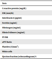
The initial electrocardiogram showed sinus tachycardia, and the initial echocardiogram evidenced reduced ejection fraction (EF 50%) due to global hypokinesia and normal coronary arteries. Because the diagnosis of MIS-C was suspected, other laboratory tests were collected (Table 2).
Table 2.
Laboratory Exams of the Patient on Admission and Discharge
TestsReference valueAdmissionDischarge
C-reactive protein (mg/dL)< 131.07< 0.4
ESR (mm/1h)< 10 mm/1stH4143
Interleucin 6 (pg/mL)< 5.942.47.4
Ferritin (ng/mL)22 - 322586.5
Fibrinogen (mg/mL)200 - 400312.52
Fibrin D dimers (mg/mL)< 1.252.531.35
PT INR1.041.0
aPTT Ratio0.860.9
Platelets (U/mm3)100000878000
White cells1790020500
Ejection fraction (echocardiogram) %> 605070
Abbreviations: ESR, erythrocyte sedimentation rate; PT, prothrombin time; aPTT, activated partial thromboplastin time; CK MB, creatine phosphokinase MB fraction.
The initial intensive care consisted of milrinone (up to 0.3 mcg/kg/min) and epinephrine (up to 0.3 mcg/kg/min) for 48 h. A broad-spectrum antibiotic treatment (100 mg/kg/day ceftriaxone and 40 mg/kg/day vancomycin) was also started due to the suspicion of toxic shock syndrome. On the second day of hospitalization, specific therapy for MIS-C, consisting of 2 g/kg human immunoglobulin and 80 mg/kg/day acetylsalicylic acid, was administered. Prophylactic enoxaparin was also added (1 mg/kg/day), and methylprednisolone pulse (27 mg/kg/day) was started according to the institutional protocol.
The patient stayed in the intensive care unit for seven days and was discharged on the following day with low-dose aspirin, corticosteroid therapy (2 mg/kg/day prednisolone), and enoxaparin.

3. Discussion

The patient presented with fever, laboratory evidence of inflammation (high interleukin 6, ESR, and C-reactive protein levels), and increased D-dimers and leukocytosis with neutrophilia. Myocarditis leading to cardiovascular system dysfunction was also evident.
The golden standard exam for the definitive diagnosis of myocarditis in pediatric patients is through myocardial biopsy, pointing inflammation findings through histological or immunohistological lesions (7). However, this exam is invasive and has high variability in sample interpretation. Such an examination is not performed in most cases, allowing a combination of clinical and imaging criteria to be valid for the diagnosis (7).
Sagar et al. (8) proposed a three-level clinical classification for myocarditis diagnosis based on levels of certainty: (1) definitive myocarditis; (2) probable acute myocarditis; and (3) possible subclinical myocarditis. For diagnosis, patients may have all or just some of the following findings: (1) elevation of cardiac biomarkers; (2) echocardiographic evidence of ventricular dysfunction without underlying structural defects, signs, and symptoms of ventricular dysfunction, electrocardiographic changes; (3) changes in myocardial magnetic resonance imaging (MRI); and (4) prodromal illness in the last two weeks (8).
As previously, our patient met the criteria for probable acute myocarditis. Moreover, the patient had positive serology for SARS-CoV-2, meeting the criteria for severe MIS-C according to the CDC and the WHO (9).
Our patient was younger than most of the other reported cases (5, 10) and presented myocardial dysfunction after infection with SARS-CoV-2. This manifestation has been described in many children diagnosed with MIS-C associated with significantly increased troponin and NT-ProBNP. As already acknowledged by Licciardi et al., infection with SARS-CoV-2 and the pro-inflammatory trend can trigger cardiac injury, post-myocardial injury, and vasculitis (11). However, the specific mechanism underlying MIS-C cardiac involvement has not been fully elucidated (2).
In a study about cardiogenic shock and hyperinflammatory syndrome in a group of young adults, the first manifestations of SARS-CoV-2 were biventricular failure and vasoplegia, which evolved into circulatory shock and multisystemic involvement, which may be similar to the involvement of our patient (12). In that study, the myocardial injury did not result from direct viral injury. The presence of high antibody titers suggested previous exposure, a few weeks before hospital admission (12).
In the present case, the patient was hypotensive and tachycardic when he was admitted to the emergency unit. Although the first echocardiogram was performed on vasoactive drugs, the left ventricle (LV) systolic performance was at the lower limit of normality. Grimaud et al. reported a case series of 20 patients with MIS-C who developed acute myocarditis. The prevalence of hypotension and tachycardia was 20%, median LV EF was 35% (25 - 55%), and LV function improved before discharge (10).
Some elements of the emerging pediatric disease MIS-C resemble those of Kawasaki's disease (KD) (10). In the case described here, the patient also met the criteria of atypical KD because he had a fever lasting over five days, bilateral not exudative conjunctival hyperemia, and polymorphic rash, corroborated by the laboratory changes (13).
Although KD can affect the myocardium, comparative articles have reported that myocardial involvement prevails when KD is related to SARS-CoV-2. This is corroborated by a study by Pouletty et al., who compared a historical cohort of KD with a cohort of KD associated with infection by SARS-CoV-2 in Paris and verified echocardiographic findings of myocarditis in 1 and 44% of the patients, respectively. On the other hand, coronary involvement was similar regardless of association with the virus, and coronary dilation occurred in 19% of the patients in both cohorts. The clinical characteristics of the two syndromes overlap probably because they have similar pathophysiological mechanisms (14). In both cases, an immune trigger is accelerated, causing an immune-mediated lesion in the heart and coronary arteries with different severity proportions. The overlap in the characteristics of the two syndromes may result from these similar pathophysiological mechanisms (15, 16). However, studies indicated that coronary changes are more likely to occur in KD, while cardiac dysfunctions are more frequent in patients with MIS-C. Remarkably, our patient did not present coronary enlargement.
Evolution to myocarditis and cardiogenic shock are possible outcomes in severe cases of MIS-C, demonstrating the severity of cardiovascular involvement. Pediatricians in emergency rooms should suspect this condition in children with cardiac dysfunction.
Although this study has shown cardiac involvement leading to cardiogenic shock in a patient with MIS-C under the average age reported in other studies, the mechanism of injury has not been fully elucidated. Thus, future studies are needed to document this mechanism and to assist in the recognition and treatment of patients with similar conditions.
Footnotes

  • Authors' Contribution: Study concept and design, K.L.C and G.M.A; Analysis and interpretation of the data, L.C.B., K.B, and K.A.M; Drafting of the manuscript, G.M.A, L.C.B., K.B, and K.A.M; Critical revision of the manuscript for important intellectual content, P.H.M and K.L.C

  • Conflict of Interests: The authors declare no conflicts of interest.

  • Ethical Approval: This article was approved by the local ethical considerations code No. 11286.

  • Funding/Support: We received no financial support for this work.

  • Informed Consent: Written informed consent was obtained from the patient’s parents for publication of the current case report.

References

  • 1.
    Rajapakse N, Dixit D. Human and novel coronavirus infections in children: A review. Paediatr Int Child Health. 2021;41(1):36-55. [PubMed ID: 32584199]. https://doi.org/10.1080/20469047.2020.1781356.
  • 2.
    Belot A, Antona D, Renolleau S, Javouhey E, Hentgen V, Angoulvant F, et al. SARS-CoV-2-related paediatric inflammatory multisystem syndrome, an epidemiological study, France, 1 March to 17 May 2020. Euro Surveill. 2020;25(22). [PubMed ID: 32524957]. [PubMed Central ID: PMC7336112]. https://doi.org/10.2807/1560-7917.ES.2020.25.22.2001010.
  • 3.
    Singh-Grewal D, Lucas R, McCarthy K, Cheng AC, Wood N, Ostring G, et al. Update on the COVID-19-associated inflammatory syndrome in children and adolescents; paediatric inflammatory multisystem syndrome-temporally associated with SARS-CoV-2. J Paediatr Child Health. 2020;56(8):1173-7. [PubMed ID: 32735721]. [PubMed Central ID: PMC7436879]. https://doi.org/10.1111/jpc.15049.
  • 4.
    Sperotto F, Friedman KG, Son MBF, VanderPluym CJ, Newburger JW, Dionne A. Cardiac manifestations in SARS-CoV-2-associated multisystem inflammatory syndrome in children: A comprehensive review and proposed clinical approach. Eur J Pediatr. 2021;180(2):307-22. [PubMed ID: 32803422]. [PubMed Central ID: PMC7429125]. https://doi.org/10.1007/s00431-020-03766-6.
  • 5.
    Belhadjer Z, Méot M, Bajolle F, Khraiche D, Legendre A, Abakka S, et al. Acute heart failure in multisystem inflammatory syndrome in children in the context of global SARS-CoV-2 pandemic. Circulation. 2020;142(5):429-36. [PubMed ID: 32418446]. https://doi.org/10.1161/CIRCULATIONAHA.120.048360.
  • 6.
    Whittaker E, Bamford A, Kenny J, Kaforou M, Jones CE, Shah P, et al. Clinical characteristics of 58 children with a pediatric inflammatory multisystem syndrome temporally associated with SARS-CoV-2. JAMA. 2020;324(3):259-69. [PubMed ID: 32511692]. [PubMed Central ID: PMC7281356]. https://doi.org/10.1001/jama.2020.10369.
  • 7.
    Canter CE, Simpson KE. Diagnosis and treatment of myocarditis in children in the current era. Circulation. 2014;129(1):115-28. [PubMed ID: 24396015]. https://doi.org/10.1161/CIRCULATIONAHA.113.001372.
  • 8.
    Sagar S, Liu PP, Cooper L. Myocarditis. Lancet. 2012;379(9817):738-47. [PubMed ID: 22185868]. [PubMed Central ID: PMC5814111]. https://doi.org/10.1016/S0140-6736(11)60648-X.
  • 9.
    CDC. Reporting multisystem inflammatory syndrome in children ( MIS-C ). USA: Centers for Disease Control and Prevention; 2020.
  • 10.
    Grimaud M, Starck J, Levy M, Marais C, Chareyre J, Khraiche D, et al. Acute myocarditis and multisystem inflammatory emerging disease following SARS-CoV-2 infection in critically ill children. Ann Intensive Care. 2020;10(1):69. [PubMed ID: 32488505]. [PubMed Central ID: PMC7266128]. https://doi.org/10.1186/s13613-020-00690-8.
  • 11.
    Licciardi F, Pruccoli G, Denina M, Parodi E, Taglietto M, Rosati S, et al. SARS-CoV-2-induced kawasaki-like hyperinflammatory syndrome: A novel COVID phenotype in children. Pediatrics. 2020;146(2). [PubMed ID: 32439816]. https://doi.org/10.1542/peds.2020-1711.
  • 12.
    Chau VQ, Giustino G, Mahmood K, Oliveros E, Neibart E, Oloomi M, et al. Cardiogenic shock and hyperinflammatory syndrome in young males with COVID-19. Circ Heart Fail. 2020;13(10). e007485. [PubMed ID: 32844662]. https://doi.org/10.1161/CIRCHEARTFAILURE.120.007485.
  • 13.
    Sotelo-Cruz N. [Atypical and incomplete Kawasaki disease in pediatric age]. Bol Med Hosp Infant Mex. 2016;73(3):147-8. Spanish. [PubMed ID: 29421201]. [PubMed Central ID: PMC7128410]. https://doi.org/10.1016/j.bmhimx.2016.05.001.
  • 14.
    Pouletty M, Borocco C, Ouldali N, Caseris M, Basmaci R, Lachaume N, et al. Paediatric multisystem inflammatory syndrome temporally associated with SARS-CoV-2 mimicking Kawasaki disease (Kawa-COVID-19): A multicentre cohort. Ann Rheum Dis. 2020;79(8):999-1006. [PubMed ID: 32527868]. [PubMed Central ID: PMC7299653]. https://doi.org/10.1136/annrheumdis-2020-217960.
  • 15.
    Jiang L, Tang K, Levin M, Irfan O, Morris SK, Wilson K, et al. COVID-19 and multisystem inflammatory syndrome in children and adolescents. Lancet Infect Dis. 2020;20(11):e276-88. [PubMed ID: 32818434]. [PubMed Central ID: PMC7431129]. https://doi.org/10.1016/S1473-3099(20)30651-4.
  • 16.
    Nakra NA, Blumberg DA, Herrera-Guerra A, Lakshminrusimha S. Multi-system inflammatory syndrome in children (MIS-C) following SARS-CoV-2 infection: Review of clinical presentation, hypothetical pathogenesis, and proposed management. Children (Basel). 2020;7(7). [PubMed ID: 32630212]. [PubMed Central ID: PMC7401880]. https://doi.org/10.3390/children7070069.

Copyright

Copyright © 2021, Author(s). This is an open-access article distributed under the terms of the Creative Commons Attribution-NonCommercial 4.0 International License (http://creativecommons.org/licenses/by-nc/4.0/) which permits copy and redistribute the material just in noncommercial usages, provided the original work is properly cited.

Similar Articles

9
Apr
2023
Report of the Association Between Multisystem Inflammatory Syndrome and Severe Myocarditis

Report of the Association Between Multisystem Inflammatory Syndrome and Severe Myocarditis

Elmira Hajiesmaeil Memar,
Aliakbar Zeinaloo,
Mohammad Reza Mirzaaghayan,
Mojtaba Gorji,
Azin Ghamari,
Mahya Ghahremanloo
,et al.

Hajiesmaeil Memar E, Zeinaloo A, Mirzaaghayan MR, Gorji M, Ghamari A, et al. Report of the Association Between Multisystem Inflammatory Syndrome and Severe Myocarditis. Inn J Pediatr. 2023;33(3):e122272. doi: https://doi.org/10.5812/ijp-122272

16
Jan
2021
Cardiac Manifestations of Multisystem Inflammatory Syndrome in Children (MIS-C) Associated with SARS-CoV-2 Infection

Cardiac Manifestations of Multisystem Inflammatory Syndrome in Children (MIS-C) Associated with SARS-CoV-2 Infection

Alireza Ghodsi,
Elnaz Mahmoudabadi,
Sara Ghahremani,
Abdolreza Malek

Ghodsi A, Mahmoudabadi E, Ghahremani S, Malek A. Cardiac Manifestations of Multisystem Inflammatory Syndrome in Children (MIS-C) Associated with SARS-CoV-2 Infection. Arch Pediatr Infect Dis. 2021;9(4):e109915. doi: https://doi.org/10.5812/pedinfect.109915

19
Aug
2022
Clinical and Laboratory Characteristics of the Multisystem Inflammatory Syndrome in Children: A Case Series of 75 Patients

Clinical and Laboratory Characteristics of the Multisystem Inflammatory Syndrome in Children: A Case Series of 75 Patients

Shabnam Hajiani Ghotbabadi,
Maryam Mollaie,
Seyedeh Sedigheh Hamzavi,
Anahita Sanaei Dashti

Hajiani Ghotbabadi S, Mollaie M, Hamzavi SS, Sanaei Dashti A. Clinical and Laboratory Characteristics of the Multisystem Inflammatory Syndrome in Children: A Case Series of 75 Patients. Arch Pediatr Infect Dis. 2022;10(4):e120863. doi: https://doi.org/10.5812/pedinfect-120863

13
Jul
2024
Comparison of Clinical Features and Outcomes of Shocks in Multisystem Inflammatory Syndrome in Children Associated with COVID-19 (MIS-C), Septic Shock, and Cardiogenic Shock

Comparison of Clinical Features and Outcomes of Shocks in Multisystem Inflammatory Syndrome in Children Associated with COVID-19 (MIS-C), Septic Shock, and Cardiogenic Shock

Kantimas Sitthikool,
Pimchanok Junsawat

Sitthikool K, Junsawat P. Comparison of Clinical Features and Outcomes of Shocks in Multisystem Inflammatory Syndrome in Children Associated with COVID-19 (MIS-C), Septic Shock, and Cardiogenic Shock. Inn J Pediatr. 2024;34(4):e145347. doi: https://doi.org/10.5812/ijp-145347

30
Jun
2025
Cardiac Manifestations of Multisystem Inflammatory Syndrome in Iranian Children (MIS-C) Associated with COVID-19; Incidence and One-Year Follow-up

Cardiac Manifestations of Multisystem Inflammatory Syndrome in Iranian Children (MIS-C) Associated with COVID-19; Incidence and One-Year Follow-up

Mina Farshidgohar,
Manoochehr Mahram

Farshidgohar M, Mahram M. Cardiac Manifestations of Multisystem Inflammatory Syndrome in Iranian Children (MIS-C) Associated with COVID-19; Incidence and One-Year Follow-up. J Inflamm Dis. 2025;29(2):e157779. doi: https://doi.org/10.69107/jid-157779

More by these authors

Gabriela Marengone AltizaniPubMedScholar
Karin BarszczPubMedScholar
Laura Cardoso BrentiniPubMedScholar
Karina Amelia MorikawaPubMedScholar
Kelly Luisa CintraPubMedScholar
Paulo Henrique MansoPubMedScholar
Share
Cited by
Metrics